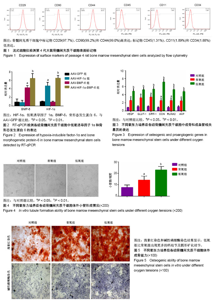
| [1] Bougioukli S, Sugiyama O, Pannell W, et al. Gene Therapy for Bone Repair Using Human Cells: Superior Osteogenic Potential of Bone Morphogenetic Protein 2-Transduced Mesenchymal Stem Cells Derived from Adipose Tissue Compared to Bone Marrow. Hum Gene Ther. 2018;29(4): 507-519.[2] Bornes TD, Adesida AB, Jomha NM. Articular Cartilage Repair with Mesenchymal Stem Cells After Chondrogenic Priming: A Pilot Study. Tissue Eng Part A. 2018;24(9-10): 761-774.[3] Wang W, Wang Y, Deng G, et al. Transplantation of Hypoxic-Preconditioned Bone Mesenchymal Stem Cells Retards Intervertebral Disc Degeneration via Enhancing Implanted Cell Survival and Migration in Rats. Stem Cells Int. 2018;2018:7564159.[4] Böhrnsen F, Schliephake H. Supportive angiogenic and osteogenic differentiation of mesenchymal stromal cells and endothelial cells in monolayer and co-cultures. Int J Oral Sci. 2016;8(4):223-230.[5] Liao H, Zhong Z, Liu Z, et al. Bone mesenchymal stem cells co-expressing VEGF and BMP-6 genes to combat avascular necrosis of the femoral head. Exp Ther Med. 2018;15(1): 954-962.[6] Muinos-López E, Ripalda-Cemboráin P, López-Martínez T, et al. Hypoxia and Reactive Oxygen Species Homeostasis in Mesenchymal Progenitor Cells Define a Molecular Mechanism for Fracture Nonunion. Stem Cells. 2016;34(9): 2342-2353.[7] Cox TR, Erler JT, Rumney RMH. Established Models and New Paradigms for Hypoxia-Driven Cancer-Associated Bone Disease. Calcif Tissue Int. 2018;102(2):163-173.[8] Stegen S, Deprez S, Eelen G, et al. Adequate hypoxia inducible factor 1α signaling is indispensable for bone regeneration. Bone. 2016;87:176-186.[9] Semenza GL. Hydroxylation of HIF-1: oxygen sensing at the molecular level. Physiology (Bethesda). 2004;19:176-182.[10] Büning H, Perabo L, Coutelle O, et al. Recent developments in adeno-associated virus vector technology. J Gene Med. 2008;10(7):717-733.[11] Luo G, Huang Y, Gu F. rhBMP2-loaded calcium phosphate cements combined with allogenic bone marrow mesenchymal stem cells for bone formation. Biomed Pharmacother. 2017; 92:536-543.[12] Westhauser F, Höllig M, Reible B, et al. Bone formation of human mesenchymal stem cells harvested from reaming debris is stimulated by low-dose bone morphogenetic protein-7 application in vivo. J Orthop. 2016;13(4):404-408.[13] Roth S, Dreixler JC, Mathew B, et al. Hypoxic-Preconditioned Bone Marrow Stem Cell Medium Significantly Improves Outcome After Retinal Ischemia in Rats. Invest Ophthalmol Vis Sci. 2016;57(7):3522-3532.[14] Sharma S, Sapkota D, Xue Y, et al. Delivery of VEGFA in bone marrow stromal cells seeded in copolymer scaffold enhances angiogenesis, but is inadequate for osteogenesis as compared with the dual delivery of VEGFA and BMP2 in a subcutaneous mouse model. Stem Cell Res Ther. 2018;9(1): 23.[15] Ramasamy SK, Kusumbe AP, Wang L, et al. Endothelial Notch activity promotes angiogenesis and osteogenesis in bone. Nature. 2014;507(7492):376-380.[16] Gómez-Puerto MC, Verhagen LP, Braat AK, et al. Activation of autophagy by FOXO3 regulates redox homeostasis during osteogenic differentiation. Autophagy. 2016;12(10): 1804-1816.[17] Liu Y, Yang X, Maureira P, et al. Permanently Hypoxic Cell Culture Yields Rat Bone Marrow Mesenchymal Cells with Higher Therapeutic Potential in the Treatment of Chronic Myocardial Infarction. Cell Physiol Biochem. 2017;44(3): 1064-1077.[18] Ciapetti G, Granchi D, Fotia C, et al. Effects of hypoxia on osteogenic differentiation of mesenchymal stromal cells used as a cell therapy for avascular necrosis of the femoral head. Cytotherapy. 2016;18(9):1087-1099.[19] Maes C, Carmeliet G, Schipani E. Hypoxia-driven pathways in bone development, regeneration and disease. Nat Rev Rheumatol. 2012;8(6):358-366.[20] Wu WJ, Zhang XK, Zheng XF, et al. SHH-dependent knockout of HIF-1 alpha accelerates the degenerative process in mouse intervertebral disc. Int J Immunopathol Pharmacol. 2013;26(3):601-609.[21] Maxwell P, Salnikow K. HIF-1: an oxygen and metal responsive transcription factor. Cancer Biol Ther. 2004;3(1): 29-35.[22] 李广周,吴伟.低氧环境对骨代谢影响的研究与进展[J].中国组织工程研究,2016,20(33):4963-4969. [23] Fan L, Li J, Yu Z, et al. The hypoxia-inducible factor pathway, prolyl hydroxylase domain protein inhibitors, and their roles in bone repair and regeneration. Biomed Res Int. 2014;2014: 239356.[24] Ma C, Wei Q, Cao B, et al. A multifunctional bioactive material that stimulates osteogenesis and promotes the vascularization bone marrow stem cells and their resistance to bacterial infection. PLoS One. 2017;12(3):e0172499.[25] Park HS, Kim JH, Sun BK, et al. Hypoxia induces glucose uptake and metabolism of adipose?derived stem cells. Mol Med Rep. 2016;14(5):4706-4714.[26] Yuan HF, Zhai C, Yan XL, et al. SIRT1 is required for long-term growth of human mesenchymal stem cells. J Mol Med (Berl). 2012;90(4):389-400.[27] Joo HY, Yun M, Jeong J, et al. SIRT1 deacetylates and stabilizes hypoxia-inducible factor-1α (HIF-1α) via direct interactions during hypoxia. Biochem Biophys Res Commun. 2015;462(4):294-300.[28] Xu Y, Wang S, Tang C, et al. Upregulation of long non-coding RNA HIF 1α-anti-sense 1 induced by transforming growth factor-β-mediated targeting of sirtuin 1 promotes osteoblastic differentiation of human bone marrow stromal cells. Mol Med Rep. 2015;12(5):7233-7238.[29] Chen MC, Hsu WL, Hwang PA, et al. Low Molecular Weight Fucoidan Inhibits Tumor Angiogenesis through Downregulation of HIF-1/VEGF Signaling under Hypoxia. Mar Drugs. 2015;13(7):4436-4451.[30] Uccella S, La Rosa S, Scaldaferri D, et al. New insights into hypoxia-related mechanisms involved in different microvascular patterns of bronchopulmonary carcinoids and poorly differentiated neuroendocrine carcinomas. Role of ribonuclease T2 (RNASET2) and HIF-1α. Hum Pathol. 2018; 79:66-76.[31] Jain T, Nikolopoulou EA, Xu Q, et al. Hypoxia inducible factor as a therapeutic target for atherosclerosis. Pharmacol Ther. 2018;183:22-33. |
.jpg)

.jpg)
.jpg)
.jpg)