中国组织工程研究 ›› 2019, Vol. 23 ›› Issue (15): 2443-2449.doi: 10.3969/j.issn.2095-4344.1183
• 组织构建综述 tissue construction review • 上一篇 下一篇
距骨软骨损伤:病因、诊断、治疗及前景
韩 宇,常 非,姜振德,冯乃波
- (吉林大学第二医院,吉林省长春市 130000)
-
收稿日期:2019-01-29出版日期:2019-05-28发布日期:2019-05-28 -
通讯作者:常非,博士,教授,吉林大学第二医院,吉林省长春市 130000 -
作者简介:韩宇,男,1993年,吉林省德惠市人,吉林大学在读硕士,主要从事足踝外科疾病、创伤的诊疗及组织工程技术研究。 -
基金资助:国家自然科学基金(81671804),项目负责人:常非;国家自然科学基金(81701811),项目参与者:常非;吉林省发改委(2018C052-4),项目负责人:常非;吉林省卫计委(20165061),项目负责人:常非;吉林省科技发展计划(20160101109JC),项目负责人:常非
Osteochondral lesions of the talus: etiology, diagnosis, treatment and prospects
Han Yu, Chang Fei, Jiang Zhende, Feng Naibo
- (Second Hospital of Jilin University, Changchun 130000, Jilin Province, China)
-
Received:2019-01-29Online:2019-05-28Published:2019-05-28 -
Contact:Chang Fei, MD, Professor, Second Hospital of Jilin University, Changchun 130000, Jilin Province, China -
About author:Han Yu, Master candidate, Second Hospital of Jilin University, Changchun 130000, Jilin Province, China -
Supported by:the Natural Science Foundation of China, No. 81671804 and 81701811 (both to CF), the Development and Reform Commission of Jilin Province, No. 2018C052-4 (to CF); the Health and Family Planning Commission of Jilin Province, No. 20165061 (to CF); the Science and Technology Development Project of Jilin Province, No. 20160101109JC (to CF)
摘要:
.jpg) 文题释义:
距骨软骨损伤:主要是由创伤等致病因素导致的涉及距骨表面软骨和/或距骨软骨下骨的各种急慢性损伤。
骨软骨移植术:是将来自自身供区或者捐献者的骨软骨块植入损伤预定损伤位置的手术方式。
文题释义:
距骨软骨损伤:主要是由创伤等致病因素导致的涉及距骨表面软骨和/或距骨软骨下骨的各种急慢性损伤。
骨软骨移植术:是将来自自身供区或者捐献者的骨软骨块植入损伤预定损伤位置的手术方式。中图分类号:
引用本文
韩 宇,常 非,姜振德,冯乃波. 距骨软骨损伤:病因、诊断、治疗及前景[J]. 中国组织工程研究, 2019, 23(15): 2443-2449.
Han Yu, Chang Fei, Jiang Zhende, Feng Naibo. Osteochondral lesions of the talus: etiology, diagnosis, treatment and prospects[J]. Chinese Journal of Tissue Engineering Research, 2019, 23(15): 2443-2449.
相较于放射线检查,MRI在分辨软骨损伤、骨挫伤等方面具有其独特的优势(图2C)[12]。此外,MRI对于早期的距骨软骨相关病变和确认关节软骨完整性中具有相当大的鉴别作用;同时,也有人利用MRI对关节进行术后评估[13]。近些年来,T2 mapping的应用使得可以对软骨的情况进行量化分析[14-16]。然而,在急性的距骨软骨损伤诊断中,MRI可能对病变的严重程度难以做出准确的评估[17]。
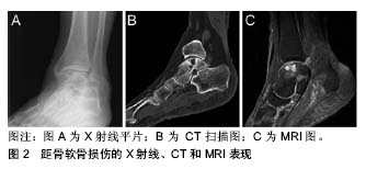
距骨软骨损伤的临床分期基本都是通过影像学检查来确定的, Berndt和Harty最早提出利用X射线平片对距骨软骨损伤进行分期(见表1)[4]。Ferkel等[3]于1990年提出了一个基于CT的分期标准(见表2),这为距骨软骨损伤的临床诊断和治疗提供了十分有力的支持;与此同时,MRI也被用于距骨软骨损伤的分期(见表3)[18]。除此以外,Pritsch和Ferkle又分别提出了关节镜3期(见表4)和扩展的关节镜分期(见表5)[19]。
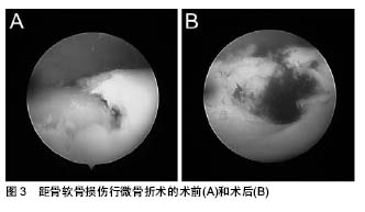
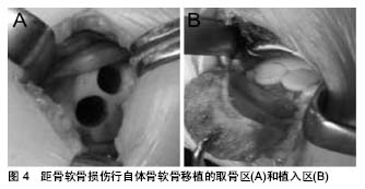
(4)骨软骨移植:对于伴有巨大软骨缺损的距骨软骨损伤,目前主要应用的是骨软骨移植技术,即在充分暴露距骨损伤区域后,清理并修整病损区域的软骨和软骨下骨,随后将移植物植入缺损区域,最终达到修复距骨软骨损伤的目的(见图4)[31-32]。
然而,目前的临床和动物实验都说明单纯的细胞输注很难修复大面积的距骨软骨损伤,此时,组织工程技术的发展为未来距骨软骨损伤的治疗指明了方向。支架、种子细胞和生长因子是组织工程修复软骨的三要素,尤其是具有生物活性3D支架,例如水凝胶和3D多孔支架等,在不断的改进和发展,这些支架提供了软骨组织产生所需的3D微环境[62-66];生长因子可以诱导增强软骨细胞分化和/或软骨形成。常见的刺激因子包括骨形态发生蛋白2、骨形态发生蛋白4、胰岛素样生长因子Ⅰ、和转化生子因子β等[67-69],还开发了具有促进软骨细胞生成和具有软骨保护作用的性质更加稳定的小分子药[70]。虽然组织工程技术在动物实验中已经取得了良好的效果,然而在临床中的实际应用尤其是长期随访,仍较为少见[71];Kaipel等[72]的研究显示,仿生支架可以有效的改善临床症状,但MRI显示其新生组织的质量却与正常组织有着一定的差距。此外,多种物理刺激也被发现具有诱导软骨生成的作用。
| [1] Leontaritis N, Hinojosa L, Panchbhavi VK. Arthroscopically detected intra-articular lesions associated with acute ankle fractures. J Bone Joint Surg Am. 2009; 91(2): 333-339.[2] Saxena A, Eakin C. Articular talar injuries in athletes: results of microfracture and autogenous bone graft. Am J Sports Med.2007; 35(10):1680-1687.[3] Ferkel RD, Zanotti RM, Komenda GA, et al.Arthroscopic treatment of chronic osteochondral lesions of the talus: long-term results. Am J Sports Med.2008;36(9): 1750-1762.[4] Berndt AL, Harty M.Transchondral fractures (osteochondritis dissecans) of the talus. J Bone Joint Surg Am. 1959;41-a: 988-1020.[5] Cheng JC, Ferkel RD. The role of arthroscopy in ankle and subtalar degenerative joint disease. Clin Orthop Relat Res. 1998; (349): 65-72.[6] Campbell CJ, Ranawat CS.Osteochondritis dissecans: the question of etiology. J Trauma.1966; 6(2): 201-221.[7] Prado MP, Kennedy JG, Raduan F,et al.Diagnosis and treatment of osteochondral lesions of the ankle: current concepts. Rev Bras Ortop. 2016;51(5): 489-500.[8] Flick AB, Gould N.Osteochondritis dissecans of the talus (transchondral fractures of the talus): review of the literature and new surgical approach for medial dome lesions. Foot Ankle.1985; 5(4): 165-185.[9] van Bergen CJ, Gerards RM, Opdam KT, et al.Diagnosing, planning and evaluating osteochondral ankle defects with imaging modalities. World J Orthop.2015; 6(11): 944-953.[10] Donnenwerth MP, Roukis TS.Outcome of arthroscopic debridement and microfracture as the primary treatment for osteochondral lesions of the talar dome. Arthroscopy.2012;28(12): 1902-1907.[11] Kirschke JS, Braun S, Baum T, et al., Diagnostic Value of CT Arthrography for Evaluation of Osteochondral Lesions at the Ankle. Biomed Res Int.2016;2016: 3594253.[12] Weber MA, Wünnemann F, Jungmann PM,et al.Modern Cartilage Imaging of the Ankle. Rofo.2017;189(10): 945-956.[13] Albano D, Martinelli N, Bianchi A,et al.Evaluation of reproducibility of the MOCART score in patients with osteochondral lesions of the talus repaired using the autologous matrix-induced chondrogenesis technique. Radiol Med.2017;122(12):909-917.[14] Van Ginckel A, Witvrouw EE.In vivo deformation of thin cartilage layers: Feasibility and applicability of T2* mapping. J Orthop Res. 2016;34(5): 771-778.[15] Tao H, Hu Y, Qiao Y, et al.T2 -Mapping evaluation of early cartilage alteration of talus for chronic lateral ankle instability with isolated anterior talofibular ligament tear or combined with calcaneofibular ligament tear. J Magn Reson Imaging.2018; 47(1):69-77.[16] Kim HS, Yoon YC.Comparison of T2 Relaxation Values in Subtalar Cartilage between Patients with Lateral Instability of the Ankle Joint and Healthy Volunteers. Eur Radiol.2018;28(10): 4151-4162.[17] Elias I, Jung JW, Raikin SM, et al.Osteochondral lesions of the talus: change in MRI findings over time in talar lesions without operative intervention and implications for staging systems. Foot Ankle Int. 2006;27(3):157-166.[18] Anderson IF, Crichton KJ, Grattan-Smith T, et al.Osteochondral fractures of the dome of the talus. J Bone Joint Surg Am.1989; 71(8): 1143-1152.[19] Cheng MR. Ferkel, and G. Applegate. Osteochondral lesions of the talus: a radiologic and surgical comparison. in Oral paper presented at the Annual Meeting of the Academy of Orthopaedic Surgeons (AAOS) New Orleans. 1995.[20] Huey DJ, Hu JC, Athanasiou KA.Unlike bone, cartilage regeneration remains elusive. Science.2012; 338(6109):917-921.[21] Verhagen RA, Struijs PA, Bossuyt PM,et al.Systematic review of treatment strategies for osteochondral defects of the talar dome. Foot Ankle Clin.2003;8(2): 233-242, viii-ix.[22] Klammer G, Maquieira GJ, Spahn S, et al.Natural history of nonoperatively treated osteochondral lesions of the talus. Foot Ankle Int.2015; 36(1): 24-31.[23] Murawski CD, Kennedy JG.Operative treatment of osteochondral lesions of the talus. J Bone Joint Surg Am. 2013;95(11): 1045-1054.[24] Grambart ST.Arthroscopic Management of Osteochondral Lesions of the Talus. Clin Podiatr Med Surg.2016; 33(4): 521-530.[25] Polat G, Er?en A, Erdil ME, et al.Long-term results of microfracture in the treatment of talus osteochondral lesions. Knee Surg Sports Traumatol Arthrosc.2016; 24(4): 1299-1303.[26] Choi WJ, Park KK, Kim BS, et al.Osteochondral lesion of the talus: is there a critical defect size for poor outcome? Am J Sports Med. 2009; 37(10): 1974-1980.[27] Shimozono Y, Coale M, Yasui Y, et al.Subchondral Bone Degradation After Microfracture for Osteochondral Lesions of the Talus: An MRI Analysis. Am J Sports Med.2018;46(3):642-648.[28] Lee KB, Park HW, Cho HJ, et al.Comparison of Arthroscopic Microfracture for Osteochondral Lesions of the Talus With and Without Subchondral Cyst. Am J Sports Med.2015;43(8): 1951-1956.[29] Kumai T, Takakura Y, Kitada C,et al.Fixation of osteochondral lesions of the talus using cortical bone pegs. J Bone Joint Surg Br.2002; 84(3): 369-374.[30] Shank JR, Benirschke SK, Swords MP.Treatment of Peripheral Talus Fractures. Foot Ankle Clin.2017;22(1): 181-192.[31] Ng A, Bernhard K. Osteochondral Autograft and Allograft Transplantation in the Talus. Clin Podiatr Med Surg. 2017; 34(4): 461-469.[32] Zhu Y, Xu X.Osteochondral Autograft Transfer Combined With Cancellous Allografts for Large Cystic Osteochondral Defect of the Talus. Foot Ankle Int.2016; 37(10): 1113-1118.[33] Leumann A, Horisberger M, Buettner O, et al.Medial malleolar osteotomy for the treatment of talar osteochondral lesions: anatomical and morbidity considerations. Knee Surg Sports Traumatol Arthrosc. 2016;24(7): 2133-2139.[34] Hannon CP, Smyth NA, Murawski CD, et al.Osteochondral lesions of the talus: aspects of current management. Bone Joint J.2014; 96-b(2): 164-171.[35] Henak CR, Ross KA, Bonnevie ED, et al.Human talar and femoral cartilage have distinct mechanical properties near the articular surface. J Biomech.2016;49(14):3320-3327.[36] Juras V, Zbýň Š, Mlynarik V, et al.The compositional difference between ankle and knee cartilage demonstrated by T2 mapping at 7 Tesla MR. Eur J Radiol.2016;85(4):771-777.[37] Hangody L.The mosaicplasty technique for osteochondral lesions of the talus. Foot Ankle Clin.2003;8(2): 259-273.[38] Hangody L, Kish G, Kárpáti Z, et al.Treatment of osteochondritis dissecans of the talus: use of the mosaicplasty technique--a preliminary report. Foot Ankle Int.1997;18(10): 628-634.[39] Sammarco GJ, Makwana NK.Treatment of talar osteochondral lesions using local osteochondral graft. Foot Ankle Int.2002;23(8): 693-698.[40] Flynn S, Ross KA, Hannon CP, et al. Autologous Osteochondral Transplantation for Osteochondral Lesions of the Talus. Foot Ankle Int.2016; 37(4):363-372.[41] Kubosch EJ, Erdle B, Izadpanah K, et al.Clinical outcome and T2 assessment following autologous matrix-induced chondrogenesis in osteochondral lesions of the talus. Int Orthop.2016; 40(1): 65-71.[42] Paul J, Sagstetter A, Kriner M, et al. Donor-site morbidity after osteochondral autologous transplantation for lesions of the talus. J Bone Joint Surg Am.2009;91(7): 1683-1688.[43] Gross AE, Agnidis Z, Hutchison CR.Osteochondral defects of the talus treated with fresh osteochondral allograft transplantation. Foot Ankle Int. 2001;22(5): 385-391.[44] Adams SB, Dekker TJ, Schiff AP, et al.Prospective Evaluation of Structural Allograft Transplantation for Osteochondral Lesions of the Talar Shoulder. Foot Ankle Int.2018;39(1): 28-34.[45] Ahmad J, Maltenfort M. Arthroscopic Treatment of Osteochondral Lesions of the Talus With Allograft Cartilage Matrix. Foot Ankle Int. 2017; 38(8): 855-862.[46] Lanham NS, Carroll JJ, Cooper MT, et al.A Comparison of Outcomes of Particulated Juvenile Articular Cartilage and Bone Marrow Aspirate Concentrate for Articular Cartilage Lesions of the Talus. Foot Ankle Spec.2017; 10(4):315-321.[47] Shimozono Y, Yasui Y, Ross AW, et al.Scaffolds based therapy for osteochondral lesions of the talus: A systematic review. World J Orthop.2017; 8(10): 798-808.[48] Gao F, Chen N, Sun W, et al.Combined Therapy with Shock Wave and Retrograde Bone Marrow-Derived Cell Transplantation for Osteochondral Lesions of the Talus. Sci Rep.2017; 7(1): 2106.[49] Shang XL, Tao HY, Chen SY, et al.Clinical and MRI outcomes of HA injection following arthroscopic microfracture for osteochondral lesions of the talus. Knee Surg Sports Traumatol Arthrosc.2016;24(4): 1243-1249.[50] Chahla J, Cinque ME, Shon JM,et al.Bone marrow aspirate concentrate for the treatment of osteochondral lesions of the talus: a systematic review of outcomes. J Exp Orthop.2016; 3(1): 33.[51] Tahta M, Akkaya M, Gursoy S, et al. Arthroscopic treatment of osteochondral lesions of the talus: Nanofracture versus hyaluronic acid-based cell-free scaffold with concentration of autologous bone marrow aspirate. J Orthop Surg (Hong Kong).2017;25(2): 2309499017717870.[52] Desando G, Bartolotti I, Vannini F, et al.Repair Potential of Matrix-Induced Bone Marrow Aspirate Concentrate and Matrix-Induced Autologous Chondrocyte Implantation for Talar Osteochondral Repair: Patterns of Some Catabolic, Inflammatory, and Pain Mediators. Cartilage.2017;8(1):50-60.[53] Kreulen C, Giza E, Walton J, et al.Seven-Year Follow-up of Matrix-Induced Autologous Implantation in Talus Articular Defects. Foot Ankle Spec.2018;11(2): 133-137.[54] Campagnoli C, Roberts IA, Kumar S,et al.Identification of mesenchymal stem/progenitor cells in human first-trimester fetal blood, liver, and bone marrow. Blood.2001;98(8): 2396-2402.[55] Bougioukli S, Sugiyama O, Pannell W, et al.Gene Therapy for Bone Repair Using Human Cells: Superior Osteogenic Potential of Bone Morphogenetic Protein 2-Transduced Mesenchymal Stem Cells Derived from Adipose Tissue Compared to Bone Marrow. Human Gene Therapy.2018; 29(4): 507-519.[56] Branly T, Bertoni L, Contentin R, et al.Characterization and use of Equine Bone Marrow Mesenchymal Stem Cells in Equine Cartilage Engineering. Study of their Hyaline Cartilage Forming Potential when Cultured under Hypoxia within a Biomaterial in the Presence of BMP-2 and TGF-beta 1. Stem Cell Rev.2017; 13(5): 611-630.[57] Kondo S, Muneta T, Nakagawa Y, et al.Transplantation of Autologous Synovial Mesenchymal Stem Cells Promotes Meniscus Regeneration in Aged Primates. J Orthop Res. 2017;35(6):1274-1282.[58] Ashton BA, Allen TD, Howlett CR,et al.Formation of bone and cartilage by marrow stromal cells in diffusion chambers in vivo. Clin Orthop Relat Res. 1980;(151):294-307.[59] Gao F, Chiu SM, Motan DA, et al.Mesenchymal stem cells and immunomodulation: current status and future prospects. Cell Death Dis. 2016;7:e2062. [60] Aggarwal S, Pittenger MF.Human mesenchymal stem cells modulate allogeneic immune cell responses. Blood. 2005;105(4): 1815-1822.[61] Vannini F, Cavallo M, Ramponi L,et al.Return to Sports After Bone Marrow-Derived Cell Transplantation for Osteochondral Lesions of the Talus. Cartilage.2017;8(1): 80-87.[62] Mehrali M, Thakur A, Pennisi CP, et al.Nanoreinforced Hydrogels for Tissue Engineering: Biomaterials that are Compatible with Load-Bearing and Electroactive Tissues. Advanced Materials. 2017; 29(8): 26.[63] Yang X, Lu Z, Wu H, et al.Collagen-alginate as bioink for three-dimensional (3D) cell printing based cartilage tissue engineering. Mater Sci Eng C Mater Biol Appl. 2018;83:195-201.[64] Dai Y, Shen T, Ma L, et al.Regeneration of osteochondral defects in vivo by a cell-free cylindrical poly(lactide-co-glycolide) scaffold with a radially oriented microstructure. J Tissue Eng Regen Med. 2018;12(3): e1647-e1661. [65] Li X, Ding J, Zhang Z, et al.Kartogenin-incorporated thermogel supports stem cells for significant cartilage regeneration. ACS Appl Mater Interfaces. 2016;8(8):5148-5159.[66] Kanatl? U, Eren A, Eren TK, et al.Single-Step Arthroscopic Repair With Cell-Free Polymer-Based Scaffold in Osteochondral Lesions of the Talus: Clinical and Radiological Results. Arthroscopy.2017; 33(9): 1718-1726.[67] Legendre F, Ollitrault D, Gomez-Leduc T, et al.Enhanced chondrogenesis of bone marrow-derived stem cells by using a combinatory cell therapy strategy with BMP-2/TGF-beta1, hypoxia, and COL1A1/HtrA1 siRNAs. Sci Rep, 2017;7(1): 3406.[68] Crecente-Campo J, Borrajo E, Vidal A, et al. New scaffolds encapsulating TGF-beta3/BMP-7 combinations driving strong chondrogenic differentiation. Eur J Pharm Biopharm.2017; 114: 69-78.[69] Gugjoo MB, Amarpal, Abdelbaset-Ismail A, et al.Mesenchymal stem cells with IGF-1 and TGF- beta1 in laminin gel for osteochondral defects in rabbits. Biomed Pharmacother.2017; 93:1165-1174.[70] Johnson K, Zhu S, Tremblay MS,et al.A stem cell-based approach to cartilage repair. Science.2012. 336(6082): 717-721.[71] Christensen BB, Foldager CB, Jensen J, et al.Poor osteochondral repair by a biomimetic collagen scaffold: 1- to 3-year clinical and radiological follow-up. Knee Surg Sports Traumatol Arthrosc.2016; 24(7): 2380-2387.[72] Kaipel M, Schreiner M, Kellner R, et al.Beneficial clinical effects but limited tissue quality following osteochondral repair with a cell-free multilayered nano-composite scaffold in the talus. Foot Ankle Surg. 2017;23(4):302-306. |
| [1] | 林清凡, 解一新, 陈婉清, 叶振忠, 陈幼芳. 人胎盘源间充质干细胞条件培养液可上调缺氧状态下BeWo细胞活力和紧密连接因子的表达[J]. 中国组织工程研究, 2021, 25(在线): 4970-4975. |
| [2] | 蒲 锐, 陈子扬, 袁凌燕. 不同细胞来源外泌体保护心脏的特点与效应[J]. 中国组织工程研究, 2021, 25(在线): 1-. |
| [3] | 张尚普, 鞠晓东, 宋恒义, 董 智, 王 晨, 孙国栋. 关节镜下带线锚钉缝线桥缝合固定治疗肩锁关节脱位[J]. 中国组织工程研究, 2021, 25(9): 1417-1422. |
| [4] | 吴 训, 孟娟红, 张建运, 王 亮. 浓缩生长因子修复兔髁突全层软骨损伤[J]. 中国组织工程研究, 2021, 25(8): 1166-1171. |
| [5] | 李嘉程, 梁学振, 刘金豹, 许 波, 李 刚. 骨性关节炎mRNA差异表达谱及竞争性内源RNA调控的网络分析[J]. 中国组织工程研究, 2021, 25(8): 1212-1217. |
| [6] | 耿秋东, 葛海雅, 王和鸣, 李 楠. 基于网络药理学探讨龟鹿二仙胶治疗骨关节炎的作用及机制[J]. 中国组织工程研究, 2021, 25(8): 1229-1236. |
| [7] | 刘翔翔, 黄云梅, 陈文列, 林如辉, 卢小冬, 李钻芳, 许亚晔, 黄美雅, 李西海. 早期骨关节炎模型大鼠半月板白区细胞的超微结构变化[J]. 中国组织工程研究, 2021, 25(8): 1237-1242. |
| [8] | 张秀梅, 翟运开, 赵 杰, 赵 萌. 类器官模型国内外数据库近10年文献研究热点分析[J]. 中国组织工程研究, 2021, 25(8): 1249-1255. |
| [9] | 侯婧瑛, 于萌蕾, 郭天柱, 龙会宝, 吴 浩. 缺氧预处理激活HIF-1α/MALAT1/VEGFA通路促进骨髓间充质干细胞生存和血管再生[J]. 中国组织工程研究, 2021, 25(7): 985-990. |
| [10] | 史洋洋, 秦英飞, 吴福玲, 何 潇, 张雪静. 胎盘间充质干细胞预处理预防小鼠毛细支气管炎[J]. 中国组织工程研究, 2021, 25(7): 991-995. |
| [11] | 梁学奇, 郭黎姣, 陈贺捷, 武 杰, 孙雅琪, 邢稚坤, 邹海亮, 陈雪玲, 吴向未. 泡状棘球绦虫原头蚴抑制骨髓间充质干细胞向成纤维细胞的分化[J]. 中国组织工程研究, 2021, 25(7): 996-1001. |
| [12] | 樊全宝, 罗惠娜, 王丙云, 陈胜锋, 崔连旭, 江文康, 赵明明, 王静静, 罗冬章, 陈志胜, 白银山, 刘璨颖, 张 晖. 低氧培养犬脂肪间充质干细胞的生物学特性[J]. 中国组织工程研究, 2021, 25(7): 1002-1007. |
| [13] | 耿 瑶, 尹志良, 李兴平, 肖东琴, 侯伟光. hsa-miRNA-223-3p调控人骨髓间充质干细胞成骨分化的作用[J]. 中国组织工程研究, 2021, 25(7): 1008-1013. |
| [14] | 伦志刚, 金 晶, 王添艳, 李爱民. 过氧化物还原酶6干预骨髓间充质干细胞增殖及体外向神经谱系诱导分化[J]. 中国组织工程研究, 2021, 25(7): 1014-1018. |
| [15] | 朱雪芬, 黄 成, 丁 健, 戴永平, 刘元兵, 乐礼祥, 王亮亮, 杨建东. 胶质细胞神经营养因子诱导骨髓间充质干细胞向功能性神经元分化的机制[J]. 中国组织工程研究, 2021, 25(7): 1019-1025. |
中国组织工程研究杂志出版内容重点:组织构建;骨细胞;软骨细胞;细胞培养;成纤维细胞;血管内皮细胞;骨质疏松;组织工程
1.3 资料提取与文献质量评价 首先阅读所有文章的标题和摘要,辨别文献质量,然后由不同作者分别阅读所有入选文献,并剔除所有重复文献,共筛选文献72篇。文献检索流程图见图1。
.jpg)
中国组织工程研究杂志出版内容重点:组织构建;骨细胞;软骨细胞;细胞培养;成纤维细胞;血管内皮细胞;骨质疏松;组织工程
中国组织工程研究杂志出版内容重点:组织构建;骨细胞;软骨细胞;细胞培养;成纤维细胞;血管内皮细胞;骨质疏松;组织工程
.jpg) 文题释义:
距骨软骨损伤:主要是由创伤等致病因素导致的涉及距骨表面软骨和/或距骨软骨下骨的各种急慢性损伤。
骨软骨移植术:是将来自自身供区或者捐献者的骨软骨块植入损伤预定损伤位置的手术方式。
中国组织工程研究杂志出版内容重点:组织构建;骨细胞;软骨细胞;细胞培养;成纤维细胞;血管内皮细胞;骨质疏松;组织工程
文题释义:
距骨软骨损伤:主要是由创伤等致病因素导致的涉及距骨表面软骨和/或距骨软骨下骨的各种急慢性损伤。
骨软骨移植术:是将来自自身供区或者捐献者的骨软骨块植入损伤预定损伤位置的手术方式。
中国组织工程研究杂志出版内容重点:组织构建;骨细胞;软骨细胞;细胞培养;成纤维细胞;血管内皮细胞;骨质疏松;组织工程
| 阅读次数 | ||||||
|
全文 |
|
|||||
|
摘要 |
|
|||||