中国组织工程研究 ›› 2018, Vol. 22 ›› Issue (8): 1217-1222.doi: 10.3969/j.issn.2095-4344.0140
• 组织构建实验造模 experimental modeling in tissue construction • 上一篇 下一篇
莫诺苷通过抑制炎症反应改善脑出血模型大鼠的神经功能
袁志俊,何晓英,袁 平,郑晓梅,李小刚
- 西南医科大学附属医院神经内科,四川省泸州市 646000
Morroniside improves the neurological function in intracerebral hemorrhage rats by inhibiting inflammatory response
Yuan Zhi-jun, He Xiao-ying, Yuan Ping, Zheng Xiao-mei, Li Xiao-gang
- Department of Neurology, the Affiliated Hospital of Southwest Medical University, Luzhou 646000, Sichuan Province, China
摘要:
文章快速阅读:
.jpg) 文题释义:
莫诺苷:是从中药山茱萸中分离提取的一种成分,据国内外研究表明它在大鼠缺血性脑损伤模型中,具有抗炎、抗氧化、抗凋亡、促进血管和神经再生、抗血小板聚集和神经保护作用。因脑出血的病理生理过程与缺血性脑损伤有许多相似之处,因此推测莫诺苷是否也可以抑制脑出血的炎症反应。
炎症反应:是临床常见的一个病理过程,可以生于机体各部位的组织和各器官。致炎因子作用于机体后,一方面引发组织细胞的损坏,使局部组织细胞显现变性、坏死;另一方面,诱导机体抗病机能增加,以益于清除致炎因子,使受损组织得到修复,从而使机体的内环境以及内环境和外环境之间达到新的均衡。
文题释义:
莫诺苷:是从中药山茱萸中分离提取的一种成分,据国内外研究表明它在大鼠缺血性脑损伤模型中,具有抗炎、抗氧化、抗凋亡、促进血管和神经再生、抗血小板聚集和神经保护作用。因脑出血的病理生理过程与缺血性脑损伤有许多相似之处,因此推测莫诺苷是否也可以抑制脑出血的炎症反应。
炎症反应:是临床常见的一个病理过程,可以生于机体各部位的组织和各器官。致炎因子作用于机体后,一方面引发组织细胞的损坏,使局部组织细胞显现变性、坏死;另一方面,诱导机体抗病机能增加,以益于清除致炎因子,使受损组织得到修复,从而使机体的内环境以及内环境和外环境之间达到新的均衡。
文题释义:
莫诺苷:是从中药山茱萸中分离提取的一种成分,据国内外研究表明它在大鼠缺血性脑损伤模型中,具有抗炎、抗氧化、抗凋亡、促进血管和神经再生、抗血小板聚集和神经保护作用。因脑出血的病理生理过程与缺血性脑损伤有许多相似之处,因此推测莫诺苷是否也可以抑制脑出血的炎症反应。
炎症反应:是临床常见的一个病理过程,可以生于机体各部位的组织和各器官。致炎因子作用于机体后,一方面引发组织细胞的损坏,使局部组织细胞显现变性、坏死;另一方面,诱导机体抗病机能增加,以益于清除致炎因子,使受损组织得到修复,从而使机体的内环境以及内环境和外环境之间达到新的均衡。摘要
背景:研究表明莫诺苷在大鼠缺血性脑损伤模型中具有抗炎、抗氧化、抗凋亡、促进血管和神经再生、抗血小板聚集和神经保护作用,但莫诺苷是否可以抑制脑出血的炎症反应尚不清楚。
目的:观察不同剂量的莫诺苷对大鼠脑出血后炎症因子白细胞介素1、肿瘤坏死因子α和炎症相关蛋白NF-κB和SUMO2/3的影响及神经功能变化。
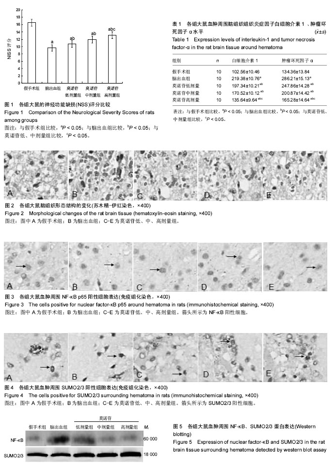
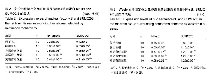
方法:将健康雄性SD大鼠随机分为假手术组、脑出血和莫诺苷低、中、高剂量组,后4组大鼠采用自体尾动脉血注入法建立脑出血模型,术后莫诺苷低、中、高剂量组分别给予莫诺苷30,90,270 mg/kg每天3次灌胃给药,连续7 d,假手术组和模型组给予等量生理盐水。给药7 d后应用NSS评分观察各组大鼠神经功能缺损症状;评分后取大鼠血肿周围脑组织,苏木精-伊红染色观察血肿周围神经细胞形态结构的变化;ELISA法检测大鼠脑组织炎症因子白细胞介素1、肿瘤坏死因子α水平;免疫组化法及Western blotting法检测通道蛋白NF-κB和SUMO2/3蛋白的表达。
结果与结论:①与脑出血组比较,莫诺苷低、中、高剂量组神经功能缺损改善,其中高剂量组改善最明显(P < 0.05);②与假手术比较,脑出血组和莫诺苷低、中、高剂量组神经功能受损,炎症因子白细胞介素1、肿瘤坏死因子α和炎症相关蛋白NF-κB、SUMO2/3表达增多(P < 0.05);③与脑出血组比较,莫诺苷低、中、高剂量组炎症因子白细胞介素1、肿瘤坏死因子α表达减少,炎症相关蛋白NF-κB降低,SUMO2/3表达增多(P < 0.05),以高剂量组作用最明显(P < 0.05)。提示:高剂量莫诺苷可通过炎症调节蛋白的变化,下调炎症因子的表达,抑制炎症反应改善脑出血大鼠的神经功能。
中国组织工程研究杂志出版内容重点:组织构建;骨细胞;软骨细胞;细胞培养;成纤维细胞;血管内皮细胞;骨质疏松;组织工程
ORCID: 0000-0001-9255-7508(袁志俊)
中图分类号:
.jpg) 文题释义:
莫诺苷:是从中药山茱萸中分离提取的一种成分,据国内外研究表明它在大鼠缺血性脑损伤模型中,具有抗炎、抗氧化、抗凋亡、促进血管和神经再生、抗血小板聚集和神经保护作用。因脑出血的病理生理过程与缺血性脑损伤有许多相似之处,因此推测莫诺苷是否也可以抑制脑出血的炎症反应。
炎症反应:是临床常见的一个病理过程,可以生于机体各部位的组织和各器官。致炎因子作用于机体后,一方面引发组织细胞的损坏,使局部组织细胞显现变性、坏死;另一方面,诱导机体抗病机能增加,以益于清除致炎因子,使受损组织得到修复,从而使机体的内环境以及内环境和外环境之间达到新的均衡。
文题释义:
莫诺苷:是从中药山茱萸中分离提取的一种成分,据国内外研究表明它在大鼠缺血性脑损伤模型中,具有抗炎、抗氧化、抗凋亡、促进血管和神经再生、抗血小板聚集和神经保护作用。因脑出血的病理生理过程与缺血性脑损伤有许多相似之处,因此推测莫诺苷是否也可以抑制脑出血的炎症反应。
炎症反应:是临床常见的一个病理过程,可以生于机体各部位的组织和各器官。致炎因子作用于机体后,一方面引发组织细胞的损坏,使局部组织细胞显现变性、坏死;另一方面,诱导机体抗病机能增加,以益于清除致炎因子,使受损组织得到修复,从而使机体的内环境以及内环境和外环境之间达到新的均衡。