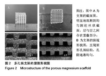
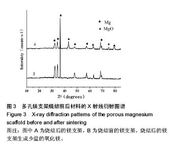
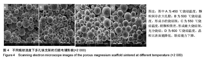
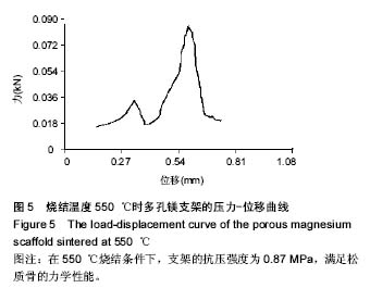
| [1]Sen MK,Miclau T.Autologous iliac crest bone graft: should it still be the gold standard for treating nonunions?Injury.2007;38 Suppl 1:S75-S80.[2]Sa MW,Kim JY.Design of multi-scaffold fabrication system for various 3D scaffolds.J Mech SciTechnol.2013;27(10):2961-2966. [3]Gaikwad VV,Patil AB,Gaikwad MV.Scaffolds for Drug Delivery in Tissue Engineering.Int J Pharm Sci Nanotechnol.2008;2(1):113-122.[4]Lin Z,Zhao Y,Wong, HM,et al.Enhanced corrosion resistance and cyto-biocompatility of plasma immersion ion implanted (PIII) WE43 magnesium scaffolds for bone tissue engineering.The 4th World Congress of the Tissue Engineering and Regenerative Medicine International Society (TERMIS), Boston, MA.,2015.[5]Lewis G. Reduction in the Corrosion Rate of Magnesium and Magnesium Alloy Specimens and Implications for Plain Fully Bioresorbable Coronary Artery Stents: A Review.World J Eng Technol. 2016;4(4):597-597.[6]张兴凯,韩伟,范德增,等.聚(乳酸一羟基乙酸)共聚物涂层的释药性能[J].功能高分子学报,2014,27(2):219-223.[7]Cox SC,Thornby JA,Gibbons GJ,et al.3D printing of porous hydroxyapatite scaffolds intended for use in bone tissue engineering applications.Mater Sci Eng C.2015;47:237-247.[8]Serre CM,Papillard M,Chavassieux P,et al.Influence of magnesium substitution on a collagen-apatite biomaterial on the production of a calcifying matrix by human osteoblasts.J Biomed Mater Res.1998;42: 626-633. [9]Wiesmnn HP,Tkotz T,Joos U,et al.Magnesium in newly formed dentin mineral of rat incisor.J Bone Miner Res.1997;12:380-383.[10]Staiger MP,Pietak AM,Huadmai J,et al.Magnesiumand its alloys as orthopedic biomaterials: a review.Biomaterials.2006;27:1728-1734. [11]SchliepHake H,Welch HA,Dullin C,et a1.Mandibular bone repair by implantation of rhBMP-2 in a slow release carrier of polylactic acid——an experimental study in rats.Biomaterials.2008;29(1):103-110.[12]Lin L,Wang Z,Zhou L,et al.The influence of prefreezing temperature on pore structure in freeze-dried beta-TCP scaffolds.Proc Inst Mech Eng H.2013,227(1):50-57.[13]Guarino V,Guaccio A,Guarnieri D,et al.Binary system thermodynamics to control pore architecture of PCL scaffold via temperature-driven pHase separation process.J Biomater Appl.2012;27(3):241-254.[14]Witte F,Ulrich H,Palm C,et al.Biodegradable magnesium scaffolds:part 1I:peri-implant bone remodeling.J Biomed Mater Res Part A,2007;81A(3):757765.[15]Zhang X,Li X,Li J,et al.Processing, microstructure and mechanical properties of biomedical magnesium with a specific two-layer structure. Prog Nat Sci MaterInt.2013;23(2):183-189.[16]Tan L,Gong M,Zheng F,et a1.Study on compression behavior of porous magnesium used as bone tissue engineering scaffolds.Biomed Mater. 2009;4(1):1-7.[17]贲玥,张乐,魏帅,等.3D打印陶瓷材料研究进展[J].材料导报, 2016,30(21): 109-118.[18]Krawczak P. Additive manufacturing of plastic and polymer composite parts: Promises and challenges of 3D-printing.Exp Polym Lett.2015; 9(11):959-959.[19]利普森.3D打印:从想象到现实[M].北京:中信出版社,2013.[20]Ahlfeld T,Akkineni AR,Förster Y,et al.Design and Fabrication of Complex Scaffolds for Bone Defect Healing: Combined 3D Plotting of a Calcium Phosphate Cement and a Growth Factor-Loaded Hydrogel. Ann Biomed Eng. 2017;45(1):1-13.[21]Farzadi A,Waran V,Solati-Hashjin M,et al.Effect of layer printing delay on mechanical properties and dimensional accuracy of 3D printed porous prototypes in bone tissue engineering.Ceram Int.2015; 41(7): 8320-8330.[22]Asadi-Eydivand M,Solati-Hashjin M,Farzad A,et al.Effect of technical parameters on porous structure and strength of 3D printed calcium sulfate prototypes.Robot Com-Int Manuf. 2016;37(C):57-67.[23]Zhang J,Zhao S,Zhu Y,et al.Three-dimensional printing of strontium-containing mesoporous bioactive glass scaffolds for bone regeneration.Acta Biomaterialia.2014;10(5):2269-2281.[24]Yang Y,Yang S,Wang Y,et al.Anti-infective efficacy, cytocompatibility and biocompatibility of a 3D-printed osteoconductive composite scaffold functionalized with quaternized chitosan.Acta Biomaterialia. 2016;46:112.[25]Standard Test Method for Density of Powder Metallurgy (PM) Materials Containing Less Than Two Percent Porosity.Astm B311-2013.[26]Pu SY.Embedded Metal Materials and their Corrosion.Beijing:Press of Beijing University of Aeronauties and Astronautics,l990:287.[27]Tsuruga E,Takita H,Itoh H,et al.Pore size of porous hydroxyapatite as the cell-substratum controls BMP-induced osteogenesis.J Biochem. 1997;121(2):317-324. [28]Clemow AJT,Weinstein AM,Klawitter JJ,et al.Interface mechanics of porous titanium implants. J Biomed Mater Res Part A. 1981;15(1): 73-82.[29]Gibson LJ.The mechanical behaviour of cancellous bone.J Biomech. 1985;18(5):317-328.[30]Levrero-Florencio F,Margetts L,Sales E,et al.Evaluating the macroscopic yield behaviour of trabecular bone using a nonlinear homogenisation approach.J Mech Behav Biomed Mater.2016;61:384. [31]Babiker H,Ding M,Overgaard S.Demineralized bone matrix and human cancellous bone enhance fixation of porous-coated titanium implants in sheep.J Tissue Eng Regene Med.2016;10(3):245.[32]Almeida HA,Bártolo PJ.Design of tissue engineering scaffolds based on hyperbolic surfaces: structural numerical evaluation.Med Eng Phys. 2014;36(8):1033.[33]Xiao D,Yang Y,Su X,et al.An integrated approach of topology optimized design and selective laser melting process for titanium implants materials.Biomed Mater Eng.2013;23(5):433-445.[34]Makar GL,Kruger J.Corrosion of Magnesium.Int Mater Rev. 1993; 38(3):138-153.[35]Kotoka R,Yamoah NK,Mensah-Darkwa K,et al. Electrochemical corrosion behavior of silver doped tricalcium pHospHate coatings on magnesium for biomedical application.Surf Coat Technol. 2016;292: 99-109.[36]Tekumalla S,Seetharaman S,Nguyen QB,et al.Influence of Cerium on the Deformation and Corrosion of Magnesium.J Eng Mater Technol. 2016;138(3):031011 .[37]Li X,Wang L,Fan Y,et al.Nanostructured scaffolds for bone tissue engineering.J Biomed Mater Res Part A.2013;101(8):2424. |
.jpg)
.jpg)

.jpg)
.jpg)