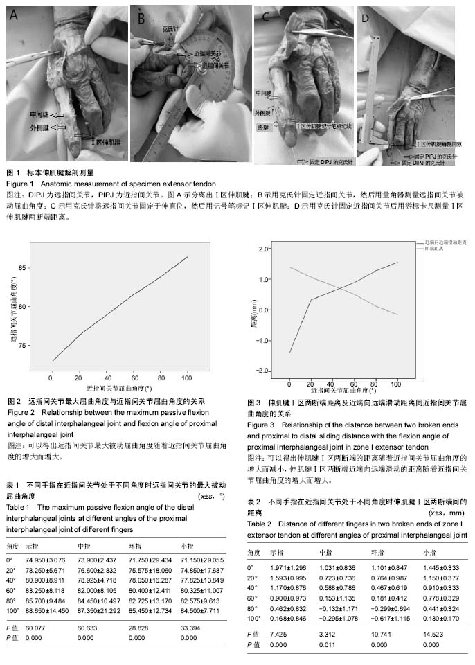
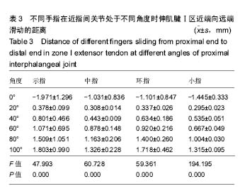
| [1] Alla SR, Deal ND, Dempsey IJ. Current concepts: mallet finger. Hand (N Y). 2014;9(2):138-144.[2] Chung DW, Lee JH. Anatomic reduction of mallet fractures using extension block and additional intrafocal pinning techniques. Clin Orthop Surg. 2012;4(1): 72-76.[3] de Jong JP, Nguyen JT, Sonnema AJ, et al. The incidence of acute traumatic tendon injuries in the hand and wrist: a 10-year population-based study. Clin Orthop Surg. 2014;6(2): 196-202.[4] Degreef I, De Smet L. Multiple simultaneous mallet fingers in goalkeeper. Hand Surg. 2009;14(2-3): 143-144.[5] Clayton RA, Court-Brown CM. The epidemiology of musculoskeletal tendinous and ligamentous injuries. Injury. 2008;39(12): 1338-1344.[6] Yeh PC, Shin SS. Tendon ruptures: mallet, flexor digitorum profundus. Hand Clin. 2012;28(3): 425-430.[7] Facca S, Nonnenmacher J, Liverneaux P. Treatment of mallet finger with dorsal nail glued splint: retrospective analysis of 270 cases. Rev Chir Orthop Reparatrice Appar Mot. 2007; 93(7): 682-689.[8] Giddins GE. The non-operative management of hand fractures. J Hand Surg Eur Vol. 2015;40(1): 33-41.[9] Valdes K, Naughton N, Algar L. Conservative treatment of mallet finger: A systematic review. J Hand Ther. 2015;28(3): 237- 246.[10] Chauhan A, Jacobs B, Andoga A, et al. Extensor tendon injuries in athletes. Sports Med Arthrosc. 2014;22(1): 45-55.[11] Williams CS 4th. Proximal interphalangeal joint fracture dislocations: stable and unstable. Hand Clin. 2012;28(3): 409-416.[12] Devan D. A novel way of treating mallet finger injuries. J Hand Ther. 2014;27(4): 325-329.[13] 孙文弢,张文龙. 外伤性锤状指研究进展[J]. 中国骨与关节损伤杂志, 2016,31(5): 559-560.[14] Lu J, Jiang J, Xu L, et al. Modification of the pull-in suture technique for mallet finger. Ann Plast Surg. 2013;70(1): 30-33.[15] Shimura H, Wakabayashi Y, Nimura A. A novel closed reduction with extension block and flexion block using Kirschner wires and microscrew fixation for mallet fractures. J Orthop Sci. 2014;19(2): 308-312.[16] Miranda BH, Murugesan L, Grobbelaar AO, et al. PBNR: Percutaneous blunt needle reduction of bony mallet injuries. Tech Hand Up Extrem Surg. 2015;19(2): 81-83.[17] Miura T. Extension block pinning using a small external fixator for mallet finger fractures. J Hand Surg Am. 2013; 38(12): 2348-2352.[18] Kakinoki R, Ohta S, Noguchi T, et al. A modified tension band wiring technique for treatment of the bony mallet finger. Hand Surg. 2013;18(2): 235-242.[19] Acar MA, Guzel Y, Gulec A, et al. Clinical comparison of hook plate fixation versus extension block pinning for bony mallet finger: a retrospective comparison study. J Hand Surg Eur. 2015;40(8): 832-839.[20] Prunieres G, Gouzou S, Facca S, et al. Treatment of unstable distal phalanx fractures by extra-articular DIP pinning: A series of 12 cases. Hand Surg Rehabil. 2016;35(5): 330-334.[21] Tocco S, Boccolari P, Landi A, et al. Effectiveness of cast immobilization in comparison to the gold-standard self-removal orthotic intervention for closed mallet fingers: a randomized clinical trial. J Hand Ther. 2013;26(3): 191-201.[22] 陈孝平,汪建平.外科学[M]. 8版.北京:人民卫生出版社, 2013: 664.[23] 高士濂.实用解剖图谱(上肢分册)[M]. 3版. 上海:上海科学技术出版社,2012:273.[24] 李雯,齐杰,蔺利剑,等. 双克氏针加压固定法治疗骨性锤状指[J]. 河北医科大学学报,2015,56(4): 472-473.[25] 闫浩,谭权昌,周胜,等. 克氏针弹性固定治疗DoyleⅠ、Ⅱ型锤状指近期疗效观察[J].中国修复重建外科杂志, 2017,31(11):1-4.[26] 施劲,王骏. 锤状指保守治疗效果及影响因素的分析[J]. 中国康复医学杂志,2017,32(7):826-828.[27] 赵占稳. 陈旧性腱性锤状指的显微外科手术治疗[J]. 中国矫形外科杂志,2008,16(24): 1898-1899.[28] 胡静波,姜德欣,李大为,等. 三种方法治疗骨性锤状指的疗效分析[J]. 浙江创伤外科, 2013,18(2): 175-176.[29] 王扬剑,魏鹏,王欣,等. 石黑法治疗伴撕脱骨折锤状指的疗效[J]. 现代实用医学,2009,21(10): 1132-1134.[30] 朱其,邱海胜,吴建伟. 微型骨锚修复止点撕脱锤状指畸形的临床应用[J]. 实用医学杂志,2010,39(20): 3759-3761.[31] 薛云皓,田光磊.172例闭合性锤状指畸形的统计学分析[J].中华手外科杂志,2014,30(2):94-97.[32] Schweitzer TP, Rayan GM. The terminal tendon of the digital extensor mechanism: Part II, kinematic study. J Hand Surg Am. 2004;29(5): 903-908.[33] Katzman BM, Klein DM, Mesa J, et al. Immobilization of the mallet finger. Effects on the extensor tendon. J Hand Surg Br. 1999;24(1): 80-84.[34] Kalzman EB. Anatomy, injuries and treatment of extensor apparatus of the hand and the digits.Clin Orthop. 1959; 13(1): 24-41.[35] Abouna JM, Brown H. The treatment of mallet finger. The results in a series of 148 consecutive cases and a review of the literature.Br J Surg. 1968;55(9):653-667.[36] Stack G.Mallet finger.Lancet. 1968;2(7581):1303-1303.[37] 孙文弢,张文龙.近指间关节活动对伸肌腱Ⅰ-Ⅱ区张力影响的解剖学研究[J].中华手外科杂志,2016,32(3):207-209.[38] 王耐,冯日祥,陈汀涝,等.双注射器针头+石膏固定治疗急性腱性锤状指的体会[J].山西医药杂志,2017,14( 15):1807-1808.[39] 王艺,孙胜男,张燕玲,等.改良支具对锤状指患者非手术治疗依从性的分析[J].实用骨科杂志,2016,22( 8):766-767.[40] 蔡利兵,董栋,费剑荣.保守治疗伸肌腱Ⅰ区断裂13例疗效分析[J].现代实用医学,2017,29(2):184-185.[41] 陈少军,丑钢.小夹板外固定配合截血膏治疗闭合性锤状指24例[J].中医外治杂志,2017,26(5): 17-18.[42] 陈少贞, 张涛, 朱庆棠, 等. 两种不同外固定方式对新鲜锤状指临床疗效的单盲随机对照研究[J].中国康复医学杂志,2012, 27(11):1006-1010.[43] 张净宇,于志亮,高顺红,等.改良克氏针固定并肌腱修复治疗儿童腱性锤状指[J].中华手外科杂志,2014,30(2):104-106. |
.jpg)


.jpg)