中国组织工程研究 ›› 2024, Vol. 28 ›› Issue (14): 2191-2196.doi: 10.12307/2024.301

• 骨组织构建 bone tissue construction • 上一篇    下一篇

基于TMT技术的激素性股骨头坏死硬化带修复模式探索及临床验证

庄至坤1,何敏聪2,3,林天烨2,3,吴荣凯1,郭金花1,吴昭克1,魏秋实2,3   

  1. 1泉州市正骨医院关节科,福建省泉州市  362000;2广东省中医骨伤研究院,广东省广州市  510000;3广州中医药大学第三附属医院关节科,广东省广州市  510000
  • 收稿日期:2023-02-22 接受日期:2023-04-14 出版日期:2024-05-18 发布日期:2023-07-28
  • 通讯作者: 魏秋实,博士,副主任医师,博士生导师,广东省中医骨伤研究院,广东省广州市 510000;广州中医药大学第三附属医院关节科,广东省广州市 510000 吴昭克,主任医师,硕士生导师,泉州市正骨医院关节科,福建省泉州市 362000
  • 作者简介:庄至坤,男,1982年生,汉族,福建省泉州市人,副主任医师,主要从事骨关节疾病的基础与临床研究。
  • 基金资助:
    国家自然科学基金面上项目(82274544),项目负责人:魏秋实;泉州市科技计划项目(2022NS011),项目负责人:庄至坤

Exploration and clinical validation of the repair mode of the sclerotic zone of steroid-induced osteonecrosis of the femoral head based on Tandem Mass Tags technology

Zhuang Zhikun1, He Mincong2, 3, Lin Tianye2, 3, Wu Rongkai1, Guo Jinhua1, Wu Zhaoke1, Wei Qiushi2, 3   

  1. 1Department of Joint Diseases, Quanzhou Orthopedic-Traumatological Hospital, Quanzhou 362000, Fujian Province, China; 2Guangdong Research Institute for Orthopedics & Traumatology of Chinese Medicine, Guangzhou 510000, Guangdong Province, China; 3Department of Joint Diseases, The Third Affiliated Hospital of Guangzhou University of Chinese Medicine, Guangzhou 510000, Guangdong Province, China
  • Received:2023-02-22 Accepted:2023-04-14 Online:2024-05-18 Published:2023-07-28
  • Contact: Wei Qiushi, MD, Associate chief physician, Doctoral supervisor, Guangdong Research Institute for Orthopedics & Traumatology of Chinese Medicine, Guangzhou 510000, Guangdong Province, China; Department of Joint Diseases, The Third Affiliated Hospital of Guangzhou University of Chinese Medicine, Guangzhou 510000, Guangdong Province, China Wu Zhaoke, Chief physician, Master’s supervisor, Department of Joint Diseases, Quanzhou Orthopedic-Traumatological Hospital, Quanzhou 362000, Fujian Province, China
  • About author:Zhuang Zhikun, Associate chief physician, Department of Joint Diseases, Quanzhou Orthopedic-Traumatological Hospital, Quanzhou 362000, Fujian Province, China
  • Supported by:
    National Natural Science Foundation of China, No. 82274544 (to WQS); Quanzhou Science and Technology Program, No. 2022NS011 (to ZZK)

摘要:


文题释义:

串联质谱标签(Tandem Mass Tags,TMT)技术:定量蛋白质组学通过对质谱进行化学标记,从而鉴定和定量蛋白质、核酸等生物大分子;相较于以往常用的凝胶与抗体定量等蛋白组学,TMT的灵敏度、通量和可重复性等优势成为了目前蛋白组学研究中的热门方法。
过氧化物酶体增殖物活化受体γ协同刺激因子1α(peroxisome proliferator-activated receptor gamma coactivator 1α,PGC-1α):作为一种多功能的转录共调节因子,具有信号诱导的特异性,能够灵敏应答于各种环境刺激和营养信号的改变,并通过选择性地结合不同转录因子,协同控制包括葡萄糖稳态、脂质代谢平衡和生物时钟稳态在内的多种生理进程。


背景:股骨头内硬化带是激素性股骨头坏死疾病发展过程中重要的影像学特征并与预后相关。过氧化物酶体增殖物活化受体γ协同刺激因子1α(peroxisome proliferator-activated receptor gamma coactivator 1α,PGC-1α)被证明具有成骨、促血管形成、抗线粒体凋亡等生物活性,与激素性股骨头坏死的骨修复可能存在紧密关联。

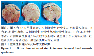
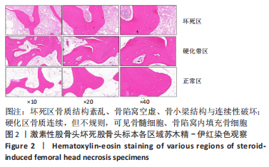
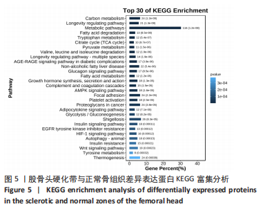
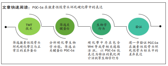
目的:筛选激素性股骨头坏死硬化带区与正常区的差异蛋白,筛选出硬化带的关键蛋白,并验证关键蛋白在激素性股骨头坏死股骨头标本中的差异表达;探索激素性股骨头坏死硬化带修复模式。
方法:取激素性股骨头坏死行人工全髋关节置换术取出的股骨头标本,通过串联质谱标签技术筛选硬化带区与正常区的差异表达基因,并进行GO与KEGG信号通路分析,构建关键靶点的蛋白互作网络、筛选关键基因。观察关键蛋白在激素性股骨头坏死硬化带中的表达,通过Western blotting、免疫组化验证关键蛋白在硬化带中的表达。

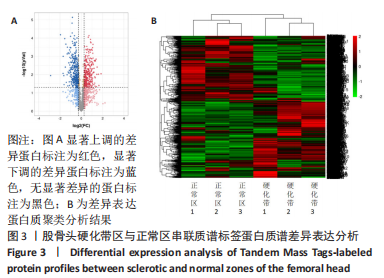
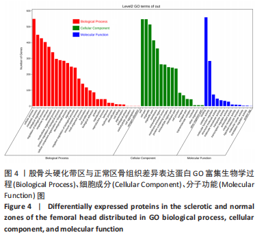
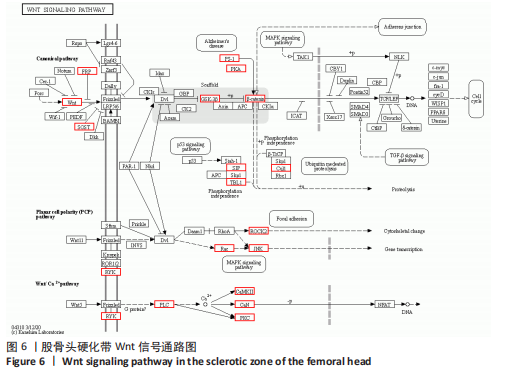
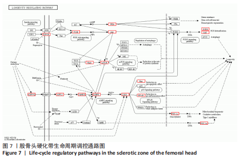
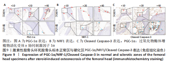
结果与结论:①通过串联质谱标签定量蛋白质谱检测发现:相比于正常区骨组织,股骨头硬化带骨组织中具有显著差异表达(Log2FC > 1.20、Log2FC < 0.84和 P < 0.05)的蛋白质有609个,其中发生上调的蛋白质290个,发生下调的蛋白质319个;②通过GO与KEGG通路富集分析,发现在前10位的富集通路中,Wnt信号通路与生命周期调控通路与骨修复关系密切;在生命周期调控通道中,PGC-1α是重要的蛋白之一;③与激素性股骨头坏死标本正常区相比,Western blotting验证了PGC-1α、NRF-1在硬化带中低表达,Cleaved Caspase-3在硬化带中高表达;④光镜下免疫组化结果显示,股骨头组织标本中硬化区及正常区的PGC-1α、NRF1及Cleaved Caspase-3阳性染色分布,可以发现骨小梁、成骨细胞和骨髓均有表达存在;硬化带区Cleaved Caspase-3表达更为明显;⑤结论:激素性股骨头坏死硬化带中存在包含Wnt等成骨相关通路活跃、以PGC-1α低表达为特征的氧化凋亡活跃等生物学行为;PGC-1α在激素性股骨头坏死硬化带中低表达可能与氧化凋亡活跃相关。

https://orcid.org/0000-0002-1444-8346(庄至坤)

中国组织工程研究杂志出版内容重点:组织构建;骨细胞;软骨细胞;细胞培养;成纤维细胞;血管内皮细胞;骨质疏松;组织工程

关键词: 串联质谱标签, 激素性股骨头坏死, PGC-1α, 线粒体氧化凋亡, 骨修复

Abstract: BACKGROUND: The sclerotic zone in the femoral head is an important imaging feature in the progression of steroid-induced femoral head necrosis, which is associated with disease prognosis. Peroxisome proliferator-activated receptor γ coactivator 1α (PGC-1α) has been shown to possess biological activities such as osteogenesis, angiogenesis and anti-mitochondrial apoptosis, which may be closely related to bone repair of steroid-induced femoral head necrosis.
OBJECTIVE: To screen for the differential proteins in the sclerotic zone of steroid-induced osteonecrosis of the femoral head versus the normal zone, to screen for hub proteins in the sclerotic zone, and to verify the differential expression of hub proteins in the femoral head specimens following steroid-induced femoral head necrosis, and to to explore the repair pattern of the sclerotic zone following steroid-induced femoral head necrosis.
METHODS: Femoral head samples were collected from patients with steroid-induced osteonecrosis of the femoral head receiving total hip arthroplasty. The differentially expressed genes in the sclerotic zone and the normal zone were screened by Tandem Mass Tags and analyzed by GO and KEGG signaling pathways to construct a protein-protein interaction network and screen hub genes. In addition, the expression of hub genes in the sclerotic zone was verified by immunohistochemistry and western blot.
RESULTS AND CONCLUSION: Quantitative protein profiling by Tandem Mass Tags revealed that 609 proteins were significantly differentially expressed (Log2FC > 1.20, Log2FC < 0.84 and P < 0.05) in the sclerotic zone of the femoral head compared with the normal zone, of which 290 proteins were upregulated and 319 proteins were downregulated. The GO and KEGG pathway enrichment analyses revealed that among the top 10 enriched pathways, Wnt signaling pathway and life-cycle regulatory pathway were closely related to bone repair; in the life-cycle regulatory pathway, PGC-1α was one of the important proteins. In addition, western blot results verified the low expression of PGC-1α and NRF1 in the sclerotic zone and high expression of Cleaved Caspase-3 in the sclerotic zone compared with the normal zone of steroid-induced femoral head necrosis specimens. Light microscopic immunohistochemical results showed the distribution of PGC-1α, NRF1 and Cleaved Caspase-3 positive expression in the sclerotic and normal zones in the femoral head tissue specimens, indicating the presence of their expression in bone trabeculae, osteoblasts and bone marrow. In contrast, the brown area of the sclerotic zone of femoral head necrosis stained darker and showed more obvious expression of Cleaved Caspase-3. To conclude, in the sclerotic zone of steroid-induced femoral head necrosis, biological behaviors including activation of osteogenesis-related pathways such as Wnt and oxidative apoptosis characterized by low expression of PGC-1 are observed. Low expression of PGC-1α in the sclerotic zone of steroid-induced femoral head necrosis may be associated with the activation of oxidative apoptosis. 

Key words: Tandem Mass Tags, steroid-induced osteonecrosis of the femoral head, peroxisome proliferative activated receptor γ coactivator 1α, mitochondrial oxidative apoptosis, bone repair

中图分类号: