中国组织工程研究 ›› 2022, Vol. 26 ›› Issue (13): 2056-2061.doi: 10.12307/2022.333

• 干细胞外泌体 Stem cell exosomes • 上一篇    下一篇

人尿源性干细胞来源外泌体的提取及鉴定

王  帅1,李超然2,章茜茹3,黄桂林4   

  1. 1遵义医科大学口腔医学院,贵州省遵义市  563000;2同济大学附属东方医院,上海市  310000;3贵州中医药大学第一附属医院,贵州省贵阳市  550002;4遵义医科大学附属口腔医院,贵州省遵义市  563003
  • 收稿日期:2020-12-14 修回日期:2020-12-18 接受日期:2021-01-23 出版日期:2022-05-08 发布日期:2021-12-20
  • 通讯作者: 王帅,博士,副教授,遵义医科大学口腔医学院,贵州省遵义市 563000 黄桂林,博士,教授,遵义医科大学附属口腔医院,贵州省遵义市 563003
  • 作者简介:王帅,男,1982年生,山东省青岛市人,汉族,2015年加拿大麦吉尔大学毕业,博士,副教授,主要从事干细胞生物学、骨组织再生以及口腔种植学的基础与临床研究。
  • 基金资助:
    国家自然科学基金(81860193),项目负责人:王帅;贵州省科技厅项目(黔科合基础【2016】1170,黔科合基础【2019】1340),项目负责人:王帅;贵州省教育厅青年科技人才成长项目(黔教合KY字【2016】198),项目负责人:王帅

Isolation and identification of human urine-derived stem cell-derived exosomes

Wang Shuai1, Li Chaoran2, Zhang Xiru3, Huang Guilin4   

  1. 1School of Stomatology, Zunyi Medical University, Zunyi 563000, Guizhou Province, China; 2Dongfang Hospital Affiliated to Tongji University, Shanghai 310000, China; 3First Affiliated Hospital of Guizhou University of Traditional Chinese Medicine, Guiyang 550002, Guizhou Province, China; 4Hospital of Stomatology, Zunyi Medical University, Zunyi 563003, Guizhou Province, China
  • Received:2020-12-14 Revised:2020-12-18 Accepted:2021-01-23 Online:2022-05-08 Published:2021-12-20
  • Contact: Wang Shuai, PhD, Associate professor, School of Stomatology, Zunyi Medical University, Zunyi 563000, Guizhou Province, China Huang Guilin, MD, Professor, Hospital of Stomatology, Zunyi Medical University, Zunyi 563003, Guizhou Province, China
  • About author:Wang Shuai, PhD, Associate professor, School of Stomatology, Zunyi Medical University, Zunyi 563000, Guizhou Province, China
  • Supported by:
    the National Natural Science Foundation of China, No. 81860193 (to WS); a grant from Department of Science and Technology of Guizhou Province, No. [2016]1170 (to WS), No. [2019]1340 (to WS); the Youth Science and Technology Talents Growth Project of Guizhou Provincial Education Department, No. [2016]198 (to WS) 

摘要:

文题释义:
人尿源性干细胞:是一种可能来源于肾脏的祖细胞/干细胞,不仅稳定地高表达间充质干细胞特异性表面标志物(CD29、CD44、CD54、CD73、CD105、CD16),还能表达胚胎干细胞特异性标志物(SSEA4、TRA-1-81、Sox2、Oct3/4)。与其他干细胞相比,其具有取材无创,提取分离步骤简便,较高的端粒酶活性和较长的端粒序列,良好的多向分化潜能以及安全性高、无致瘤性等优点。 
外泌体:是细胞经过“内吞-融合-外排”等一系列调控过程形成直径40-100 nm的膜性脂质小囊泡,其内含有功能性蛋白质、mRNA、microRNA及长链非编码RNA等物质,在细胞间的信息传递过程中发挥重要作用,其载有的mRNA可在靶细胞内翻译出相应蛋白质,载有的microRNA可通过降解靶细胞内mRNA或抑制mRNA翻译来调控目的蛋白的表达。研究证实,外泌体参与机体的各种生理或病理过程,如免疫调节、肿瘤转移、遗传信息交换、细胞增殖及凋亡、血管新生等。 

背景:近年来研究发现,干细胞所发挥的组织修复与再生功能在很大程度上是通过旁分泌作用实现的,外泌体作为其中一种重要的旁分泌因子,已经被证实在心脏、神经、骨、视网膜损伤修复及再生方面发挥重要作用,研究人员也在寻找更好的干细胞来源外泌体。 
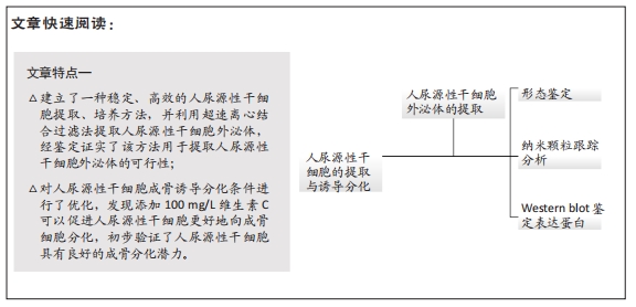
目的:提取并鉴定人尿源性干细胞来源外泌体。 
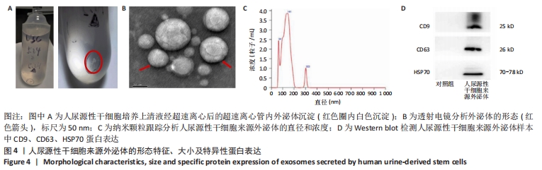
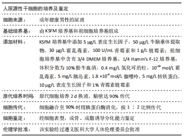
方法:收集健康成年男性新鲜尿液,采用贴壁培养法分离得到人尿源性干细胞,对其进行培养、传代。流式细胞术鉴定其表面标记物,体外定向诱导其向成骨细胞、脂肪细胞分化,分别采用茜素红、Von Kossa和油红O染色进行鉴定。采用改良超高速离心法从人尿源性干细胞培养上清中提取外泌体,透射电镜观察其形态,纳米颗粒跟踪分析其大小及浓度,蛋白质免疫印迹法检测其特异性蛋白CD9、CD63和HSP70的表达。
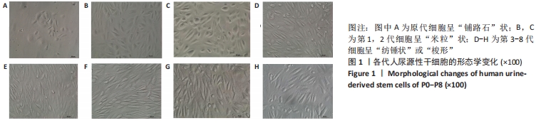
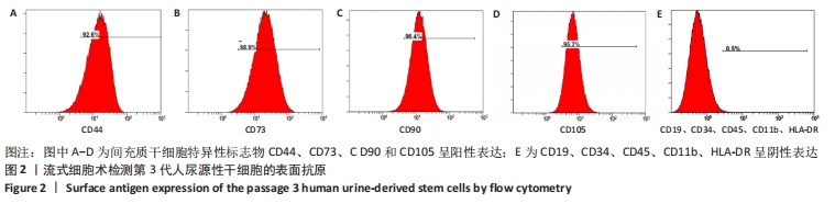
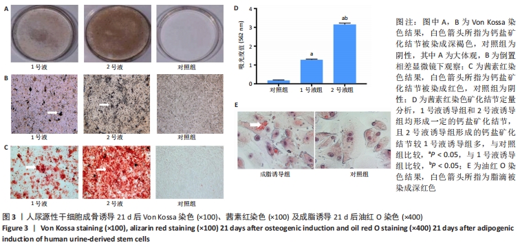
结果与结论:①人尿源性干细胞培养3-5 d后开始贴壁生长,细胞在原代时呈“铺路石”样,第1代和第2代细胞呈“米粒”样,传至第3代后逐渐呈“纺锤形”或“梭形”;流式细胞仪检测结果显示人尿源性干细胞阳性表达间充质干细胞的特异表面标志物CD44、CD73、CD90和CD105;人尿源性干细胞经成骨、成脂诱导培养后能成功分化为成骨细胞、脂肪细胞;②经过低速、高速、超高速序列离心方法,从人尿源性干细胞的条件培养基中提取外泌体,透射电镜观察呈圆形或者椭圆形膜性囊泡样,形似“茶托”,大小主要集中在64-141 nm之间,蛋白质免疫印迹分析显示人尿源性干细胞来源外泌体表达CD9、CD63和HSP70蛋白;③该研究证实了改良超高速离心法提取人尿源性干细胞外泌体的可行性。 
缩略语:人尿源性干细胞来源外泌体:exosomes secreted by human urine-derived stem cells,hUSCs-Exo

https://orcid.org/0000-0001-6805-7239(王帅);https://orcid.org/0000-0003-3224-7094(黄桂林)

中国组织工程研究杂志出版内容重点:干细胞;骨髓干细胞;造血干细胞;脂肪干细胞;肿瘤干细胞;胚胎干细胞;脐带脐血干细胞;干细胞诱导;干细胞分化;组织工程

关键词: 干细胞, 人尿源性干细胞, 外泌体, 超高速离心, 成骨分化, 粒径, 纳米

Abstract: BACKGROUND: Recent studies have found that the tissue repair and regeneration function of stem cells are largely realized by paracrine function. Exosomes, as one of important paracrine factors, have been proven to play an important role in the repair and regeneration of heart, nerve, bone and retina injury. Researchers are looking for a better source of stem cells-derived exosomes.
OBJECTIVE: To isolate and identify exosomes secreted by human urine-derived stem cells. 
METHODS: Human urine-derived stem cells were isolated from urine of healthy male adults and then cultured and passaged. The surface markers were identified by flow cytometry. Human urine-derived stem cells were induced to differentiate into osteoblasts and adipocytes, and identified by alizarin red, Von Kossa and oil red O staining, respectively. Modified ultra-high speed centrifugation was used to isolate exosomes secreted by human urine-derived stem cells from human urine-derived stem cells conditioned medium. Transmission electron microscopy was used to analyze their morphology. Nanoparticle Tracking Analysis was used to analyze the size and concentration. Western blot assay was used to detect the expression of specific protein CD9, CD63 and HSP70. 
RESULTS AND CONCLUSION: (1) Adherent cell growth was identified in human urine-derived stem cells after 3-5 days of culturing. They resembled “slabstone” in the primary passage, and then resembled “rice” in the first and second passages, and in the third passage, they gradually turned into a “spindle-like shape” or “shuttle-like shape”. Flow cytometry results showed the positive expression of human urine-derived stem cells with mesenchymal stem cell specific surface markers (CD44, CD73, CD90 and CD105). After osteogenic and adipogenic induction in vitro, human urine-derived stem cells successfully differentiated into osteoblasts and adipocytes. (2) After low speed, high speed, ultra-high speed centrifugation, exosomes secreted by human urine-derived stem cells were extracted from conditioned medium of human urine-derived stem cells. Observation by transmission electron microscope showed round or oval membranous vesicles, resembling “saucer”, mainly concentrated with the size of 64-141 nm. Western blot assay showed the protein expression of CD9, CD63 and HSP70 in the exosomes secreted by human urine-derived stem cells. (3) This study confirmed the feasibility of the improved ultra-high speed centrifugation method to extract the exosomes secreted by human urine-derived stem cells.

Key words: stem cells, human urine-derived stem cells, exosomes, ultrahigh speed centrifugation, osteogenic differentiation, particle size, nanoparticle

中图分类号: