中国组织工程研究 ›› 2021, Vol. 25 ›› Issue (27): 4283-4288.doi: 10.12307/2021.183
• 骨与关节生物力学 bone and joint biomechanics • 上一篇 下一篇
有限元模型建立青少年骶椎腰化的验证分析
吴雪海,王 星,张少杰,徐雪彬,史 君,王铁英,李志军
- 1赤峰市第二医院骨科,内蒙古自治区赤峰市 024000;内蒙古医科大学基础医学院,2解剖学教研室,3生理学教研室,内蒙古自治区呼和浩特市 010110
-
收稿日期:2020-05-19修回日期:2020-05-20接受日期:2020-10-16出版日期:2021-09-28发布日期:2021-04-10 -
通讯作者:李志军,硕士,教授,博士生导师,内蒙古医科大学基础医学院解剖学教研室,内蒙古自治区呼和浩特市 010110 王铁英,主任医师,赤峰市第二医院骨科,内蒙古自治区赤峰市 024000 -
作者简介:吴雪海,男,1985年生,内蒙古自治区赤峰市人, 蒙古族,2018年内蒙古医科大学毕业,硕士,主治医师,主要从事骨外科方向的研究。 王星,男,1979年生,内蒙古自治区乌拉特中旗人,汉族,北京中医药大学中医学院在读博士,讲师,主要从事脊柱脊髓的数字与临床应用解剖学方面的研究。 -
基金资助:国家自然科学基金项目(81860382),项目负责人:王星;国家自然科学基金项目(81860383,81560348),项目负责人:李志军;国家自然科学基金项目(81660358),项目负责人:张少杰;内蒙古自然科学基金项目(2020MS03061),项目负责人:王星;内蒙古自然科学基金项目(2019MS08017),项目负责人:张少杰;内蒙古自治区科技发展计划项目(2019GG158),项目负责人:王星;内蒙古医科大学科技百万项目(YKD2017KJBW009),项目负责人:王星;内蒙古医科大学科技百万项目(2015YKDKJBW03),项目负责人:张少杰
Establishment and validation of a finite element model of sacral lumbarization in adolescents
Wu Xuehai, Wang Xing, Zhang Shaojie, Xu Xuebin, Shi Jun, Wang Tieying, Li Zhijun
- 1Department of Orthopedics, Chifeng City Second Hospital, Chifeng 024000, Inner Mongolia Autonomous Region, China; 2Human Anatomy Teaching and Research,
3Physiology Teaching Laboratory, Basic Medical College of Inner Mongolia Medical University, Hohhot 010110, Inner Mongolia Autonomous Region, China
-
Received:2020-05-19Revised:2020-05-20Accepted:2020-10-16Online:2021-09-28Published:2021-04-10 -
Contact:Li Zhijun, Master, Professor, Doctoral supervisor, Human Anatomy Teaching and Research, Basic Medical College of Inner Mongolia Medical University, Hohhot 010110, Inner Mongolia Autonomous Region, China Wang Tieying, Chief physician, Department of Orthopedics, Chifeng City Second Hospital, Chifeng 024000, Inner Mongolia Autonomous Region, China -
About author:Wu Xuehai, Master, Attending physician, Department of Orthopedics, Chifeng City Second Hospital, Chifeng 024000, Inner Mongolia Autonomous Region, China Wang Xing, Doctoral candidate, Lecturer, Human Anatomy Teaching and Research, Basic Medical College of Inner Mongolia Medical University, Hohhot 010110, Inner Mongolia Autonomous Region, China -
Supported by:the National Natural Science Foundation of China, No. 81860382 (to WX), No. 81860383, 81560348 (to LZJ), No. 81660358 (to ZSJ); the Natural Science Foundation of Inner Mongolia Autonomous Region, No. 2020MS03061 (to WX), No. 2019MS08017 (to ZSJ); the Science and Technology Development Program of Inner Mongolia Autonomous Region, No. 2019GG158 (to WX); the Science and Technology Project of Inner Mongolia Medical University, No. YKD2017KJBW009 (to WX), No. 2015YKDKJBW03 (to ZSJ)
摘要:
文题释义:
腰骶移行椎:指腰椎数目变化及其伴随的L5 和S1 的形态结构变化。一般情况下整个脊柱的椎骨总数不变,只是各节段数目有所增减,可分为骶椎腰化和腰椎骶化。
有限元模型分析法:是近年来研究人体生物力学的有效方法,其基本原理是把连续的物体离散为一组有限个,并按一定方式相互联结在一起的单元组合体,然后求其相互之间的关系及特征。
背景:腰椎骶化会使椎体节段发生变化,从而导致在运动中引起腰痛。流行病学研究显示,青少年腰椎骶化的发病率逐年升高。
目的:建立青少年骶椎腰化患者的三维有限元模型,分析骶椎腰化患者在模拟正常运动条件下腰椎力学特性及应力应变规律,为临床中骶椎腰化引起的腰痛治疗提供力学依据。
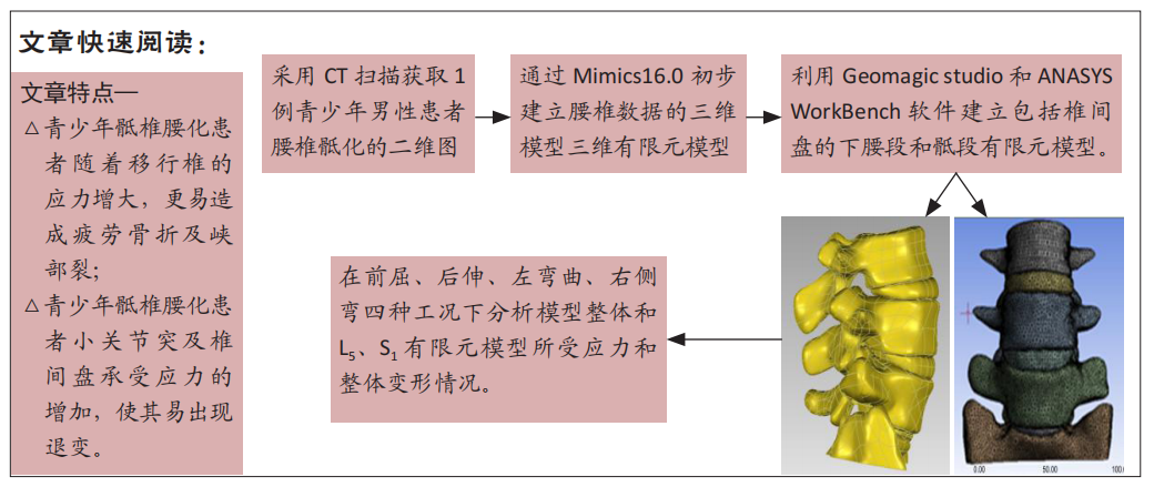
方法:选择1例骶椎腰化16岁男性青少年患者的CT影像,利用 mimics 16.0初步建立腰椎数据的三维模型、导入Geomagic studio和 ANASYS WorkBench 软件采用三维数字医学建模方法,建立包括椎间盘的下腰段和骶段有限元模型。通过施加2 N·mm的力矩模拟加载正常人体载荷后前屈、后伸、左侧弯、右侧弯运动对模型进行生物力学测试,分析各工况条件下移位椎的力学变化特性。试验于2018-03-05经内蒙古医科大学伦理委员会批准,批准号YKD2018015。
结果与结论:在伸屈活动时L5及S1椎弓根承受应力显著大于侧弯活动,移行的S1椎弓根应力变化方向与正常成人L5椎弓根峡部应力变化方向相同。青少年骶椎腰化患者,移行椎在腰椎活动中,S1椎弓根峡部所承受的应力明显大于L5椎弓根峡部,其小关节突及椎间盘承受应力更大。提示对于青少年骶椎腰化患者,在腰椎活动中随着移行椎的应力增大,更易造成疲劳骨折及峡部裂,而小关节突及椎间盘承受应力的增加,使其易出现退变。
http://orcid.org/0000-0003-1684-4518 (Li Zhijun)
中国组织工程研究杂志出版内容重点:人工关节;骨植入物;脊柱;骨折;内固定;数字化骨科;组织工程
中图分类号:
引用本文
吴雪海, 王 星, 张少杰, 徐雪彬, 史 君, 王铁英, 李志军. 有限元模型建立青少年骶椎腰化的验证分析[J]. 中国组织工程研究, 2021, 25(27): 4283-4288.
Wu Xuehai, Wang Xing, Zhang Shaojie, Xu Xuebin, Shi Jun, Wang Tieying, Li Zhijun. Establishment and validation of a finite element model of sacral lumbarization in adolescents[J]. Chinese Journal of Tissue Engineering Research, 2021, 25(27): 4283-4288.
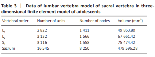
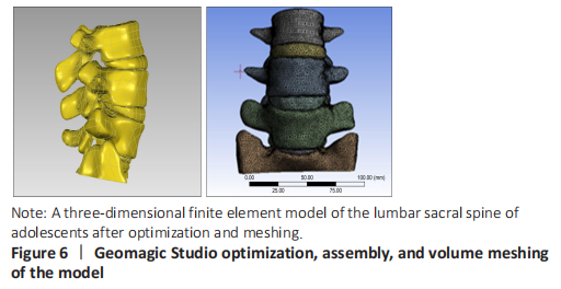
In this study, a three-dimensional finite element model of lumbar sacral vertebrae in adolescents was established based on the data of patients with lumbar sacral vertebrae, including 22 815 elements and 12 785 nodes (Table 3). The model is highly simulated, the geometric structure is good, the appearance of the model is close to the structure of sacral lumbar data, and has a high degree of reduction (Table 3 and Figure 6).
Stress analysis of lumbar vertebrae of sacral vertebrae under different working conditions in three-dimensional finite element model of adolescents
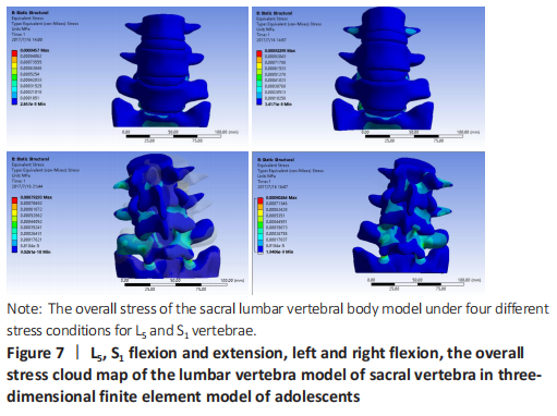
When the torque of 2 N·mm was applied to the lumbar vertebra finite element model of sacral vertebrae, the stresses of L5 and S1 under lumbar vertebrae of sacral vertebrae were as follows: during flexion, the stress was mainly concentrated in the facet joint of S1Unix 2, followed by the posterior structure of each vertebra. During the extension, the stress was mainly concentrated in the S1ax 2 facet joint, and decreased in turn from S1 to upward, which was basically consistent with the flexion movement. In the left and right side flexion, the stress is mainly concentrated in the S1hip 2 facet joint, followed by the superior articular process of S1, and decreases upward from S1. For the symmetry of human body development, the stress and strain of the model under the right bending condition is the same as that of the left bending. In the axial torsion of the left and right sides, the stress is greater in the anterior part of the C2-3 vertebral body, and the stress is also obvious in the facet joint and pedicle of the opposite side of the vertebral body. For L5 and S1 vertebrae, the maximum stress appeared in the inferior articular process of the vertebrae under flexion and extension, followed by the isthmus of the pedicle of the vertebrae, and in the condition of left and right lateral bending, the maximum stress appeared in the inferior articular process of the vertebrae, followed by the superior articular process of the vertebrae, and then in the isthmus of the pedicle of the vertebrae (Figure 7).
Strain and displacement analysis of lumbar vertebrae of sacral vertebrae under different working conditions in adolescents three-dimensional finite element model
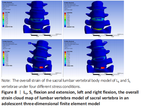
In the stress analysis of the model, the maximum strain was located in the intervertebral disc tissue under different working conditions, and the strain law was that S1 decreased upward, and the maximum displacement was located at the anterior upper edge of L4 during flexion. Under the left bending condition, the maximum strain of the model was S1Scan2, and there was little difference in stress between L4-5 and L5/S1 (Figure 7). To analyze the displacement changes of the model under different working conditions, during flexion and extension, the maximum displacement is located at the upper edge of L4, and the displacement decreases successively from L4 to the sacrum. Under the condition of left and right side bending, the maximum displacement is located at the front and upper edge of L4, and the displacement decreases successively from L4 to downward (Figure 8).
| [1] MITRA R, CARLISLE M. Bertolotti’s syndrome: a case report. Pain Pract. 2009;9(2): 152-154. [2] LV YP, Lin W, Ma XM. The localization of iliolumbar ligaments in lumbar column segmentation. Fangshe Xue Shijian. 2016;31(11):1080-1083. [3] JANCUSKA JM, SPIVAK JM, BENDO JA. A Review of Symptomatic Lumbosacral Transitional Vertebrae: Bertolotti’s Syndrome. Int J Spine Surg. 2015;9:42. [4] ZHAO S. Imaging study of percutaneous intervertebral foramen approach in lumbosacral 1. Suzhou: Suzhou University. 2015. [5] GONG H, SLUNSKY P, KLASS LG, et al. Brunnberg L. Prevalence of lumbosacral transitional vertebrae in dogs in Berlin. Pol J Vet Sci. 2020;23(2):261-265. [6] SEKHARAPPA V, AMRITANAND R, KRISHNAN V, et al. Lumbosacral transition vertebra: prevalence and its significance. Asian Spine J. 2014;8(1):51-58. [7] TANG M, YANG XF, YANG SW, et al. Lumbosacral transitional vertebra in a population-based study of 5860 individuals: prevalence and relationship to low back pain. Eur J Radiol. 2014;83(9):1679-1682. [8] MAHATO NK, DHASON R, RAM DR. Quantifying Range of Motion and Stress Patterns at the Transitional Lumbosacral Junction: Pilot Study Using a Computational Model for Load-Bearing at Accessory L5-S1 Articulation. Int J Spine Surg. 2019;13(1):17-23. [9] MAHATO NK. Trabecular bone structure in lumbosacral transitional vertebrae: distribution and densities across sagittal vertebral body segments. Spine J. 2013 Aug;13(8):932-937. [10] JARAMILLO HE, GÓMEZ L, GARCÍA JJ. A finite element model of the L4-L5-S1 human spine segment including the heterogeneity and anisotropy of the discs. Acta Bioeng Biomech. 2015;17(2):15-24. [11] LIN ZY, XIE YS, XIE PS, et al. Construction and accuracy test of lumbar intelligent medical image modeling system. Zhongguo Linchuang Jiepou Xue Zazhi. 2020; 38(4):444-449. [12] SHIRAZI-ADL A, AHMED AM, SHRIVASTAVA SC. Mechanical response of a lumbar motion segment in axial torque alone and combined with compression. Spine (Phila Pa 1976). 1986;11(9):914-927. [13] SCHMIDT H, HEUER F, DRUMM J, et al. Application of a calibration method provides more realistic results for a finite element model of a lumbar spinal segment. Clin Biomech (Bristol, Avon). 2007;22(4):377-384. [14] LIU YK, RAY G, HIRSCH C. The resistance of the lumbar spine to direct shear. Orthop Clin North Am. 1975;6(1):33-49. [15] HU Y, XIE H, YANG SH. Utilization of three- dimensional finite element method in spinal biomechanics. Yiyong Shengwu Lixue. 2006;21(3):246-250. [16] TOKGOZ N, UCAR M, ERDOGAN AB, et al. Are spinal or paraspinal anatomic markers helpful for vertebral numbering and diagnosing lumbosacral transitional vertebrae?. Korean J Radiol. 2014;15(2):258-266. [17] GIRDLER S, CHO B, MIKHAIL CM, et al. Emerging Techniques in Diagnostic Imaging for Idiopathic Scoliosis in Children and Adolescents: A Review of the Literature. World Neurosurg. 2020;136:128-135. [18] JANCUSKA JM, SPIVAK JM, BENDO JA. A Review of Symptomatic Lumbosacral Transitional Vertebrae: Bertolotti’s Syndrome. Int J Spine Surg. 2015;9:42. [19] MAHATO NK. Implications of structural variations in the human sacrum: why is an anatomical classification crucial?. Surg Radiol Anat. 2016;38(8):947-954. [20] LUOMA K, VEHMAS T, KERTTULA L, et al. Chronic low back pain in relation to Modic changes, bony endplate lesions, and disc degeneration in a prospective MRI study. Eur Spine J. 2016;25(9):2873-2881. [21] HOU LS, BAI XD, GE F, et al. Castellvi’s classification type-Ⅱa lumbarization combined with L5-S2 disc herniation: a case report. Dier Junyi Daxue Xuebao. 2018;39(10):1177-1179. [22] HANHIVAARA J, MÄÄTTÄ JH, NIINIMÄKI J, et al. Lumbosacral transitional vertebrae are associated with lumbar degeneration: retrospective evaluation of 3855 consecutive abdominal CT scans. Eur Radiol. 2020;30(6):3409-3416. [23] DANG L, CHEN ZQ LIU XG, et al. Etiology of lumbar disc herniation in adolescents: excessive load on the lumbar spine and aberrant configurations of the lumbosacral region. Zhongguo Jizhu Jisui Zazhi. 2015;25(11):991-996. [24] FUJIWARA A, TAMAI K, KURIHASHI A, et al. Relationship between morphology of iliolumbar ligament and lower lumbar disc degeneration. J Spinal Disord. 1999; 12(4):348-352. [25] AHN SS, CHIN DK, KIM SH, et al. The Clinical Significance of Lumbosacral Transitional Vertebrae on the Surgical Outcomes of Lumbar Discectomy: A Retrospective Cohort Study of Young Adults. World Neurosurg. 2017;99:745-750. [26] BERLEMANN U, JESZENSZKY DJ, BÜHLER DW, et al. Facet joint remodeling in degenerative spondylolisthesis: an investigation of joint orientation and tropism. Eur Spine J. 1998;7(5):376-380. [27] HAMMERBERG KW. New concepts on the pathogenesis and classification of spondylolisthesis. Spine (Phila Pa 1976). 2005;30(6 Suppl):S4-S11. [28] JIANG HC, WANG JX, YANG XD, et al. Analysis of the related factors of lumbar spondylolysis. Zhongguo Linchuang Jiepou Xue Zazhi. 2019;37(5):583-585,589. [29] KANEMATSU R, HANAKITA J, TAKAHASHI T, et al. Extraforaminal entrapment of the fifth lumbar spinal nerve by nearthrosis in patients with lumbosacral transitional vertebrae. Eur Spine J. 2020;29(9):2215-2221. [30] ALBANO D, MESSINA C, GAMBINO A, et al. Segmented lordotic angles to assess lumbosacral transitional vertebra on EOS. Eur Spine J. 2020;29(10):2470-2476. |
| [1] | 王建平, 张晓辉, 余进伟, 魏绍亮, 张新民, 许幸新, 曲海军. 三维图像配准及坐标变换膝关节运动分析在机构学中的应用[J]. 中国组织工程研究, 2022, 26(在线): 1-5. |
| [2] | 许新忠, 吴钟汉, 余水生, 赵 耀, 徐春归, 张 鑫, 郑嵋戈, 荆珏华. 斯氏针置入股骨头不同方式的生物力学分析[J]. 中国组织工程研究, 2022, 26(9): 1313-1317. |
| [3] | 魏国强, 李云峰, 王 一, 牛晓芬, 车丽芳, 王海燕, 李志军, 史国鹏, 白 灵, 莫 凯, 张晨晨, 许阳阳, 李筱贺. 非均匀材料股骨在不同负荷情况下的生物力学分析[J]. 中国组织工程研究, 2022, 26(9): 1318-1322. |
| [4] | 吕倩忆, 陈芯仪, 郑慧娥, 何灏龙, 李棋龙, 陈楚淘, 田浩梅. 不同角度肘按法对正常人腰椎椎体及后部结构的应力和位移分析[J]. 中国组织工程研究, 2022, 26(9): 1346-1350. |
| [5] | 张玉芳, 吕 蒙, 梅 钊. 青少年脊柱侧弯全脊柱生物力学模型的构建及验证[J]. 中国组织工程研究, 2022, 26(9): 1351-1356. |
| [6] | 张吉超, 董跃福, 牟志芳, 张 震, 李冰言, 徐祥钧, 李佳意, 任 梦, 董万鹏. 骨关节炎患者在不同步态角度下膝关节内部生物力学变化的有限元分析[J]. 中国组织工程研究, 2022, 26(9): 1357-1361. |
| [7] | 白子兴, 曹旭含, 孙承颐, 杨艳军, 陈 思, 温建民, 林新晓, 孙卫东. 步态周期中踝关节有限元模型的构建及生物力学分析[J]. 中国组织工程研究, 2022, 26(9): 1362-1366. |
| [8] | 刘 峰, 冯 毅. 步态周期下不同克氏针张力带治疗髌骨横行骨折的有限元分析[J]. 中国组织工程研究, 2022, 26(9): 1367-1371. |
| [9] | 潘保顺, 方 镇, 高明杰, 方贵明 , 陈金水. 基于影像学数据设计带融合器的寰枢椎后路内固定系统[J]. 中国组织工程研究, 2022, 26(9): 1372-1376. |
| [10] | 姚晓玲, 彭建城, 许岳荣, 杨志东, 张顺聪. 可变角度零切迹前路椎间融合内固定系统治疗脊髓型颈椎病:30个月随访[J]. 中国组织工程研究, 2022, 26(9): 1377-1382. |
| [11] | 王 帅, 王连成, 张书豪, 李富丽, 董佳兴, 张亚杰. 青少年特发性脊柱侧凸患者凸凹侧椎旁肌肌电比值与Cobb角、顶椎偏距、冠状面平衡距离的相关性[J]. 中国组织工程研究, 2022, 26(9): 1402-1406. |
| [12] | 金 涛, 刘 林, 朱晓燕, 史宇悰, 牛建雄, 张同同, 吴树金, 杨青山. 骨关节炎与线粒体异常[J]. 中国组织工程研究, 2022, 26(9): 1452-1458. |
| [13] | 肖 豪, 刘 静, 周 君. 脉冲电磁场治疗绝经后骨质疏松症的研究进展[J]. 中国组织工程研究, 2022, 26(8): 1266-1271. |
| [14] | 刘冬铖, 赵继军, 周子红, 吴沼锋, 俞颖豪, 陈宇浩, 冯德宏. 开放楔形胫骨高位截骨手术不同力线矫正参考方法的比较[J]. 中国组织工程研究, 2022, 26(6): 827-831. |
| [15] | 温明韬, 梁学振, 李嘉程, 许 波, 李 刚 . 两种方式固定Sanders Ⅱ型跟骨骨折后的力学稳定性[J]. 中国组织工程研究, 2022, 26(6): 838-842. |
For patients with lumbar sacral vertebrae, the analysis of the maximum load of each vertebral segment and the trend of mechanical changes is the basis of clinical treatment of low back pain caused by lumbar vertebrae, but at present, there are few mechanical simulation studies. Studies on the mechanics of lumbar pedicle in adolescent patients with transitional vertebrae are even less[10-11]. In this study, the CT images of adolescent patients with lumbar sacralization were selected to obtain three-dimensional finite element models of lower lumbar vertebrae of adolescent sacral lumbar vertebrae, and their stress and strain distribution and mechanical characteristics were analyzed, so as to provide carrier and basis for further study of mechanical changes and bone fatigue coefficient of lower lumbar vertebrae in transitional vertebrae patients.
中国组织工程研究杂志出版内容重点:人工关节;骨植入物;脊柱;骨折;内固定;数字化骨科;组织工程
A three-dimensional finite element analysis.
Time and settings
The experiment was conducted in 2019 at the Digital Medical Center of Inner Mongolia Medical University.
Subjects
One patient with lumbar sacral vertebrae was selected, a 16-year-old male of Han nationality, with a body mass of 60 kg, height of 1.75 m and body mass index (19.59 kg/m2). The volunteer had no previous history of lumbar trauma and related diseases. We obtain written informed consent from patient and his guardian. The research was approved by the Ethics Committee of Inner Mongolia Medical University (approval number YKD2018015) on March 5, 2018.
Methods
Acquisition of two-dimensional data
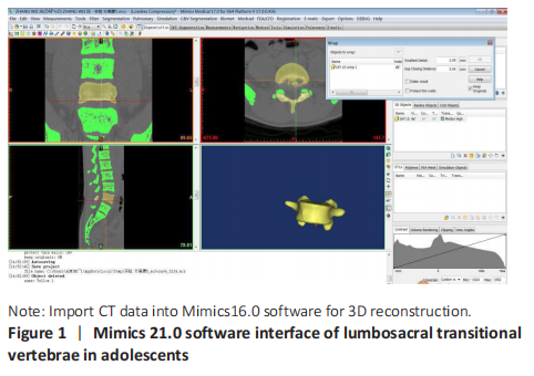
In this study, the Lightspeed dual-source 64-slice spiral CT (GE, USA) was used to scan the upper edge of the L1 vertebra to the lower edge of the L5 vertebra using the volume scan mode. Lie on your back with your head forward. Scanning parameters: slice thickness 5 mm, layer spacing 5 mm, scanning field 20 cm2, matrix 512 × 512, standard algorithm reconstruction. At the end of the scan, the data is divided into layer thickness 0.625 mm, layer spacing 0.625 mm and saved in DICOM format (Figure 1).
3D reconstruction of 2D data
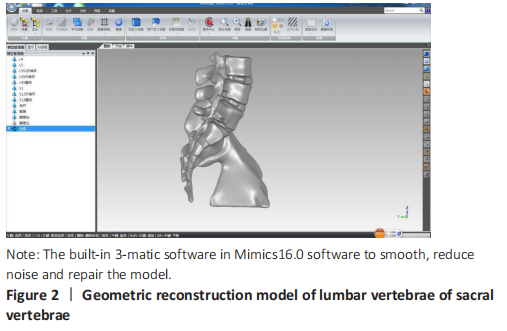
Import the original DICOM data into Mimics16.0 (Materialise Belgium), through the steps of threshold setting, image segmentation, region growth, image filling and so on, the L4-sacrum is segmented and smoothed, and then the Mask, of each vertebra is obtained through the Calculate3D module to get the 3D image of the vertebra. Then use the 3-matic software in Mimics16.0 software to optimize the model and export it in STL format (Figure 2).
Reconstruction of 3D solid model
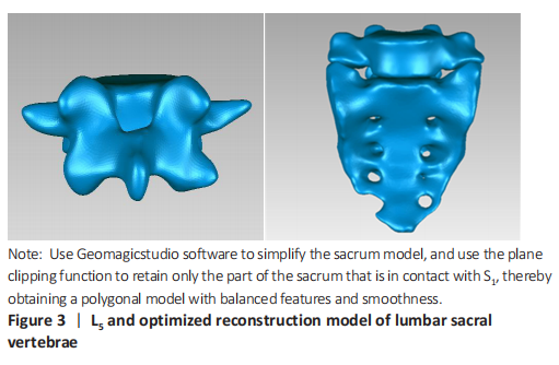
After Mimics image extraction and 3-matic model repair, the complete vertebral models of L5 and sacrum were obtained. At present, the appearance of each model is close to the real vertebra, with regular vertebrae, transverse process, spinous process, upper and lower articular process and other main structures. After introducing the model into Geomagicstudio (Geomagic, USA) the model unit is first selected, which is millimeter by default, and the system automatically prompts for grid doctor examination. After this processing, the polygon model of feature and smoothness balance is obtained. Simplify the sacrum model, use the plane clipping function, retain only the part of the sacrum in contact with S1, and carefully close the clipping plane, using the grid doctor to check to avoid causing the model to be unclosed (Figure 3).
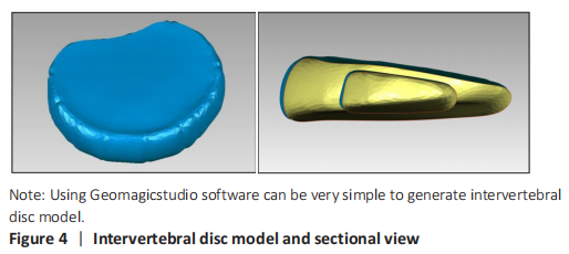
Making of lumbar intervertebral disc model
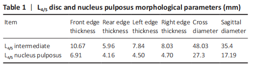
The real lumbar vertebra is a complex motion system composed of vertebrae, intervertebral discs, ligaments, muscles and so on. If you want to carry out finite element analysis, the most simplified model should at least include vertebrae and intervertebral discs. It is very easy to make intervertebral disc models with Geomagic Studio software. The shape of the intervertebral disc model made in the experiment is lifelike, showing a disk shape similar to the solid, the leading edge is thick, the trailing edge is flat, and the edge is smooth without edges and corners. The nucleus pulposus is located slightly behind the center of the intervertebral disc on the cross section and in the middle of the intervertebral disc on the coronal plane. After measurement, the relevant parameters of intervertebral disc and nucleus pulposus are consistent with the real human body. After assembling, the intervertebral disc and adjacent vertebrae can be closely attached, the relative position of intervertebral disc and vertebra is correct, and there are no pathological manifestations such as intervertebral disc herniation, which can meet the needs of further experiments (Table 1 and Figure 4).
Assembly and volume meshing of lumbosacral transitional vertebrae
Through the previously described work, a high-quality triangular model of the lumbar spine and intervertebral disc was obtained, but it is not a CAD model and it needs to be further transformed into a NURBS surface solid model and exported in IGES, sat, X_T, and other formats in order for the data to be smoothly imported into the WorkBench finite element analysis software (ANSYS, USA). The specific workflow is as follows, using the L4/5 disc as an example:
(1) After importing the model into Geomagic Studio, the Exact Surfaces Phase is clicked and Exact Surface is clicked.
(2) Click the Detect Curvature method and select Auto Estimate and Simplify Contour.
(3) Using the Construct Patch tool, the area was re-divided. There were occasional errors such as intersecting paths, different sizes of surface patches in the structure, or mismatch between the distribution pattern and the actual shape of the model, and further manual editing of the surface patches was required.
(4) Use the Move Panel function under the Move option to edit the patch.
(5) Construct the grid, click on the Construct Grid button, click on Repair Intersection Area, and check the geometry option, and use a resolution of 20 to get the model image that covers the grid.
(6) Click on the fitted surface to transform the model formed by the grid into a NURBS surface and optimize the smoothness option to further smooth the model. Then, save the NURBS surface in IGES or STEP format for the next step.
Import of finite element model into WorkBench and calculations
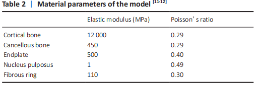
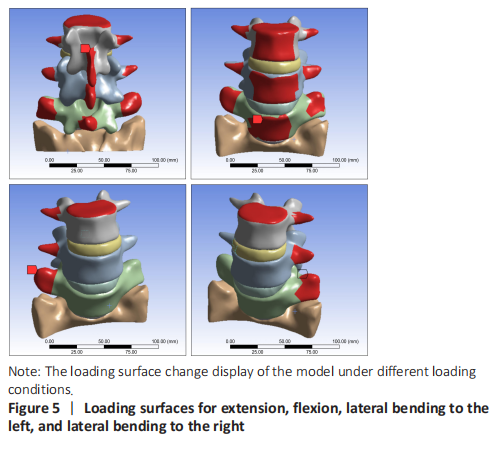
The assembled model was imported into WorkBench as described previously[11-13]. The corresponding material properties were assigned to the vertebrae, the annulus fibrosus, and the nucleus pulposus. Next, the assembled model and properties were assigned, the mesh was defined, and the contact surface, boundary conditions, and load were determined. For loading, a 2 N·mm force was applied for the analysis and calculation under the conditions of flexion, extension, lateral bending to the left, and lateral bending to the right. The stress and overall deformation of the model as a whole and the L5 and S1 finite element models were calculated when the flexion, extension, and bending moments were 2 N·mm (Table 2 and Figure 5).
Main outcome measures
To analyze the mechanical properties and stress-strain rules of lumbar vertebrae under simulated normal exercise conditions.
In 1975, LIU et al.[14] constructed the three-dimensional finite element model of vertebral body for the first time in the study of direct shear resistance of lumbar vertebrae, which marked the beginning of the application of three-dimensional finite element method in the study of spinal biomechanics. With the in-depth study of spinal diseases, three-dimensional finite element method has been used and developed to a great extent. Clinicians can collect data from practical problems, use three-dimensional finite element method to design and construct a simulation model, analyze the model, and determine the treatment plan and effect evaluation combined with the actual situation[15-16]. In lumbar diseases, lumbosacral transitional vertebra is a common spinal abnormality, including lumbar sacralization and sacral lumbar vertebrae, which are two distinct developmental abnormalities[17]. Estimates of the prevalence of lumbosacral transitional vertebrae in the general population vary widely throughout the literature, ranging from 4.0% to 31.9%, with an average of 12.3%. However, lumbosacral transitional vertebrae can affect the degeneration of intervertebral disc above lumbosacral segment and affect the function of innervating muscles or nerves[18-20]. The degenerative changes above the abnormal joints are caused by overactivity and abnormal torque above the lumbosacral transitional vertebrae and restricted movement between L5 and S1. Facet joint asymmetry (such as CastellviIIA) is mainly related to low back pain, leading to the possibility of discogenic pain[21-22]. From the point of view of human mechanics, the changes of lumbosacral transitional vertebrae on the structure of lumbar motion segments are completely different, one is the increase of lumbar motion segments, and the other is the decrease of lumbar motion segments. Theoretically, it should be two different mechanical models. In the related works, we can see that most of the studies on lumbosacral transitional vertebrae basically refer to lumbar sacralization, and the related research works on low back pain induced by this have been more mature. DANG et al.[23] reported that disc herniation in patients without transitional vertebrae or with lumbar sacralization mainly occurred in L4/L5. However, the disc herniation in patients with lumbar spondylolisthesis occurs in L5xS1, and the excessive limitation of L5/S1 lumbar joint will lead to stress conduction, which makes the intervertebral disc and vertebrae of L4cyan5 easily damaged, while insufficient restriction will lead to L5/S1 stress increase and damage[24]. According to the study of AHN et al.[25], patients with lumbar disc herniation with LSTV have a gap in postoperative pain symptoms and recovery of living ability compared with non-LSTV patients. And lumbar back pain increased in varying degrees
12-24 months after lumbar disc-related surgery, so the study of transitional vertebrae has a very important clinical significance. Abnormal anatomical morphology of lumbosacral vertebrae also plays an important role in the occurrence of degenerative spondylolisthesis. BERLEMANN et al.[26] suggested that abnormal spinal arrangement and abnormal direction of lower lumbar endplate may be high risk factors for lumbar spondylolisthesis. HAMMERBERG [27] found that lumbar degenerative spondylolisthesis is closely related to the increase of pelvic incidence angle. In addition, the abnormality of joint paroxysm, the degeneration of articular process and the change of direction and asymmetry of articular process are all related to the occurrence of lumbosacral transitional vertebrae[28-30]. In comparison, there are few references for the mechanical causes of pain induced by lumbar sacral vertebrae. The finite element model of lumbar vertebrae of sacral vertebrae established in this study provides a mechanical reference for clinical low back pain caused by this cause by analyzing the stress, strain and displacement of L5/S1 vertebrae.
Characteristics and mechanical analysis of finite element model of lumbosacral transitional vertebrae
In this study, finite element analysis was used to simulate the mechanical analysis of a patient with lumbar vertebrae under four working conditions of flexion, extension, left bending and right bending. It was found that the maximum load on the lumbar spine of the patients with lumbar vertebra increased gradually to the lumbar pedicle isthmus of S1, so the stress of the pedicle isthmus of lumbar vertebra was significantly higher than that of lumbar 5 pedicle isthmus during lumbar movement. It is more likely to cause fatigue fracture and isthmus fissure, and its facet process and intervertebral disc bear greater stress, so it is more prone to degeneration. The stress of L5 and S1 pedicle isthmus and articular process on the same side of the movement direction was significantly higher than that of the contralateral pedicle isthmus during the left and right bending movement. Therefore, compared with the normal human body, the stress of the lumbar pedicle isthmus is greater than that of the normal human body, and the stress of each pedicle of the complete sacral lumbar vertebra model is also significantly higher than that of the lateral bending movement; and the stress change direction of S1 pedicle isthmus is the same as that of L5 pedicle isthmus in normal people. For the adolescent patients with thorough lumbar vertebrae, the stress in the isthmus of the pedicle of the transitional vertebra is significantly higher than that of the L5 pedicle isthmus, which is more likely to lead to fatigue fracture and isthmus, and the facet process bears greater stress and is more prone to degeneration. The corresponding abnormal S1 and S2 intervertebral disc becomes the most strained part of all lumbar and sacral intervertebral discs, in addition, it is generally poorly developed. Pathological changes such as disc degeneration and protrusion are more likely to occur in lumbosacral movement. Therefore, long-term engaged in weightlifting, volleyball, swimming and other sports activities or manual labor that requires repeated extension and flexion of the lumbar spine, due to frequent reciprocating one-way lateral bending of the lumbar spine, it is more likely to occur superior contralateral lumbar articular process degeneration and pedicle injury. It is also prone to transitional spondylolysis, and its pathogenic factors are consistent with lumbar spondylolysis in normal people. If the course of isthmus fracture is similar to that of normal L5 isthmus fissure, the same internal fixation can be used as L5 isthmus fissure, which is of guiding significance for clinical treatment.
Deficiency and prospect of the research
In this experiment, the finite element analysis can be used to make an effective mechanical analysis of the lumbar sacral vertebrae of a teenager, in order to find its stress characteristics and stress changes, but it is not representative. Only the use of this technique is only the mechanical analysis of adolescent sacral lumbar vertebrae, which provides a theoretical basis for clinical precision and personalized treatment in the later stage.
中国组织工程研究杂志出版内容重点:人工关节;骨植入物;脊柱;骨折;内固定;数字化骨科;组织工程
文题释义:#br# 腰骶移行椎:指腰椎数目变化及其伴随的L5 和S1 的形态结构变化。一般情况下整个脊柱的椎骨总数不变,只是各节段数目有所增减,可分为骶椎腰化和腰椎骶化。#br# 有限元模型分析法:是近年来研究人体生物力学的有效方法,其基本原理是把连续的物体离散为一组有限个,并按一定方式相互联结在一起的单元组合体,然后求其相互之间的关系及特征。
| 阅读次数 | ||||||
|
全文 |
|
|||||
|
摘要 |
|
|||||