Chinese Journal of Tissue Engineering Research ›› 2014, Vol. 18 ›› Issue (38): 6090-6098.doi: 10.3969/j.issn.2095-4344.2014.38.005
Previous Articles Next Articles
Platelet-derived growth factor-B gene transfection reduces ischemia and hypoxia-induced myocardial apoptosis
Chen Bang-dang1, Chen Xiao-cui1, Ma Yi-tong1, 2, Yang Yi-ning1, 2, Ma Xiang2, Liu Fen1
- 1Xinjiang Key Laboratory of Cardiovascular Disease, Clinical Medical Research Institute, First Affiliated Hospital of Xinjiang Medical University, Urumqi 830054, Xinjiang Uygur Autonomous Region, China; 2Heart Center, First Affiliated Hospital of Xinjiang Medical University, Urumqi 830054, Xinjiang Uygur Autonomous Region, China
-
Received:2014-08-04Online:2014-09-10Published:2014-09-10 -
Contact:Ma Yi-tong, M.D., Chief physician, Professor, Doctoral supervisor, Xinjiang Key Laboratory of Cardiovascular Disease, Clinical Medical Research Institute, First Affiliated Hospital of Xinjiang Medical University, Urumqi 830054, Xinjiang Uygur Autonomous Region, China; Heart Center, First Affiliated Hospital of Xinjiang Medical University, Urumqi 830054, Xinjiang Uygur Autonomous Region, China -
About author:Chen Bang-dang, Master, Research assistant, Xinjiang Key Laboratory of Cardiovascular Disease, Clinical Medical Research Institute, First Affiliated Hospital of Xinjiang Medical University, Urumqi 830054, Xinjiang Uygur Autonomous Region, China -
Supported by:the Natural Science Foundation of Xinjiang Uygur Autonomous Region, No. 2011211B34; the National Natural Science Foundation of China, No. 81160026; the Youth Foundation of First Affiliated Hospital of Xinjiang Medical University, No. 2011QN04
CLC Number:
Cite this article
Chen Bang-dang, Chen Xiao-cui, Ma Yi-tong, Yang Yi-ning, Ma Xiang, Liu Fen.
share this article
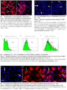
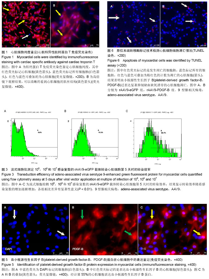
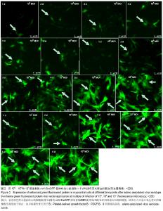
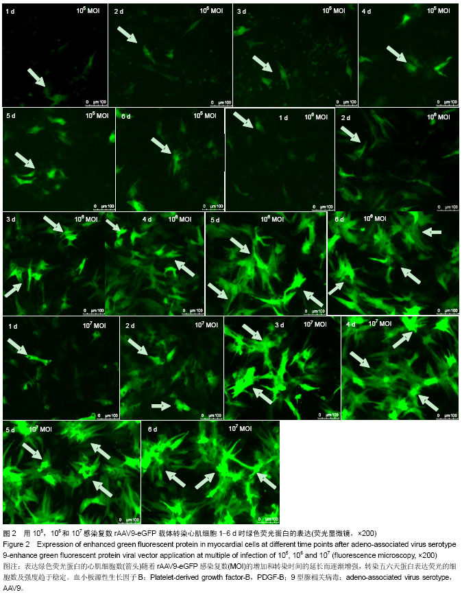
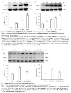
2.1 心肌细胞的培养及纯度鉴定 取培养2 d的细胞,用心肌特异性肌钙蛋白T进行免疫荧光鉴定,于荧光倒置显微镜下观察计数,红色荧光标记的是心肌细胞,蓝色荧光标记视野下所有的细胞核,计数后,心肌细胞的纯度可达到98%以上(图1A),400倍荧光显微镜下可以清晰看到心肌细胞的肌丝结构(图1B),说明采用胰蛋白酶结合胶原酶消化心肌组织、二次差速贴壁及添加BrdU的分离培养方法可获得高纯度心肌细胞,有效抑制非心肌细胞污染。 2.2 rAAV9-eGFP载体转染效率 用感染复数为1×10⁵、1×10⁶和1×10⁷ MOI转染心肌细胞后,倒置荧光显微镜下观察发现eGFP的表达随着感染复数的增加和转染时间的延长而逐渐增强(图2)。在病毒转染1 d后可见少量心肌细胞表达,转染两三天表达eGFP阳性的细胞逐渐增加,随着时间的延长,eGFP阳性的细胞逐渐增加,转染五六天荧光表达的细胞数及强度趋于稳定,转染第9天仍有表达,但随着细胞的衰老表达强度略有减弱。 收集1×10⁵、1×10⁶和1×10⁷ MOI的rAAV9-eGFP转染第5天的心肌细胞,流式细胞仪测定其转染效率分别为(8.23±0.49)%、(19.60±0.65)%、(60.90±1.13)%,见图3,各组相比差异有显著性意义(P < 0.01),表明rAAV9-eGFP载体对心肌细胞有较高的亲和力,可高效转染乳鼠心肌细胞。 2.3 Western Blot检测PDGF-B蛋白表达结果 分别收集1×10⁵、1×10⁶和1×10⁷ MOI的rAAV9-PDGF-B转染第5天时的心肌细胞,提取细胞总蛋白,Western Blot检测发现PDGF-B蛋白的表达随着感染复数的增加而逐渐增强(图4A,B),与内参GAPDH蛋白相比,其表达分别为(31.51±4.80)%,(54.86±5.22)%,(86.51±5.56)%,各组相比差异有显著性意义(P < 0.01)。 1×10⁷ MOI的rAAV9-PDGF-B转染1-5 d的心肌细胞,PDGF-B蛋白的表达水平随着转染时间的延长而逐渐增强(图4C,D),与内参GAPDH相比,其表达分别为(28.45±2.88)%,(46.25±2.84)%,(57.79±2.93)%,(71.92± 2.25)%,(86.51± 5.56)%,各组相比差异有显著性意义(P < 0.05),表明rAAV9-PDGF-B载体转染心肌细胞后可高效正确表达PDGF-B蛋白。 2.4 免疫荧光鉴定PDGF-B蛋白表达结果 取转染后第5天的心肌细胞进行检测,在倒置荧光显微镜下观察,红色荧光为表达PDGF-B的心肌细胞,用DAPI(蓝色)复染细胞核,经计算约55%的心肌细胞表达PDGF-B蛋白(图5)。 2.5 心肌细胞凋亡检测结果 取缺血缺氧培养24 h的心肌细胞,用TUNEL染色检测细胞凋亡数目,其中红色标记凋亡的细胞核,蓝色标记所有的细胞核,红色与蓝色可叠加为粉红色的计数为凋亡的细胞(图6),计数后,rAAV9- PDGF-B组心肌细胞凋亡率为(20.10±2.50)%,而rAAV9-eGFP组为(30.00±3.40)%,PDGF-B的过表达可使心肌细胞凋亡率减少33%,两组相比差异有显著性意义 (P < 0.05),说明PDGF-B蛋白表达可有效抵抗缺血缺氧诱导心肌细胞凋亡的作用。 2.6 细胞凋亡蛋白检测结果 Western blot检测各组细胞Bax表达,经分析正常对照组Bax表达为(9.54±1.01)%,eGFP组为(20.90±2.67)%、PDGF-B组为(12.10±2.08)%。与正常对照相比,PDGF-B组Bax水平无明显差异,与eGFP组相比Bax水平显著下调(P < 0.05)。 Caspase-3表达变化趋势与Bax基本一致,其表达对照组为(7.85±0.66)%,eGFP组为(12.99±1.92)%,而PDGF-B组为(8.82±0.67)%,与eGFP组相比Caspase-3的表达明显下调(P < 0.05)(图7)。结果表明,PDGF-B蛋白表达可有效抑制促凋亡蛋白Bax和Caspase-3的表达。"
"
"

| [1] Yi GY, Zhang X, Zhang J, et al. Factors related to the use of reperfusion strategies in elderly patients with acute myocardial infarction. J Cardiothorac Surg. 2014;9(1):111. [2] Nascimento BR, de Sousa MR, Demarqui FN, et al. Risks and benefits of thrombolytic, antiplatelet, and anticoagulant therapies for ST segment elevation myocardial infarction: systematic review. ISRN Cardiol. 2014:1-11. [3] Tao Z, Chen B, Tan X, et al. Coexpression of VEGF and angiopoietin-1 promotes angiogenesis and cardiomyocyte proliferation reduces apoptosis in porcine myocardial infarction (MI) heart. PNAS. 2011;108(5):2064-2069. [4] Wang G, Hamid T, Keith RJ, et al. Cardioprotective and antiapoptotic effects of heme oxygenase-1 in the failing heart. Circulation. 2010;121(17):1912-1925. [5] Vantler M, Karikkineth BC, Naito H, et al. PDGF-BB protects cardiomyocytes from apoptosis and improves contractile function of engineered heart tissue. J Mol Cell Cardiol. 2010; 48(6):1316-1323. [6] Lange S, Heger J, Euler G, et al. Platelet-derived growth factor BB stimulates vasculogenesis of embryonic stem cell-derived endothelial cells by calcium- mediated generation of reactive oxygen species. Cardiovasc Res. 2009;81(1): 159-168. [7] Scimia MC, Gumpert AM, Koch WJ. Cardiovascular gene therapy for myocardial infarction. Expert Opin Biol Ther. 2014; 14(2):183-115. [8] Korpisalo P, Ylä-Herttuala S. Stimulation of functional vessel growth by gene therapy. Integr Biol (Camb). 2010;2(2-3): 102-112. [9] Prasad KM, Xu Y, Yang Z, et al. Robust cardiomyocyte- specific gene expression following systemic injection of AAV: in vivo gene delivery follows a Poisson distribution. Gene Ther. 2011;18(1):43-52. [10] Pacak CA, Mah CS, Thattaliyath BD, et al. Recombinant adeno-associated virus serotype 9 leads to preferential cardiac transduction in vivo. Circ Res. 2006;99(4):e3-e9. [11] Inagaki K, Fuess S, Storm TA, et al. Robust systemic transduction with AAV9 vectors in mice: efficient global cardiac gene transfer superior to that of AAV8. Mol Ther. 2006;14(1):45-53. [12] 向阳,马依彤,杨毅宁,等.重组9型腺相关病毒载体转染小鼠心脏及对心功能的影响[J].中华微生物学和免疫学杂志,2009,29(10): 899-902. [13] Pulicherla N, Shen S, Yadav S, et al. Engineering liver-detargeted AAV9 vectors for cardiac and musculoskeletal gene transfer. Mol Ther. 2011;19(6): 1070-1078. [14] Bostick B, Ghosh A, Yue Y, et al. Systemic AAV9 transduction in mice is influenced by animal age but not by the route of administration. Gene Therapy. 2007;14(22): 1605-1609. [15] The Ministry of Science and Technology of the People’s Republic of China. Guidance Suggestions for the Care and Use of Laboratory Animals. 2006-09-30. [16] Sreejit P, Kumar S, Verma RS. An improved protocol for primary culture of cardiomyocyte from neonatal mice. In Vitro Cell Dev Biol Anim. 2008;44(3-4):45-50. [17] Lu DF, Yao Y, Su ZZ, et al. Downregulation of HDAC1 is involved in the cardiomyocyte differentiation from mesenchymal stem cells in a myocardial microenvironment. PLoS One. 2014;9(4):e93222. [18] Excoffon KJ, Gerber JT, Dickey DD. et al. Directed evolution of adeno-associated virus to an infectious respiratory virus. PNAS. 2009;106(10):3865-3870. [19] 杜雷,马依彤,杨毅宁,等.9型重组腺相关病毒转染大鼠心脏成纤维细胞的体外研究[J].心血管康复医学杂志,2010,19(2): 121-125. [20] Martherus RS, Zeijlemaker VA, Ayoubi TA. Electrical stimulation of primary neonatal rat ventricular cardiomyocytes using pacemakers. Biotechniques. 2010;48(1):65-67. [21] 刘霞,王常勇.新生大鼠心肌细胞的分离和培养[J].西北农林科技大学学报,2005,33(6):36-39. [22] 邸美仙,李应东.乳鼠心肌细胞的培养[J].中华中医药学刊, 2005, 25(4):691-692. [23] Nickson P, Toth A, Erhardt P, et al. PUMA is critical for neonatal cardiomyocyte apoptosis induced by endoplasmic reticulum stress. Cardiovasc Res. 2007;73(1):48-56. [24] Hajjar RJ. Potential of gene therapy as a treatment for heart failure. J Clin Invest. 2013;123(1):53-61. [25] Yang L, Xiao X. Creation of a cardiotropic adeno-associated virus: the story of viral directed evolution. Virol J. 2013;10:50. [26] Hadad I, Veithen A, Springael JY, et al. Stroma cell-derived factor-1alpha signaling enhances calcium transients and beating frequency in rat neonatal cardiomyocytes. PLoS One. 2013;8(2):e56007. [27] Nguyen PD, Hsiao ST, Sivakumaran P, et al. Enrichment of neonatal rat cardiomyocytes in primary culture facilitates long-term maintenance of contractility in vitro. Am J Physiol cell Physiol. 2012;303(12):C1220-C1228. [28] Louch WE, Sheehan KA, Wolska BM. Methods in cardiomyocyte isolation, culture, and gene transfer. J Mol Cell Cardiol. 2011;51(3):288-298. [29] 高霞,马依彤,杨毅宁,等.9型重组腺相关病毒转染体外培养大鼠心肌细胞的影响[J].细胞与分子免疫学杂志,2010,26(01): 18-20. [30] 姚永钊,马依彤,赵爱超,等.腺相关病毒介导血小板源性生长因 子-B基因转染心肌细胞的研究[J].中华生物医学工程杂志, 2013, 19(5):377-381. [31] Bakar E, Ulucam E, Cerkezkayabekir A. Protective effects of proanthocyanidin and vitamin E against toxic effects of formaldehyde in kidney tissue. Biotech Histochem. 2014; 1-10. [32] Yang L, Yang Q, Zhang K, et al. Neuroprotective Effects of Daphnetin against NMDA Receptor-Mediated Excitotoxicity. Molecules. 2014;19(9):14542-14555. [33] Yang SD, Bai ZL, Zhang F, et al. Levofloxacin increases the effect of serum deprivation on anoikis of rat nucleus pulposus cells via Bax/Bcl-2/caspase-3 pathway. Toxicol Mech Methods. 2014:1-21. [34] Zhang J, Shen MH, Ruan SM. Beta-HIVS combined cisplatin inhibited activities of human ovarian cancer cell line SKOV3 in vitro. Zhongguo Zhong Xi Yi Jie He Za Zhi. 2014;34(8): 987-990. [35] Zhao JY, Liu LY, Shen AL, et al. Effect of bear bile powder on STAT3 pathway in hepatocellular carcinoma xenograft. Zhongguo Zhong Xi Yi Jie He Za Zhi. 2014 Aug;34(8):976-81. Chinese. [36] Song E, Fu J, Xia X, et al. Bazhen Decoction Protects against Acetaminophen Induced Acute Liver Injury by Inhibiting Oxidative Stress, Inflammation and Apoptosis in Mice. PLoS One. 2014;9(9):e107405. [37] Kukreja RC, Xi L. eNOS phosphorylation: a pivotal molecular switch in vasodilatation and cardioprotection. J Mol Cell Cardiol. 2007;42(2):280-282. [38] Bajt ML, Farhood A, Lemasters JJ. et al. Mitochondrial bax translocation accelerates DNA fragmentation and cell necrosis in a murine model of acetaminophen hepatotoxicity. J Pharmacol Exp Ther. 2008;324(1):8-14. |
| [1] | Zhang Tongtong, Wang Zhonghua, Wen Jie, Song Yuxin, Liu Lin. Application of three-dimensional printing model in surgical resection and reconstruction of cervical tumor [J]. Chinese Journal of Tissue Engineering Research, 2021, 25(9): 1335-1339. |
| [2] | Geng Qiudong, Ge Haiya, Wang Heming, Li Nan. Role and mechanism of Guilu Erxianjiao in treatment of osteoarthritis based on network pharmacology [J]. Chinese Journal of Tissue Engineering Research, 2021, 25(8): 1229-1236. |
| [3] | Pei Lili, Sun Guicai, Wang Di. Salvianolic acid B inhibits oxidative damage of bone marrow mesenchymal stem cells and promotes differentiation into cardiomyocytes [J]. Chinese Journal of Tissue Engineering Research, 2021, 25(7): 1032-1036. |
| [4] | Zeng Yanhua, Hao Yanlei. In vitro culture and purification of Schwann cells: a systematic review [J]. Chinese Journal of Tissue Engineering Research, 2021, 25(7): 1135-1141. |
| [5] | Li Shibin, Lai Yu, Zhou Yi, Liao Jianzhao, Zhang Xiaoyun, Zhang Xuan. Pathogenesis of hormonal osteonecrosis of the femoral head and the target effect of related signaling pathways [J]. Chinese Journal of Tissue Engineering Research, 2021, 25(6): 935-941. |
| [6] | Xu Yinqin, Shi Hongmei, Wang Guangyi. Effects of Tongbi prescription hot compress combined with acupuncture on mRNA expressions of apoptosis-related genes,Caspase-3 and Bcl-2, in degenerative intervertebral discs [J]. Chinese Journal of Tissue Engineering Research, 2021, 25(5): 713-718. |
| [7] | Zhang Wenwen, Jin Songfeng, Zhao Guoliang, Gong Lihong. Mechanism by which Wenban Decoction reduces homocysteine-induced apoptosis of myocardial microvascular endothelial cells in rats [J]. Chinese Journal of Tissue Engineering Research, 2021, 25(5): 723-728. |
| [8] | Liu Qing, Wan Bijiang. Effect of acupotomy therapy on the expression of Bcl-2/Bax in synovial tissue of collagen-induced arthritis rats [J]. Chinese Journal of Tissue Engineering Research, 2021, 25(5): 729-734. |
| [9] | Xie Chongxin, Zhang Lei. Comparison of knee degeneration after anterior cruciate ligament reconstruction with or without remnant preservation [J]. Chinese Journal of Tissue Engineering Research, 2021, 25(5): 735-740. |
| [10] | Xu Dongzi, Zhang Ting, Ouyang Zhaolian. The global competitive situation of cardiac tissue engineering based on patent analysis [J]. Chinese Journal of Tissue Engineering Research, 2021, 25(5): 807-812. |
| [11] | Wu Zijian, Hu Zhaoduan, Xie Youqiong, Wang Feng, Li Jia, Li Bocun, Cai Guowei, Peng Rui. Three-dimensional printing technology and bone tissue engineering research: literature metrology and visual analysis of research hotspots [J]. Chinese Journal of Tissue Engineering Research, 2021, 25(4): 564-569. |
| [12] | Chang Wenliao, Zhao Jie, Sun Xiaoliang, Wang Kun, Wu Guofeng, Zhou Jian, Li Shuxiang, Sun Han. Material selection, theoretical design and biomimetic function of artificial periosteum [J]. Chinese Journal of Tissue Engineering Research, 2021, 25(4): 600-606. |
| [13] | Liu Fei, Cui Yutao, Liu He. Advantages and problems of local antibiotic delivery system in the treatment of osteomyelitis [J]. Chinese Journal of Tissue Engineering Research, 2021, 25(4): 614-620. |
| [14] | Li Xiaozhuang, Duan Hao, Wang Weizhou, Tang Zhihong, Wang Yanghao, He Fei. Application of bone tissue engineering materials in the treatment of bone defect diseases in vivo [J]. Chinese Journal of Tissue Engineering Research, 2021, 25(4): 626-631. |
| [15] | Zhang Zhenkun, Li Zhe, Li Ya, Wang Yingying, Wang Yaping, Zhou Xinkui, Ma Shanshan, Guan Fangxia. Application of alginate based hydrogels/dressings in wound healing: sustained, dynamic and sequential release [J]. Chinese Journal of Tissue Engineering Research, 2021, 25(4): 638-643. |
| Viewed | ||||||
|
Full text |
|
|||||
|
Abstract |
|
|||||