Chinese Journal of Tissue Engineering Research ›› 2022, Vol. 26 ›› Issue (31): 5008-5013.doi: 10.12307/2022.955
Previous Articles Next Articles
Effect and mechanism of conditioned medium from hypoxia preconditioned human Wharton’s Jelly derived mesenchymal stem cells on myocardial ischemia/reperfusion injury
Li Yan1, 2, Zhang Yan2, Zhang Ningkun2, Chen Yu1, 2
- 1Naval Clinical College, Anhui Medical University; The Fifth Clinical Medicine, Anhui Medical University, Hefei 230032, Anhui Province, China; 2The Sixth Medical Center of PLA General Hospital, Beijing 100048, China
-
Received:2021-12-17Accepted:2022-01-25Online:2022-11-08Published:2022-04-24 -
Contact:Chen Yu, MD, Chief physician, Professor, Master’s supervisor, Naval Clinical College, Anhui Medical University; The Fifth Clinical Medicine, Anhui Medical University, Hefei 230032, Anhui Province, China; The Sixth Medical Center of PLA General Hospital, Beijing 100048, China -
About author:Li Yan, Master candidate, Naval Clinical College, Anhui Medical University; The Fifth Clinical Medicine, Anhui Medical University, Hefei 230032, Anhui Province, China; The Sixth Medical Center of PLA General Hospital, Beijing 100048, China -
Supported by:National Natural Science Foundation of China, No. 81370238 (to CY)
CLC Number:
Cite this article
Li Yan, Zhang Yan, Zhang Ningkun, Chen Yu. Effect and mechanism of conditioned medium from hypoxia preconditioned human Wharton’s Jelly derived mesenchymal stem cells on myocardial ischemia/reperfusion injury[J]. Chinese Journal of Tissue Engineering Research, 2022, 26(31): 5008-5013.
share this article
Add to citation manager EndNote|Reference Manager|ProCite|BibTeX|RefWorks

2.2 H9C2细胞的形态及建模情况 H9C2细胞贴壁生长,呈椭圆形、三角形、梭形等多形性,两端有角长出,有折光性,予以低氧培养6 h后细胞形态无明显变化,可见少量漂浮细胞,见图2A。H9C2细胞加入WJMSC条件上清后镜下观察细胞生长快,形态无明显变化,见图2B。试剂盒检测显示模型组H9C2细胞上清中乳酸脱氢酶活力为(266.00±28.58) U/L,较常氧对照组[(63.67±9.02) U/L]明显升高(P < 0.001),表明缺血再灌注损伤模型构建成功;加入常氧WJMSC条件上清后H9C2细胞上清中乳酸脱氢酶活力为(212.3±14.29) U/L,加入低氧WJMSC条件上清后乳酸脱氢酶活力为(160.7±11.06) U/L,较模型组均明显降低(P < 0.05和P < 0.001),说明加入WJMSC条件上清液后H9C2细胞损伤减少,见图2C。"


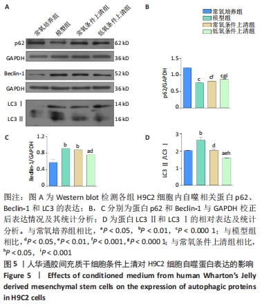
2.4 WJMSC条件上清对缺血再灌注损伤H9C2细胞自噬的调节 Western blot检测显示,与常氧培养组相比,模型组p62蛋白表达降低,Beclin-1、LC3Ⅱ/Ⅰ表达升高(P < 0.01或P < 0.000 1),表明经缺氧复氧及无血清处理后H9C2细胞自噬水平升高;与模型组相比,常氧条件上清组、低氧条件上清组p62蛋白表达升高(P < 0.001或P < 0.000 1),LC3Ⅱ/Ⅰ表达降低(P < 0.05或P < 0.01),自噬水平降低,而Beclin-1蛋白表达差异无显著性意义;低氧条件上清组较常氧条件上清组p62蛋白表达升高,LC3Ⅱ/Ⅰ表达降低,差异均有显著性意义(P < 0.001或P < 0.05),见图5。"
| [1] YELLON DM, HAUSENLOY DJ. Myocardial reperfusion injury. N Engl J Med. 2007;357(11):1121-1135. [2] ABBASZADEH H, GHORBANI F, DERAKHSHANI M, et al. Regenerative potential of Wharton’s jelly-derived mesenchymal stem cells: A new horizon of stem cell therapy. J Cell Physiol. 2020;235(12):9230-9240. [3] RANJBARAN H, ABEDIANKENARI S, MOHAMMADI M, et al. Wharton’s Jelly Derived-Mesenchymal Stem Cells: Isolation and Characterization. Acta Med Iran. 2018;56(1):28-33. [4] MARINO L, CASTALDI MA, ROSAMILIO R, et al. Mesenchymal Stem Cells from the Wharton’s Jelly of the Human Umbilical Cord: Biological Properties and Therapeutic Potential. Int J Stem Cells. 2019;12(2):218-226. [5] 张宁坤,陈宇,王志国,等.华通胶源间充质干细胞经冠状动脉移植治疗慢性缺血性心肌病的实验研究[J].解放军医学杂志,2015, 40(11):885-891. [6] LI L, LI L, ZHANG Z, et al. Hypoxia promotes bone marrow-derived mesenchymal stem cell proliferation through apelin/APJ/autophagy pathway. Acta Biochim Biophys Sin (Shanghai). 2015;47(5):362-367. [7] LIU H, SUN X, GONG X, et al. Human umbilical cord mesenchymal stem cells derived exosomes exert antiapoptosis effect via activating PI3K/Akt/mTOR pathway on H9C2 cells. J Cell Biochem. 2019;120(9):14455-14464. [8] SCIARRETTA S, MAEJIMA Y, ZABLOCKI D, et al. The Role of Autophagy in the Heart. Annu Rev Physiol. 2018;80:1-26. [9] GAO LR, ZHANG NK, DING QA, et al. Common expression of stemness molecular markers and early cardiac transcription factors in human Wharton’s jelly-derived mesenchymal stem cells and embryonic stem cells. Cell Transplant. 2013;22(10):1883-1900. [10] ISHIUCHI N, NAKASHIMA A, DOI S, et al. Hypoxia-preconditioned mesenchymal stem cells prevent renal fibrosis and inflammation in ischemia-reperfusion rats. Stem Cell Res Ther. 2020;11(1):130. [11] ZHANG Z, YANG C, SHEN M, et al. Autophagy mediates the beneficial effect of hypoxic preconditioning on bone marrow mesenchymal stem cells for the therapy of myocardial infarction. Stem Cell Res Ther. 2017;8(1):89. [12] QIAN L, SHI J, ZHANG C, et al. Downregulation of RACK1 is associated with cardiomyocyte apoptosis after myocardial ischemia/reperfusion injury in adult rats. In Vitro Cell Dev Biol Anim. 2016;52(3):305-313. [13] 中国心血管健康与疾病报告编写组.中国心血管健康与疾病报告2020概要[J].中国循环杂志,2021,36(6):521-545. [14] ARSLAN F, LAI RC, SMEETS MB, et al. Mesenchymal stem cell-derived exosomes increase ATP levels, decrease oxidative stress and activate PI3K/Akt pathway to enhance myocardial viability and prevent adverse remodeling after myocardial ischemia/reperfusion injury. Stem Cell Res. 2013;10(3):301-312. [15] WANG M, YAN L, LI Q, et al. Mesenchymal stem cell secretions improve donor heart function following ex vivo cold storage. J Thorac Cardiovasc Surg. 2020;(20):32487-32489. [16] LEE TL, LAI TC, LIN SR, et al. Conditioned medium from adipose-derived stem cells attenuates ischemia/reperfusion-induced cardiac injury through the microRNA-221/222/PUMA/ETS-1 pathway. Theranostics. 2021;11(7):3131-3149. [17] NAZARINIA D, SHARIFI M, DOLATSHAHI M, et al. FoxO1 and Wnt/β-catenin signaling pathway: Molecular targets of human amniotic mesenchymal stem cells-derived conditioned medium (hAMSC-CM) in protection against cerebral ischemia/reperfusion injury. J Chem Neuroanat. 2021;112:101918. [18] ZHANG Q, LIU X, PIAO C, et al. Effect of conditioned medium from adipose derived mesenchymal stem cells on endoplasmic reticulum stress and lipid metabolism after hepatic ischemia reperfusion injury and hepatectomy in swine. Life Sci. 2022;289:120212. [19] 张蘋,郭莹,高亚杰,等.低氧预处理人脐带间充质干细胞促进其源性外泌体对心肌梗死后心肌损伤的修复[J].中国组织工程研究, 2019,23(17):2630-2636. [20] LEE JH, YOON YM, LEE SH. Hypoxic Preconditioning Promotes the Bioactivities of Mesenchymal Stem Cells via the HIF-1alpha-GRP78-Akt Axis. Int J Mol Sci. 2017;18(6):1320. [21] HU X, XU Y, ZHONG Z, et al. A Large-Scale Investigation of Hypoxia-Preconditioned Allogeneic Mesenchymal Stem Cells for Myocardial Repair in Nonhuman Primates. Circulation Research. 2016;118(6):970-983. [22] OBRADOVIC H, KRSTIC J, TRIVANOVIC D, et al. Improving stemness and functional features of mesenchymal stem cells from Wharton’s jelly of a human umbilical cord by mimicking the native, low oxygen stem cell niche. Placenta. 2019;82:25-34. [23] MENG SS, XU XP, CHANG W, et al. LincRNA-p21 promotes mesenchymal stem cell migration capacity and survival through hypoxic preconditioning. Stem Cell Res Ther. 2018;9(1):280. [24] ZHAO D, LIU L, CHEN Q, et al. Hypoxia with Wharton’s jelly mesenchymal stem cell coculture maintains stemness of umbilical cord blood-derived CD34(+) cells. Stem Cell Res Ther. 2018;9(1):158. [25] YU H, XU Z, QU G, et al. Hypoxic Preconditioning Enhances the Efficacy of Mesenchymal Stem Cells-Derived Conditioned Medium in Switching Microglia toward Anti-inflammatory Polarization in Ischemia/Reperfusion. Cell Mol Neurobiol. 2021;41(3):505-524. [26] LI X, HE S, MA B. Autophagy and autophagy-related proteins in cancer. Mol Cancer. 2020;19(1):12. [27] YOSHII SR, MIZUSHIMA N. Monitoring and Measuring Autophagy. Int J Mol Sci. 2017;18(9):1865. [28] MARTANO M, ALTAMURA G, POWER K, et al. Beclin 1, LC3 and P62 Expression in Equine Sarcoids. Animals (Basel). 2021;12(1):20. [29] LI Q, HAN Y, DU J, et al. Alterations of apoptosis and autophagy in developing brain of rats with epilepsy: Changes in LC3, P62, Beclin-1 and Bcl-2 levels. Neurosci Res. 2018;130:47-55. [30] LI Y, LIANG P, JIANG B, et al. CARD9 promotes autophagy in cardiomyocytes in myocardial ischemia/reperfusion injury via interacting with Rubicon directly. Basic Res Cardiol. 2020;115(3):29. [31] KANG X, LI C, XIE Y, et al. Hippocampal ornithine decarboxylase/spermidine pathway mediates H2S-alleviated cognitive impairment in diabetic rats: Involving enhancment of hippocampal autophagic flux.J Adv Res. 2021;27:31-40. [32] GUAN G, YANG L, HUANG W, et al. Mechanism of interactions between endoplasmic reticulum stress and autophagy in hypoxia/reoxygenationinduced injury of H9c2 cardiomyocytes. Mol Med Rep. 2019;20(1):350-358. [33] CHEN G, WANG M, RUAN Z, et al. Mesenchymal stem cell-derived exosomal miR-143-3p suppresses myocardial ischemia-reperfusion injury by regulating autophagy. Life Sci. 2021;280:119742. |
| [1] | Wang Shuo, Liu Wenying, Lü Chaofan, Li Jiacong, Geng Yi, Zhao Yungang. Cardioprotective effect of 3-nitro-N-methyl salicylamide on the isolated rat heart under cold ischemia preservation [J]. Chinese Journal of Tissue Engineering Research, 2022, 26(8): 1194-1201. |
| [2] | Yin Tingting, Du Dayong, Jiang Zhixin, Liu Yang, Liu Qilin, Li Yuntian. Granulocyte colony-stimulating factors improve myocardial fibrosis in rats with myocardial infarction [J]. Chinese Journal of Tissue Engineering Research, 2022, 26(5): 730-735. |
| [3] | Dong Miaomiao, Lai Han, Li Manling, Xu Xiuhong, Luo Meng, Wang Wenhao, Zhou Guoping. Effect of electroacupuncture on expression of nucleotide binding oligomerization domain-like receptor protein 3/cysteinyl aspartate specific proteinase 1 in rats with cerebral ischemia/reperfusion injury [J]. Chinese Journal of Tissue Engineering Research, 2022, 26(5): 749-755. |
| [4] | Yang Qian, Zhang Yiou, Jia Lili, Xie Jun, Feng Mali, Li Tingkai. Establishment and disease progression in a rat myocardial infarction model [J]. Chinese Journal of Tissue Engineering Research, 2022, 26(23): 3733-3737. |
| [5] | Zhang Yu, Tian Shaoqi, Zeng Guobo, Hu Chuan. Risk factors for myocardial infarction following primary total joint arthroplasty [J]. Chinese Journal of Tissue Engineering Research, 2021, 25(9): 1340-1345. |
| [6] | Nie Huijuan, Huang Zhichun. The role of Hedgehog signaling pathway in transforming growth factor beta1-induced myofibroblast transdifferentiation [J]. Chinese Journal of Tissue Engineering Research, 2021, 25(5): 754-760. |
| [7] | Lang Limin, He Sheng, Jiang Zengyu, Hu Yiyi, Zhang Zhixing, Liang Minqian. Application progress of conductive composite materials in the field of tissue engineering treatment of myocardial infarction [J]. Chinese Journal of Tissue Engineering Research, 2021, 25(22): 3584-3590. |
| [8] | Sun Weixing, Zhao Yongchao, Zhao Ranzun. Mesenchymal stem cell transplantation in the treatment of myocardial infarction: problems, crux and new breakthrough [J]. Chinese Journal of Tissue Engineering Research, 2021, 25(19): 3103-3109. |
| [9] | Chen Siyu, Li Yannan, Xie Liying, Liu Siqi, Fan Yurong, Fang Changxing, Zhang Xin, Quan Jiayu, Zuo Lin. Thermosensitive chitosan-collagen composite hydrogel loaded with basic fibroblast growth factor retards ventricular remodeling after myocardial infarction in mice [J]. Chinese Journal of Tissue Engineering Research, 2021, 25(16): 2472-2478. |
| [10] | Shang Qingqing, Zhou Jianye. Combination of hyaluronic acid hydrogel and bone marrow mesenchymal stem cells promotes cardiac function after myocardial infarction in rats (II) [J]. Chinese Journal of Tissue Engineering Research, 2020, 24(34): 5559-5563. |
| [11] |
Shang Zhizhong, Jiang Yanbiao, Yao Lan, Wang Anan, Wang Hongxia, Tian Yuanxin, Liu Dengrui, Ma Bin .
Feasibility of stem cell therapy for renal ischemia/reperfusion injury: a systematic review based on animal experiments [J]. Chinese Journal of Tissue Engineering Research, 2020, 24(25): 4094-4100. |
| [12] |
Chen Siyu, Zhang Tao, Yin Wenjuan, Cai Lei, Li Yannan, Xie Liying, Zuo Lin.
Changes of cardiac function in rats with myocardial
infarction after umbilical cord-derived mesenchymal stem cell transplantation:
a meta-analysis |
| [13] | He Jigang, Xie Qiaoli, Wang Zihao, Yan Dan, Zhang Hongbo. Time-volume variation in miR-330-3p expression in GATA-4-overexpressing bone marrow-derived mesenchymal stem cell exosomes [J]. Chinese Journal of Tissue Engineering Research, 2019, 23(29): 4617-4622. |
| [14] | Long Yanfang1, 2, Wang Xinlei2, Wang Mingpu3, Tang Xingjiang2. Effects of androgen on the expression of Bcl-2, Bax and Cyt-C in brain tissue of adult rat models of middle cerebral artery occlusion [J]. Chinese Journal of Tissue Engineering Research, 2019, 23(27): 4344-4349. |
| [15] | Zhang Pin, Guo Ying, Gao Yajie, Wang Zhendong, Li Baiyi, Zhang Xiaomin, Niu Yuhu, Liu Zhizhen, Ma Lihui, Niu Bo, Guo Rui. Exosomes derived from human umbilical cord mesenchymal stem cells promote myocardial repair after myocardial infarction under hypoxia [J]. Chinese Journal of Tissue Engineering Research, 2019, 23(17): 2630-2636. |
| Viewed | ||||||
|
Full text |
|
|||||
|
Abstract |
|
|||||
