Chinese Journal of Tissue Engineering Research ›› 2020, Vol. 24 ›› Issue (13): 2126-2132.doi: 10.3969/j.issn.2095-4344.2049
Changes of cardiac function in rats with myocardial
infarction after umbilical cord-derived mesenchymal stem cell transplantation:
a meta-analysis
Chen Siyu1, Zhang Tao2, Yin Wenjuan3, Cai Lei2, Li Yannan1, Xie Liying1, Zuo Lin1
- 1Key Laboratory of Cellular Physiology of Ministry of Education, Shanxi Provincial Key Laboratory, Department of Physiology, Shanxi Medical University, Taiyuan 030001, Shanxi Province, China; 2Department of Applied Psychology, 3School of Basic Medical Sciences, Shanxi Medical University, Taiyuan 030001, Shanxi Province, China
-
Received:2019-08-26Revised:2019-08-31Accepted:2019-10-15Online:2020-05-08Published:2020-03-11 -
Contact:Zuo Lin, MD, Associate professor, Key Laboratory of Cellular Physiology of Ministry of Education, Shanxi Provincial Key Laboratory, Department of Physiology, Shanxi Medical University, Taiyuan 030001, Shanxi Province, China -
About author:Chen Siyu, Master candidate, Key Laboratory of Cellular Physiology of Ministry of Education, Shanxi Provincial Key Laboratory, Department of Physiology, Shanxi Medical University, Taiyuan 030001, Shanxi Province, China -
Supported by:the Shanxi Provincial Youth Foundation, No. 201601D202106; the Scientific Research Fund for Returned Overseas Students in Shanxi Province, No. 2014-036; the Key Subject Construction of Shanxi “1331 Project” (1331KSC), No. XK201708
CLC Number:
Cite this article
Chen Siyu, Zhang Tao, Yin Wenjuan, Cai Lei, Li Yannan, Xie Liying, Zuo Lin.
Changes of cardiac function in rats with myocardial
infarction after umbilical cord-derived mesenchymal stem cell transplantation:
a meta-analysis
share this article
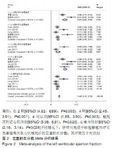
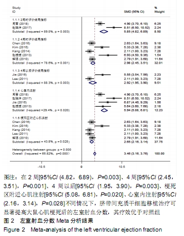
2.3 Meta分析结果 2.3.1 左室射血分数Meta分析结果 9篇文献报道了脐带间充质干细胞对大鼠心肌梗死后左室射血分数的影响[24-32],根据2周、4周和4周以后3个评价结局指标时间以及心室内注射和梗死心肌周围注射2种注射部位,将纳入的9篇文献分为5个亚组,进行了组间的差异对比分析。由于各研究结果之间存在异质性(I2 > 50%),故采用随机效应模型进行Meta分析。结果显示:①2周时,脐带间充质干细胞组的左室射血分数显著高于对照组,差异有显著性意义[SMD=5.85,95%CI (4.82,6.89),P=0.003],I2=89.0%,组内异质性高;②4周时,脐带间充质干细胞组的左室射血分数显著高于对照组,差异有显著性意义[SMD=2.98,95%CI(2.45,3.51),P=0.001],I2=78.6%,组内异质性较高;③4周之后,脐带间充质干细胞组的左室射血分数显著高于对照组,差异有显著性意义[SMD=2.92,95%CI (1.95,3.90),P=0.003],I2=88.3%,组内异质性较高;④心室内注射,脐带间充质干细胞组的左室射血分数显著高于对照组,差异有显著性意义[SMD=5.94,95%CI (5.08,6.81),P=0.020],I2=29.4%,亚组内异质性低;⑤梗死区附近心肌注射,脐带间充质干细胞组的左室射血分数显著高于对照组,差异有显著性意义[SMD=2.65,95%CI (2.16,3.14),P=0.028],I2=43.8%,亚组内异质性较低,见图2。 "
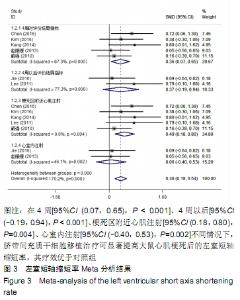
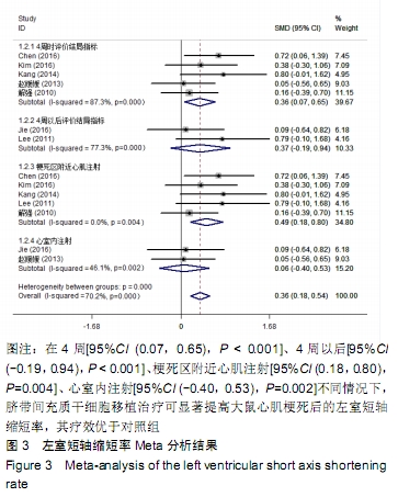
2.3.2 左室短轴缩短率Meta分析结果 7篇文献报道了脐带间充质干细胞对大鼠心肌梗死后左室短轴缩短率的影 响[26-32],根据4周和4周以后2个评价结局指标时间以及心室内注射和梗死心肌周围注射2种注射部位,将纳入的7篇文献分为4个亚组,进行了组间的差异对比分析。因为各研究结果之间存在异质性(I2 > 50%),故采用随机效应模型进行Meta分析。结果显示:①4周时,脐带间充质干细胞组的左室短轴缩短率显著高于对照组,差异有显著性意义[SMD=0.36,95%CI (0.07,0.65),P < 0.001],I2=87.3%,组内异质性高;②4周以后,脐带间充质干细胞组的左室短轴缩短率显著高于对照组,差异有显著性意义[SMD=0.37,95%CI (-0.19,0.94),P < 0.001],I2=77.3%,组内异质性较高;③梗死区附近心肌注射,脐带间充质干细胞组的左室短轴缩短率显著高于对照组,差异有显著性意义[SMD=0.49,95%CI (0.18,0.80),P=0.004],I2=0.0%,亚组内异质性小;④心室内注射,脐带间充质干细胞组的左室短轴缩短率显著高于对照组,差异有显著性意义[SMD=0.06,95%CI (-0.40,0.53),P=0.002],I2=46.1%,亚组内异质性较小,见图3。 "
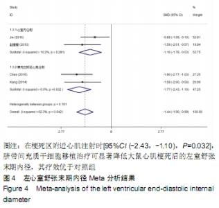
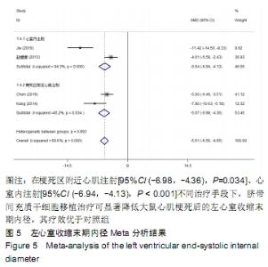
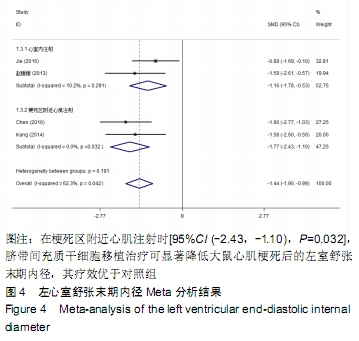
2.3.3 左室舒张末期内径和左室收缩末期内径Meta分析结果 5篇文献报道了脐带间充质干细胞对大鼠心肌梗死后左室舒张末期内径和左室收缩末期内径的影响[26-27,29-30,32],根据心室内注射和梗死心肌周围注射2种注射部位,将纳入的4篇文献分为2个亚组,进行了组间的差异对比。由于各研究关于左室舒张末期内径和左室收缩末期内径的异质性检验结果显示异质性较高(I2 > 50%),故使用随机效应模型进行分析。结果显示:①梗死区周围心肌组织注射脐带间充质干细胞移植治疗对大鼠心肌梗死后的左室舒张末期内径有显著减小作用,其疗效优于对照组,两组间差异有显著性意义[SMD=-1.77,95%CI (-2.43,-1.10),P=0.032],I2=0.0%,组内异质性较小;而心室内注射时两组差异无显著性意义[SMD=-1.16,95%CI (-1.78,-0.53),P=0.291],I2=10.2%,组内异质性较小,见图4;②梗死区附近心肌注射脐带间充质干细胞移植治疗的左室收缩末期内径显著小于对照组,差异有显著性意义[SMD=-5.67,95%CI (-6.98,-4.36),P=0.034],I2=45.2%,亚组内异质性低;心室内注射脐带间充质干细胞移植治疗的左室收缩末期内径显著高于对照组,差异有显著性意义[SMD=-5.54,95%CI (-6.94,-4.13),P < 0.001],I2= 94.3%,组内异质性高,见图5。 "
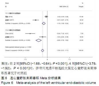
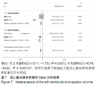
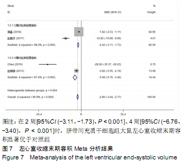
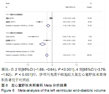
2.3.4 左室舒张末期容积和左室收缩末期容积Meta分析结果 4篇文献报道了脐带间充质干细胞对大鼠心肌梗死后左室舒张末期容积和左室收缩末期容积的影响[24-26,31],根据结局指标评价时间不同,将纳入的4篇文献分为2个亚组,进行了组间的差异对比。由于各研究关于左室舒张末期容积和左室收缩末期容积的异质性检验结果显示异质性较高(I2 > 50%),故使用随机效应模型进行分析。结果显示:①2周时,脐带间充质干细胞组的左室舒张末期容积显著优于对照组,差异有显著性意义[SMD=-1.16,95%CI (-1.68,-0.64),P < 0.001],I2=96.9%,但亚组内异质性高;4周时,脐带间充质干细胞组的左室舒张末期容积相对于对照组显著改善,差异有显著性意义[SMD=-2.85,95%CI (-3.78,-1.92),P < 0.001],I2=93.9%,亚组内异质性较高,见图6;②2周时,脐带间充质干细胞组的左室收缩末期容积显著小于对照组,差异有显著性意义[SMD=-2.42,95%CI (-3.11,-1.73),P < 0.001],I2=98.0%,亚组内异质性高;4周时,脐带间充质干细胞组的左室收缩末期容积相对于对照组显著改善,差异有显著性意义[SMD=-5.08,95%CI (-6.76,-3.40),P < 0.001],I2=97.4%,亚组内异质性高,见图7。 "
| [1] HUU AL, PRAKASH S, SHUM-TIM D. Cellular cardiomyoplasty: current state of the field. Regen Med. 2012;7(4):571-582. [2] BENJAMIN EJ, BLAHA MJ, CHIUVE SE, et al. Heart Disease and Stroke Statistics-2017 Update: A Report From the American Heart Association. Circulation. 2017;135(10):e146-e603. [3] BURNS RJ, GIBBONS RJ, YI Q, et al. The relationships of left ventricular ejection fraction, end-systolic volume index and infarct size to six-month mortality after hospital discharge following myocardial infarction treated by thrombolysis. J Am Coll Cardiol. 2002;39(1):30-36. [4] 胡盛寿,高润霖,刘力生,等.《中国心血管病报告2018》概要[J].中国循环杂志,2019,34(3):209-220. [5] LIU H, PAUL C, XU M. Optimal Environmental Stiffness for Stem Cell Mediated Ischemic Myocardium Repair. Methods Mol Biol. 2017;1553:293-304. [6] TURKSEN K. Adult stem cells and cardiac regeneration. Stem Cell Rev Rep. 2013;9(5):537-540. [7] KOUDSTAAL S, JANSEN OF LORKEERS SJ, GAETANI R, et al. Concise review: heart regeneration and the role of cardiac stem cells. Stem Cells Transl Med. 2013;2(6):434-443. [8] MU J, LI X, YUAN S, et al. Directional differentiation of human embryonic stem cells into cardiomyocytes by direct adherent culture. Journal of Histotechnology. 2014;37(4):125-131. [9] LI J, ZHU K, WANG Y, et al. Combination of IGF‑1 gene manipulation and 5‑AZA treatment promotes differentiation of mesenchymal stem cells into cardiomyocyte‑like cells. Mol Med Rep. 2015;11(2):815-820. [10] JUMABAY M, MATSUMOTO T, YOKOYAMA S, et al. Dedifferentiated fat cells convert to cardiomyocyte phenotype and repair infarcted cardiac tissue in rats. J Mol Cell Cardiol. 2009; 47(5):565-575. [11] WEGENER M, BADER A, GIRI S. How to mend a broken heart: adult and induced pluripotent stem cell therapy for heart repair and regeneration. Drug Discov Today. 2015;20(6):667-685. [12] HONG KU, BOLLI R. Cardiac stem cell therapy for cardiac repair. Curr Treat Options Cardiovasc Med. 2014;16(7):324. [13] NATSUMEDA M, FLOREA V, CASTELLANOS AM, et al. The Combination of Allogeneic Mesenchymal Stem Cells and Cardiac Stem Cells Produce Synergistic Effects in Cardiac Regeneration. Circulation. 2016;134:A19681. [14] KIM JT, CHUNG HJ, SEO JY, et al. A fibrin-supported myocardial organ culture for isolation of cardiac stem cells via the recapitulation of cardiac homeostasis. Biomaterials. 2015;48:66-83. [15] ZHENG SX, WENG YL, ZHOU CQ, et al. Comparison of cardiac stem cells and mesenchymal stem cells transplantation on the cardiac electrophysiology in rats with myocardial infarction. Stem Cell Rev Rep. 2013;9(3):339-349. [16] MUSIALEK P, MAZUREK A, JAROCHA D, et al. Myocardial regeneration strategy using Wharton's jelly mesenchymal stem cells as an off-the-shelf 'unlimited' therapeutic agent: results from the Acute Myocardial Infarction First-in-Man Study. Postepy Kardiol Interwencyjnej. 2015;11(2):100-107. [17] XIONG ZH, WEI J, LU MQ, et al. Protective effect of human umbilical cord mesenchymal stem cell exosomes on preserving the morphology and angiogenesis of placenta in rats with preeclampsia. Biomed Pharmacother. 2018;105:1240-1247. [18] SHELTON E, ALLEGRETTI JR, STEVENS B, et al. Efficacy of Vedolizumab as Induction Therapy in Refractory IBD Patients: A Multicenter Cohort. Inflamm Bowel Dis. 2015;21(12):2879-2885. [19] WAN M, CUI S, WEI W, et al. Bi-component synergic effect in lily-like CdS/Cu,7,S,4,QDs for dye degradation. Rsc Adv. 2019; 9(5): 2441-2450. [20] REYNOLDS K, GO AS, LEONG TK, et al. Trends in Incidence of Hospitalized Acute Myocardial Infarction in the Cardiovascular Research Network (CVRN). Am J Med. 2017;130(3):317-327. [21] JUN Y, CHUNJU Y, QI A, et al. The effects of compound danshen dripping pills and human umbilical cord blood mononuclear cell transplant after acute myocardial infarction. Exp Clin Transplant. 2014;12(2):123-128. [22] LI T, MA Q, NING M, et al. Cotransplantation of human umbilical cord-derived mesenchymal stem cells and umbilical cord blood-derived CD34⁺ cells in a rabbit model of myocardial infarction. Mol Cell Biochem. 2014;387(1-2):91-100. [23] LUNDH A, GØTZSCHE PC. Recommendations by Cochrane Review Groups for assessment of the risk of bias in studies. BMC Med Res Methodol. 2008;8:22. [24] 周蔷,邢燕.疏血通注射液联合心室内注射人脐带间充质干细胞对急性心肌梗死大鼠心功能的影响及机制探讨[J].河北中医,2018, 40(11):1709-1713. [25] 赵璐洋,孙瑛,李连冲.心室内注射人脐带间充质干细胞改善心肌梗死大鼠心功能及作用机制[J].中国组织工程研究, 2017,21(25):4026-4031. [26] CHEN G, YUE A, YU H, et al. Mesenchymal Stem Cells and Mononuclear Cells From Cord Blood: Cotransplantation Provides a Better Effect in Treating Myocardial Infarction. Stem Cells Transl Med. 2016;5(3):350-357. [27] MA J, ZHAO Y, SUN L, et al. Exosomes Derived from Akt-Modified Human Umbilical Cord Mesenchymal Stem Cells Improve Cardiac Regeneration and Promote Angiogenesis via Activating Platelet-Derived Growth Factor D. Stem Cells Transl Med. 2017;6(1):51-59. [28] KIM SW, JIN HL, KANG SM, et al. Therapeutic effects of late outgrowth endothelial progenitor cells or mesenchymal stem cells derived from human umbilical cord blood on infarct repair. Int J Cardiol. 2016;203:498-507. [29] KANG BJ, KIM H, LEE SK, et al. Umbilical-cord-blood-derived mesenchymal stem cells seeded onto fibronectin-immobilized polycaprolactone nanofiber improve cardiac function. Acta Biomater. 2014;10(7):3007-3017. [30] 赵媛媛,曹文明,马洁, 等.构建大鼠急性心肌梗死模型并初步探讨人脐带间充质干细胞的修复作用[J].临床检验杂志,2013, 31(12):919-922. [31] LEE EJ, CHOI EK, KANG SK, et al. N-cadherin determines individual variations in the therapeutic efficacy of human umbilical cord blood-derived mesenchymal stem cells in a rat model of myocardial infarction. Mol Ther. 2012;20(1):155-167. [32] 解强,江宏伟,白雪涛, 等.人脐血干细胞移植治疗大鼠心肌梗死的实验研究[J].成都医学院学报,2010,5(1):39-42. [33] KELLE S, ROES SD, KLEIN C, et al. Prognostic value of myocardial infarct size and contractile reserve using magnetic resonance imaging. J Am Coll Cardiol. 2009;54(19):1770-1777. [34] VELAGALETI RS, PENCINA MJ, MURABITO JM, et al. Long-term trends in the incidence of heart failure after myocardial infarction. Circulation. 2008;118(20):2057-2062. [35] WOLLERT KC, DREXLER H. Cell therapy for the treatment of coronary heart disease: a critical appraisal. Nat Rev Cardiol. 2010;7(4):204-215. [36] WANG G, ZHAO Q, CHENG Q, et al. Comparison short time discharge with long time discharge following uncomplicated percutaneous coronary intervention for Non-ST elevation myocardial infarction patients. BMC Cardiovasc Disord. 2019; 19(1):109. [37] WRITING GROUP MEMBERS, LLOYD-JONES D, ADAMS RJ, et al. Heart disease and stroke statistics--2010 update: a report from the American Heart Association. Circulation. 2010;121(7):e46-e215. [38] HASIN T, GERBER Y, WESTON SA, et al. Heart Failure After Myocardial Infarction Is Associated With Increased Risk of Cancer. J Am Coll Cardiol. 2016;68(3):265-271. [39] 王巍,李肖甫,李中健.不同孕周人脐带间充质干细胞移植改善心肌梗死模型心脏功能的比较[J].中国组织工程研究,2016,20(6):799-806. [40] TAKIZAWA S, NAGATA E, NAKAYAMA T, et al. Recent Progress in Endothelial Progenitor Cell Culture Systems: Potential for Stroke Therapy. Neurol Med Chir (Tokyo). 2016;56(6):302-309. |
| [1] | Zhang Yu, Tian Shaoqi, Zeng Guobo, Hu Chuan. Risk factors for myocardial infarction following primary total joint arthroplasty [J]. Chinese Journal of Tissue Engineering Research, 2021, 25(9): 1340-1345. |
| [2] | Zhang Chao, Lü Xin. Heterotopic ossification after acetabular fracture fixation: risk factors, prevention and treatment progress [J]. Chinese Journal of Tissue Engineering Research, 2021, 25(9): 1434-1439. |
| [3] | Chen Junming, Yue Chen, He Peilin, Zhang Juntao, Sun Moyuan, Liu Youwen. Hip arthroplasty versus proximal femoral nail antirotation for intertrochanteric fractures in older adults: a meta-analysis [J]. Chinese Journal of Tissue Engineering Research, 2021, 25(9): 1452-1457. |
| [4] | Chen Jinping, Li Kui, Chen Qian, Guo Haoran, Zhang Yingbo, Wei Peng. Meta-analysis of the efficacy and safety of tranexamic acid in open spinal surgery [J]. Chinese Journal of Tissue Engineering Research, 2021, 25(9): 1458-1464. |
| [5] | Hu Kai, Qiao Xiaohong, Zhang Yonghong, Wang Dong, Qin Sihe. Treatment of displaced intra-articular calcaneal fractures with cannulated screws and plates: a meta-analysis of 15 randomized controlled trials [J]. Chinese Journal of Tissue Engineering Research, 2021, 25(9): 1465-1470. |
| [6] | Huang Dengcheng, Wang Zhike, Cao Xuewei. Comparison of the short-term efficacy of extracorporeal shock wave therapy for middle-aged and elderly knee osteoarthritis: a meta-analysis [J]. Chinese Journal of Tissue Engineering Research, 2021, 25(9): 1471-1476. |
| [7] | Wang Yongsheng, Wu Yang, Li Yanchun. Effect of acute high-intensity exercise on appetite hormones in adults: a meta-analysis [J]. Chinese Journal of Tissue Engineering Research, 2021, 25(8): 1305-1312. |
| [8] | Lun Zhigang, Jin Jing, Wang Tianyan, Li Aimin. Effect of peroxiredoxin 6 on proliferation and differentiation of bone marrow mesenchymal stem cells into neural lineage in vitro [J]. Chinese Journal of Tissue Engineering Research, 2021, 25(7): 1014-1018. |
| [9] | Kong Desheng, He Jingjing, Feng Baofeng, Guo Ruiyun, Asiamah Ernest Amponsah, Lü Fei, Zhang Shuhan, Zhang Xiaolin, Ma Jun, Cui Huixian. Efficacy of mesenchymal stem cells in the spinal cord injury of large animal models: a meta-analysis [J]. Chinese Journal of Tissue Engineering Research, 2021, 25(7): 1142-1148. |
| [10] | Huang Dengcheng, Wang Zhike, Cao Xuewei. Intravenous, topical tranexamic acid alone or their combination in total knee arthroplasty: a meta-analysis of randomized controlled trials [J]. Chinese Journal of Tissue Engineering Research, 2021, 25(6): 948-956. |
| [11] | Li Yan, Wang Pei, Deng Donghuan, Yan Wei, Li Lei, Jiang Hongjiang. Electroacupuncture for pain control after total knee arthroplasty: a meta-analysis [J]. Chinese Journal of Tissue Engineering Research, 2021, 25(6): 957-963. |
| [12] | He Xiangzhong, Chen Haiyun, Liu Jun, Lü Yang, Pan Jianke, Yang Wenbin, He Jingwen, Huang Junhan. Platelet-rich plasma combined with microfracture versus microfracture in the treatment of knee cartilage lesions: a meta-analysis [J]. Chinese Journal of Tissue Engineering Research, 2021, 25(6): 964-969. |
| [13] | Hua Haotian, Zhao Wenyu, Zhang Lei, Bai Wenbo, Wang Xinwei. Meta-analysis of clinical efficacy and safety of antibiotic artificial bone in the treatment of chronic osteomyelitis [J]. Chinese Journal of Tissue Engineering Research, 2021, 25(6): 970-976. |
| [14] | Zhan Fangbiao, Cheng Jun, Zou Xinsen, Long Jie, Xie Lizhong, Deng Qianrong. Intraoperative intravenous application of tranexamic acid reduces perioperative bleeding in multilevel posterior spinal surgery: a meta-analysis [J]. Chinese Journal of Tissue Engineering Research, 2021, 25(6): 977-984. |
| [15] | Nie Huijuan, Huang Zhichun. The role of Hedgehog signaling pathway in transforming growth factor beta1-induced myofibroblast transdifferentiation [J]. Chinese Journal of Tissue Engineering Research, 2021, 25(5): 754-760. |
| Viewed | ||||||
|
Full text |
|
|||||
|
Abstract |
|
|||||
