Chinese Journal of Tissue Engineering Research ›› 2014, Vol. 18 ›› Issue (10): 1554-1559.doi: 10.3969/j.issn.2095-4344.2014.10.012
Previous Articles Next Articles
Human umbilical cord mesenchymal stem cells are induced in vitro to differentiate into fibroblasts
Yang Yi, Luo Xin, Jiang Xue-feng, Shuai Han-lin, Song Hong, Zhang Jing-li, Lan Jian-fa
- Department of Gynecology and Obstetrics, First Affiliated Hospital of Jinan University, Guangzhou 510630, Guangdong Province, China
-
Online:2014-03-05Published:2014-03-05 -
Contact:Luo Xin, M.D., Chief physician, Professor, Department of Gynecology and Obstetrics, First Affiliated Hospital of Jinan University, Guangzhou 510630, Guangdong Province, China -
About author:Yang Yi, Studying for master’s degree, Department of Gynecology and Obstetrics, First Affiliated Hospital of Jinan University, Guangzhou 510630, Guangdong Province, China -
Supported by:the National Natural Science Foundation of China, No. 81070459; the Science and Technology Fund of Guangdong Science and Technology Bureau, No. 2007B06041054; the Natural Science Foundation of Guangdong Province, No. 8451063201000290; the Medical Scientific Research Foundation of Guangdong Province, No. A2008364, A2007338
CLC Number:
Cite this article
Yang Yi, Luo Xin, Jiang Xue-feng, Shuai Han-lin, Song Hong, Zhang Jing-li, Lan Jian-fa. Human umbilical cord mesenchymal stem cells are induced in vitro to differentiate into fibroblasts[J]. Chinese Journal of Tissue Engineering Research, 2014, 18(10): 1554-1559.
share this article
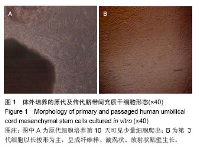
2.1 脐带间充质干细胞的分离、传代培养结果 贴壁法培养的原代细胞7-12 d后可见少量细胞爬出(图1A),3周左右细胞达80%融合,将细胞消化至T25中继续培养。随着传代次数的增加,细胞渐渐得到纯化,形态较均一。传代至P3时,细胞以长梭形为主,呈成纤维样、漩涡状、放射状贴壁生长(图1B),且增殖速度明显加快,三四天可传代1次。"


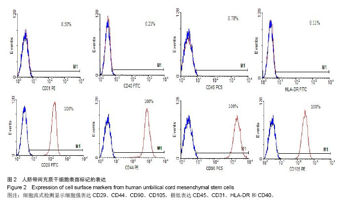
2.2 脐带间充质干细胞的鉴定结果 间充质干细胞能表达多种细胞表面标志,但并没有特异性的表面标志物,目前没有统一标准。研究表明,间充质干细胞表面标记物阳性表达的有CD29、CD90、CD44、CD105、CD59等,造血细胞表面标记CD31和CD45和免疫排斥相关标记HLA-DR、CD40等均表达阴性。实验所得细胞经流式细胞仪检测显示结果与文献报道一样(图2)。"

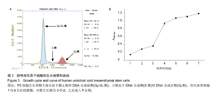
2.3 脐带间充质干细胞的生长周期和曲线 细胞生长周期显示细胞大部分处于静止期和DNA合成前期(G0-G1期),少数处于DNA合成期(S期)和DNA合成后期(G2期),说明细胞增殖能力很强(图3A)。通过绘制细胞生长曲线,发现细胞生长曲线大致呈“S”形,传代培养细胞1-3 d生长较缓慢,对数生长期为3-5 d,之后进入平台期,横坐标为细胞培养时间(d),纵坐标为细胞吸光度值(A450 nm,图3B)。"

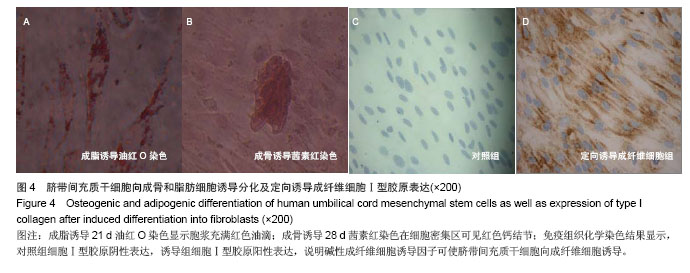
2.4 细胞向成骨和脂肪细胞诱导的形态变化及其功能 脐带间充质干细胞在成脂细胞诱导液中随着时间的延长,细胞形态逐渐变成短梭形,连续诱导21 d后油红O染色显示胞浆中充满红色的油滴(图4A),对照组未见明显脂滴形成。脐带间充质干细胞在成脂细胞诱导液中细胞逐渐变成多边形,可见细胞部分呈聚集生长。连续诱导28 d后行茜素红染色,可在细胞密集区见红色的钙结节(图4B),对照组细胞未见钙结节。 2.5 定向诱导为成纤维细胞并合成Ⅰ型胶原免疫组织化学染色结果 实验中诱导组在培养基中添加碱性成纤维细胞生长因子连续培养3 d后行Ⅰ型胶原免疫组织化学染色,成纤维细胞诱导组的大部分细胞染色阳性,对照组阴性表达(图4C,D)。"
| [1]Zickri MB, El Aziz DH, Metwally HG. Histological experimental study on the effect of stem cell therapy on adriamycin induced chemobrain. Int J Stem Cells. 2013;6(2):104-112.[2]Nirmal RS, Nair PD. Significance of soluble growth factors in the chondrogenic response of humanumbilical cord matrix stemcells in a porous three dimensional scaffold. Eur Cell Mater.2013;26:234-251.[3]Ruan ZB, Zhu L, Yin YG, et al.The mechanism underlying the differentiation of human umbilical cord-derived mesenchymal stem cells into myocardial cells induced by 5-azacytidine. Indian J Med Sci. 2010;64(9):402-407.[4]Chen X, Zhang F, He X,et al.Chondrogenic differentiation of umbilical cord-derived mesenchymal stem cells in type I collagen-hydrogel for cartilage engineering. Injury.2013; 44(4):540-549.[5]Xu Y, Meng H, Li C,et al.Umbilical cord-derived mesenchymal stem cells isolated by a novel explantation technique can differentiate into functional endothelial cells and promote revascularization. Stem Cells Dev. 2010; 19(10):1511-1522.[6]Jackson L, Jones DR, Scotting P, et al.Adult mesenchymal stem cells: differentiation potential and therapeutic applications. J Postqrad Med. 2007; 53(2):121-127.[7]Passier R, van-Laake LW, Mummery CL.Stem-cell-based therapy and lessons from theheart. Nature.2008;453(7193): 322-329.[8]Zhang Y, Cai W, Huang Q, et al.Mesenchymal Stem Cells Alleviate Bacteria-Induced Liver Injury in Mice by Inducing Regulatory Dendritic Cells.Hepatology. 2014;59(2):671-682.[9]Ohmura Y, Tanemura M, Kawaguchi N, et al.Combined transplantation of pancreatic islets and adipose tissue-derived Stem Cells enhances the survival and insulin function of islet grafts in diabetic mice. Transplantation.2010;90(12): 1366-1373.[10]Sacco A, Mourkioti F, Tran R, et al. Short Telomeres and Stem Cell Exhaustion Model Duchenne Muscular Dystrophy in mdx/mTR Mice.Cell. 2010;143(7):1059-1071.[11]Jameel Mohammad Nuruqadr, Zhang Jianyi.Stem cell threapy for Ischemic Heart Disease. Antioxidants and Redox Signaling. 2010;13(12):1879-1897.[12]徐丽南,姚志成,林楠,等.人脐血间充质干细胞对T淋巴细胞旁分泌的免疫调节作用[J].中国组织工程研究与临床康复,2011, 15(40):7463-7466.[13]刘伟,刘萌,祝劲松,等.人骨髓间充质干细胞的体外培养、鉴定及成骨分化[J].中国组织工程研究,2012,16(14):2515-2519.[14]Camassola M, de Macedo Braqa LM, Chaqastelles PC, et al. Methodology, biology and clinical applications of human mesenchymal stem cells. Methods Mol Biol. 2012;879:491- 504.[15]潘指挥,王丽,刘瑞风,等.皮肤间充质干细胞的原代培养[J].中国皮肤性病学杂志,2012,26(2):114-117.[16]Friedenstein AJ, Gorskaja JF, Kulagina NN. Fibroblast precursors in normal and irradiatedmouse hematopoietic organs. Exp Hematol.1976;4(5):267-274.[17]Caplan AI. Mesenchymal stem cells. J Orthop Res.1991;9 (5):641-650.[18]罗新,高绿芬,陈翠平. 17雌二醇对骨髓间充质干细胞增殖及成肌分化作用的研究[J].中国妇产科临床杂志.2010,11(5):369-373.[19]Pittenger MF, Maekay AM, Beck SC,et al. Multilineage potential of adult human mesenchymal stem cells. Science. 1999;284(5411):143-147.[20]Sobolewski K,Bańkowski E,Chyczewski L, et al. Collagen andglycosaminoglycans of Wharton’s jelly. Biol Neonate. 1997;71(1):11-21.[21]任瑞芳,任琛琛,赵冰,等.体外分离共培养诱导大鼠骨髓间充质干细胞定向分化为子宫韧带成纤维细胞的研究[J].实用妇产科杂志,2011,27(2):110-113.[22]代媛媛,原工杰,余桂戎,等.间接共培养诱导大鼠骨髓间充质干细胞向牙周膜成纤维样细胞分化[J].临床口腔医学杂志,2010, 26(5): 275-277.[23]Vanjak-Novakovic G, Altman G, Horan R, et al. Tissue engineering of ligaments. Annu Rev Biomed Enq. 2004; 6:131-156.[24]罗新.女性盆底解剖结构的新概念[J].中国实用妇科与产科杂志, 2006,22(1):78-80.[25]张艳君,梅虎,蒋稼欢,等.共培养下后交叉韧带成纤维细胞中赖氨酰氧化酶的基因表达[J].中国组织工程研究,2013,17(20): 3692-3698.[26]Dominici M,Le Blanc K, Mueller I, et al.Minimal criteria for definingmultipotent mesenchymal stromal cells. The International Societyfor Cellular Therapy position statement. Cytotherapy.2006;8 (4) :315-317.[27]Burdick JA, Vunjak-Novakovic G.Engineered Microenvironments for Controlled Stem Cell Differentiation. Tissue Eng Part A.2009;15(2):205-219.[28]Brafman David A. Constructing stem cell microenvironments using bioengineering approaches. Physiological Genomics. 2013;45(23):1123-1135.[29]何绍清,罗振宇,刘秋英,等.人脐带间充质干细胞分离培养及向脂肪和成骨细胞的分化[J].中国组织工程研究与临床康复,2010, 14(14):2492-2495.[30]Lee CH, Moioli EK, Mao JJ. Fibroblastic differentiation of human mesenchymal stem cells using connective tissue growthfactor.Conf Proc IEEE Eng Med Biol Soc.2006; 1: 775-778.[31]Liu HF, Fan HB, Wang Y. The interaction between a combinedknitted silk scaffold and microporous silk sponge with human mesenchymal stem cells for ligament tissue engineering. Biomaterials.2008;29(6):662-674.[32]杜尧,李晓声,张娜娟.大鼠骨髓间充质干细胞及大隐静脉来源细胞向组织工程化韧带种子细胞诱导的比较[J].中国组织工程研究与临床康复,2010,14(19):3427-3430.[33]孙智晶,郎景和,朱兰,等.胶原状态与压力性尿失禁的病因学研究[J].中国妇产科临床杂志,2004,5(1):6-8.[34]Liapis A ,Bakas P ,Pafiti A, et al. Changes in the quantity of collagen type I in women with genuine stress incontinence. Urological Research. 2000;28(5):323-326.[35]罗新,姚润斯,宋泓,等.人脐带间充质干细胞移植治疗大鼠压力性尿失禁的研究[J].中华妇产科杂志,2013,48(8):1-5.[36]Kyung CS, Sun-Ouck K, Soo YJ,et al.Effects of Mesenchymal Stem Cells on StressIncontinence in a Rat Model. Korean J Urol.2008;49:432-438.[37]Wu G,Song Y,Zheng X,et al.Adipose-derived stromal cell transplantation for treatment ofstress urinary incontinence. Tissue Cell.2011;43(4):246-253.[38]徐惠成,王延洲,王丹,等.肌源性干细胞治疗压力性尿失禁的实验研究[J].第三军医大学学报,2007,29(2):104.[39]石海燕,蓝建发,罗新.间充质干细胞治疗压力性尿失禁从动物实验到临床应用的转化.中华腔镜泌尿外科杂志:电子版,2013, 7(5):1-4. |
| [1] | Lin Qingfan, Xie Yixin, Chen Wanqing, Ye Zhenzhong, Chen Youfang. Human placenta-derived mesenchymal stem cell conditioned medium can upregulate BeWo cell viability and zonula occludens expression under hypoxia [J]. Chinese Journal of Tissue Engineering Research, 2021, 25(在线): 4970-4975. |
| [2] | Pu Rui, Chen Ziyang, Yuan Lingyan. Characteristics and effects of exosomes from different cell sources in cardioprotection [J]. Chinese Journal of Tissue Engineering Research, 2021, 25(在线): 1-. |
| [3] | Zhang Xiumei, Zhai Yunkai, Zhao Jie, Zhao Meng. Research hotspots of organoid models in recent 10 years: a search in domestic and foreign databases [J]. Chinese Journal of Tissue Engineering Research, 2021, 25(8): 1249-1255. |
| [4] | Hou Jingying, Yu Menglei, Guo Tianzhu, Long Huibao, Wu Hao. Hypoxia preconditioning promotes bone marrow mesenchymal stem cells survival and vascularization through the activation of HIF-1α/MALAT1/VEGFA pathway [J]. Chinese Journal of Tissue Engineering Research, 2021, 25(7): 985-990. |
| [5] | Shi Yangyang, Qin Yingfei, Wu Fuling, He Xiao, Zhang Xuejing. Pretreatment of placental mesenchymal stem cells to prevent bronchiolitis in mice [J]. Chinese Journal of Tissue Engineering Research, 2021, 25(7): 991-995. |
| [6] | Liang Xueqi, Guo Lijiao, Chen Hejie, Wu Jie, Sun Yaqi, Xing Zhikun, Zou Hailiang, Chen Xueling, Wu Xiangwei. Alveolar echinococcosis protoscolices inhibits the differentiation of bone marrow mesenchymal stem cells into fibroblasts [J]. Chinese Journal of Tissue Engineering Research, 2021, 25(7): 996-1001. |
| [7] | Fan Quanbao, Luo Huina, Wang Bingyun, Chen Shengfeng, Cui Lianxu, Jiang Wenkang, Zhao Mingming, Wang Jingjing, Luo Dongzhang, Chen Zhisheng, Bai Yinshan, Liu Canying, Zhang Hui. Biological characteristics of canine adipose-derived mesenchymal stem cells cultured in hypoxia [J]. Chinese Journal of Tissue Engineering Research, 2021, 25(7): 1002-1007. |
| [8] | Geng Yao, Yin Zhiliang, Li Xingping, Xiao Dongqin, Hou Weiguang. Role of hsa-miRNA-223-3p in regulating osteogenic differentiation of human bone marrow mesenchymal stem cells [J]. Chinese Journal of Tissue Engineering Research, 2021, 25(7): 1008-1013. |
| [9] | Lun Zhigang, Jin Jing, Wang Tianyan, Li Aimin. Effect of peroxiredoxin 6 on proliferation and differentiation of bone marrow mesenchymal stem cells into neural lineage in vitro [J]. Chinese Journal of Tissue Engineering Research, 2021, 25(7): 1014-1018. |
| [10] | Zhu Xuefen, Huang Cheng, Ding Jian, Dai Yongping, Liu Yuanbing, Le Lixiang, Wang Liangliang, Yang Jiandong. Mechanism of bone marrow mesenchymal stem cells differentiation into functional neurons induced by glial cell line derived neurotrophic factor [J]. Chinese Journal of Tissue Engineering Research, 2021, 25(7): 1019-1025. |
| [11] | Duan Liyun, Cao Xiaocang. Human placenta mesenchymal stem cells-derived extracellular vesicles regulate collagen deposition in intestinal mucosa of mice with colitis [J]. Chinese Journal of Tissue Engineering Research, 2021, 25(7): 1026-1031. |
| [12] | Pei Lili, Sun Guicai, Wang Di. Salvianolic acid B inhibits oxidative damage of bone marrow mesenchymal stem cells and promotes differentiation into cardiomyocytes [J]. Chinese Journal of Tissue Engineering Research, 2021, 25(7): 1032-1036. |
| [13] | Guan Qian, Luan Zuo, Ye Dou, Yang Yinxiang, Wang Zhaoyan, Wang Qian, Yao Ruiqin. Morphological changes in human oligodendrocyte progenitor cells during passage [J]. Chinese Journal of Tissue Engineering Research, 2021, 25(7): 1045-1049. |
| [14] | Wang Zhengdong, Huang Na, Chen Jingxian, Zheng Zuobing, Hu Xinyu, Li Mei, Su Xiao, Su Xuesen, Yan Nan. Inhibitory effects of sodium butyrate on microglial activation and expression of inflammatory factors induced by fluorosis [J]. Chinese Journal of Tissue Engineering Research, 2021, 25(7): 1075-1080. |
| [15] | Wang Xianyao, Guan Yalin, Liu Zhongshan. Strategies for improving the therapeutic efficacy of mesenchymal stem cells in the treatment of nonhealing wounds [J]. Chinese Journal of Tissue Engineering Research, 2021, 25(7): 1081-1087. |
| Viewed | ||||||
|
Full text |
|
|||||
|
Abstract |
|
|||||