Chinese Journal of Tissue Engineering Research ›› 2021, Vol. 25 ›› Issue (8): 1249-1255.doi: 10.3969/j.issn.2095-4344.3073
Previous Articles Next Articles
Research hotspots of organoid models in recent 10 years: a search in domestic and foreign databases
Zhang Xiumei1, 2, Zhai Yunkai3, Zhao Jie4, Zhao Meng2
- 1Institute of Scientific and Technical Information of China, Beijing 100038, China; 2WanFang Data Co., Ltd., Beijing 100038, China; 3School of Management Engineering, Zhengzhou University, Zhengzhou 450001, Henan Province, China; 4The First Affiliated Hospital of Zhengzhou University, Zhengzhou 450052, Henan Province, China
-
Received:2020-06-12Revised:2020-06-18Accepted:2020-07-08Online:2021-03-18Published:2020-12-14 -
Contact:Zhang Xiumei, Institute of Scientific and Technical Information of China, Beijing 100038, China; WanFang Data Co., Ltd., Beijing 100038, China -
About author:Zhang Xiumei, Institute of Scientific and Technical Information of China, Beijing 100038, China; WanFang Data Co., Ltd., Beijing 100038, China -
Supported by:the National Key R & D Program of China, No. 2017YFC0909900
CLC Number:
Cite this article
Zhang Xiumei, Zhai Yunkai, Zhao Jie, Zhao Meng. Research hotspots of organoid models in recent 10 years: a search in domestic and foreign databases[J]. Chinese Journal of Tissue Engineering Research, 2021, 25(8): 1249-1255.
share this article
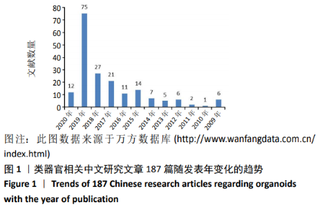
2.1 基于万方数据库187篇类器官研究文章的热点分析 2.1.1 万方数据库类器官研究文章在2019年研究呈爆发持增长 从2009至2019年,中国万方数据库收录了类器官相关中文研究187篇,随年代逐渐增加,其中2019年文章最多为75篇,而2010年文章最少仅1篇,2020年截止到2020-05-14仅检索到12篇。中国类器官研究起步较晚,从2015及2016年开始逐渐增多,2019年类器官研究呈爆发式增长,说明了过去1年中国学者在类器官相关领域中研究较为活跃。见图1。"
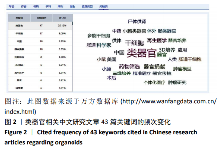
2.1.2 万方数据库的187篇类器官研究文章的关键词分析 干细胞3D培养及药物筛选应用是中国类器官研究领域的热点 在检索到的全部关键词中,出现频率最高的是类器官47次,其次是干细胞17次,器官捐献10次,药物筛选10次,3D培养6次。说明中国学者在将不同类型干细胞的进行体外3D培养建立类器官模型方面研究较多,主要用于药物体外筛选。在类器官种类上主要是肠类器官较多,在用途上主要是精准医疗、肿瘤研究和个体化医疗占主导,见图2。"


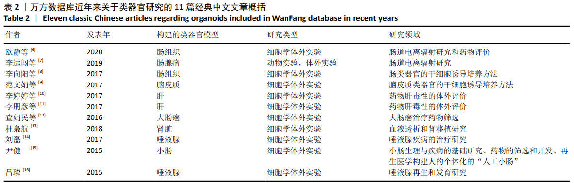
2.1.3 收集被引次数和下载次数相对较高的11篇经典类器官研究文章显示中国研究者从事类器官研究的特点 在11篇经典研究中[6-16],有3篇文章被引次数相对较高[9-11],李婷婷等[10]采用液滴重叠法构建肝类器官3D培养模型,主要用于药物肝毒性的体外评价,被引4次;范文娟等[9]利用小鼠诱导性多能干细胞建立大脑皮质类器官并分析其生物学特性,建立了一种脑皮质类器官的干细胞诱导培养方法,被引3次;李朋彦等[11]采用HepaRG细胞建立肝类器官3D培养模型,主要用于药物肝毒性的体外评价,被引2次。这些研究所构建的类器官模型种类主要包括肠、脑、肝、肾和唾液腺,见表2。"
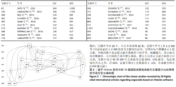
2.2 基于Web of Science数据库(核心集)检索出2 450篇类器官研究的热点文章分析 2.2.1 在检索到的2 450篇文章以Histcite软件分析后筛选20篇类器官研究高被引文章[3,17-35] 生成的可视化引文编年图,见图3。 进一步分析图3中各文献的5条引文终点文 献[17-18, 22,25,29],可显示出国际研究者对类器官的研究方向主要集中在肠、脑、前列腺、肾和胃5种类器官的构建方法,分别是各领域的开山之作。 (1) SATO等[17](图3中65号文章:本地被引343次,全数据库被引1 050次)开发了一种可用于模拟人类胃肠道的感染、炎症或肿瘤组织的类器官;此研究在小鼠小肠培养体系的基础上,对小鼠和人结肠培养体系进行了优化。 (2) LANCASTER等[18](图3中109号文章:本地被引343次,全数据库被引1 050次)开发了一种人类多能干细胞衍生的三维大脑类器官培养系统,可显示人类皮质发育的特征。此研究开发了一种人类多能干细胞衍生的三维类器官培养系统,称为大脑类器官,它可开发出各种不同的并相互依赖的大脑区域。此研究还证明了患者类器质的早期神经元分化,这种变化有助于解释疾病的表型。总之,这些数据表明,即使在最复杂的人体组织中,三维类器官也能再现人体的发育和疾病状态。 (3) GAO等[22](图3中172号文章:本地被引150次,全数据库被引501次)利用三维器官系统,报道了从活检标本和循环肿瘤细胞中长期培养前列腺癌的成功案例。实验结果概括了前列腺癌亚型的分子多样性,包括TMPRSS2-ERG融合、SPOP突变、SPINK1过度表达和CHD1缺失。全外显子组测序显示出较低的突变负担,与基因组学研究一致,但关于FOXA1和PIK3R1的突变,以及DNA修复和染色质修饰途径在晚期疾病研究中已有报道。p53和RB肿瘤抑制通路功能的丧失是类器官模型中最常见的特征。此研究所描述的方法可培养出大量的患者源性前列腺癌株系,因此这些基因和药理学研究都是可行的。 (4) TAKASATO等[25](图3中282号文章:本地被引131次,全数据库被引413次)构建了含有肾单位的肾类器官,探讨集合管对肾间充质祖细胞优先诱导的发育机制。此研究建立了包含肾单位和肾间质和内皮细胞包围的集合管网络的肾单位。在这些类器官中,单个肾单位分裂成远端和近端小管、早期Henle环和含肾小球的足细胞,形成足突并开始血管化。当肾脏类器官的转录谱与人类胎儿组织比较时,它们与妊娠早期人类肾脏的一致性最高。此类肾脏器官代表了未来可应用于人体器官的强大模型,其主要用途包括肾毒性筛选、疾病建模和作为治疗细胞来源。 (5) MCCRACKEN等[29](图3中196号文章:本地被引117次,全数据库被引333次)报告了一种通过人类多能干细胞的定向分化方法在体外构建三维人胃组织类器官模型,可用于阐明人类胃部发育和疾病的潜在机制。此研究利用hGO培养物来识别调节早期内胚层模式和转录因子NEUROG3上游胃内分泌细胞分化的新信号机制。用hGOs模拟人类疾病的发病机制,发现幽门螺杆菌感染导致毒力因子CagA与c-Met受体迅速相关,可激活信号传导通路,诱导上皮细胞增殖。这些结果描述了一个新的和强有力的体外系统,用于阐明人体胃发育和疾病的潜在机制。"
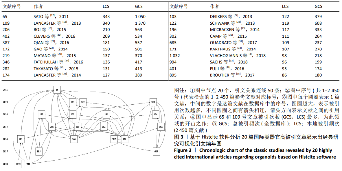

2.2.2 从检索到2 450篇文献及其10 974条参考文献归纳出11篇该领域的经典文章 (1) SATO等[36]于2009年用源于小鼠肠道的成体干细胞培育出首个微型肠道类器官,真正开启类器官培养的时代。 (2)在随后的几年里,其他科学家构建出了大脑、胰腺、前列腺癌和直肠等类器官体外模型,类器官研究热度逐渐提升。 (3)归纳出11篇此领域的经典文献[17-19,21-23,25-26,36-38],见表3。"
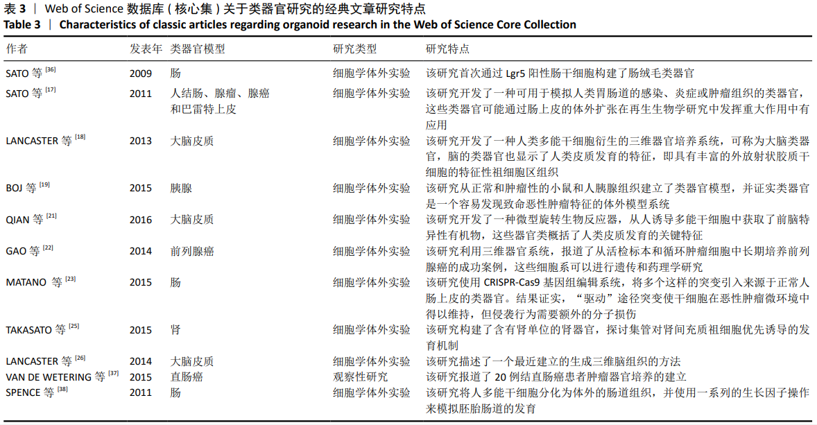
2.3 检索中国和北美临床注册中心结果分析类器官的临床研究前景 2.3.1 中国临床试验注册中心数据 中国临床试验注册中心对类器官研究在2017至2020年注册12项,其中2项最终关注的随机对照试验(干预性研究)正在进行中,其他10项研究均为观察性研究。 (1) 中山大学附属第七医院在2020年注册了3项研究(注册号:ChiCTR2000029049,ChiCTR2000028889,ChiCTR2000028856);同济大学附属第十人民医院分别在2018年和2019年注册了2项研究(注册号:ChiCTR1900023682,ChiCTR1800017767),说明此2个机构研究在中国类器官临床试验研究方面较为活跃积极。 (2)在中国临床试验中心注册的类器官模型研究方案中,方向为胃癌的有3项,胰腺癌的有2项,子宫内膜癌的有2项,直肠癌、鼻咽癌、乳腺癌和卵巢癌的各有1项,多种恶性肿瘤研究有1项,共计12项注册项目,注册时间为2017至2020年,见表4。"


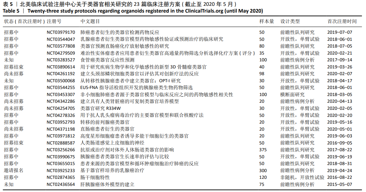
2.3.2 北美临床试验注册中心数据 检索到与类器官研究有关的北美临床注册项目23项,首次注册时间2015至2019年。 (1)此23项方案中,包括前瞻性队列研究有9项,开放性,单臂试验有8项,前瞻性病例分析4项,横断面研究和非随机开放性试验1项,尚未发现正在进行的随机对照试验。 (2)在此23项类器官注册项目中,除包括胃癌、胰腺癌、直肠癌和乳腺癌类器官模型外,还包括肺癌、食管癌、骨髓瘤、头颈部鳞状细胞癌、肾脏癌、星形细胞瘤和肝胰腺癌类器官模型,器官研究领域更为广泛,见表5。"
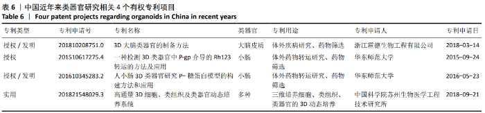
2.3.3 类器官临床注册方案在中国华西和美国北美临床试验数据库注册项目的对比分析 (1) 类器官临床研究领域中,美国类器官研究领域更广泛,研究相对较多,开展时间更早(美国2015年,中国2017年),但多为队列研究和单臂试验,也缺乏随机对照试验。 (2) 中国近年来在类器官领域已经取得了一些成果,一些受人瞩目的临床研究试验方案正在进行中,其中已经有2个随机对照试验方方案正在进行中,分别是同济大学附属第十人民医院进行的类器官模型联合微流芯片技术构建卵巢癌药筛体系及临床转化应用(注册号:ChiCTR1800017767),复旦大学附属眼耳鼻喉科医院进行的“鼻咽癌肿瘤类器官建立和个体化精准治疗研究”(注册号:ChiCTR1900024322),其未来临床研究结果值得期待。 2.4 基于SooPAT中国专利数据库2015年以来55项有权专利中可见中国类器官研究领域的最新专利成果 (1)在SooPAT中国专利数据库检索到55项有权专利,经筛选标题后纳入与类器官研究十分相关的专利文章4项,见表6。 (2) 浙江霍德生物工程有限公司(专利申请号:201810208751.0,授权/发明专利)从得到的高度纯化的神经球开始,用较为简单的方法可以控制并培养得到大小及结构均一的3D大脑类器官,适用于体外疾病研究、药物筛选等。 (3) 华东师范大学有2项(专利申请号:201510617275.4,授权专利;专利申请号:201610345283.2,授权/发明专利),浙江霍德生物工程有限公司(专利申请号:201810208751.0,授权/发明专利)和中国科学院苏州生物医学工程技术研究所(专利申请号:201821548029.3,实用专利)各有1项专利,说明中国学者的类器官相关专利还比较少。 (4)中国科学院苏州生物医学工程技术研究所(专利申请号:201821548029.3,实用新型专利)公开了一种高通量3D细胞、类组织及类器官动态培养系统,可自动化完成3D动态培养,安全可靠、高效便捷。"

| [1] 高坚钧,秦伟,王浩,等.类器官技术在肿瘤研究中的应用与展望[J].中国组织工程研究,2019,23(7):1136-1141. [2] DROST J, CLEVERS H. Organoids in cancer research. Nat Rev Cancer. 2018; 18(7):407-418. [3] VLACHOGIANNIS G, HEDAYAT S, VATSIOU A, et al. Patient-derived organoids model treatment response of metastatic gastrointestinal cancers. Science. 2018;359(6378):920-926. [4] LI M, IZPISUA BELMONTE JC. Organoids-preclinical models of human disease. N Engl J Med. 2019;380(6):569-579. [5] 许丹,侯跃芳.国外医学信息学研究领域可视化引文分析[J].医学信息学杂志,2011,32(10):12-19. [6] 欧静,徐珍妮,刘登群,等.3D培养体系中不同肠上皮细胞株形成肠类器官潜能的比较及应用[J].第三军医大学学报,2020,42(1):31-38. [7] 李远闯,富国祥,潘梦雪,等.小鼠肠腺瘤类器官培养及其辐射敏感性[J].复旦学报(医学版),2019,46(2):193-198. [8] 李向阳,赵鑫,相小松,等.诱导型多能干细胞在体外三维环境中诱导分化出肠道类器官[J].中国组织工程研究,2017,21(25):4057-4061. [9] 范文娟,王倩,孙仪征,等.起源于小鼠诱导性多能干细胞的大脑皮质类器官的建立及其生物学特性[J].解剖学报,2017,48(4):387-396. [10] 李婷婷,李瑞红,刘振兴,等.基于类器官3D培养的何首乌易感物质肝毒性评价[J].药学学报,2017,52(7):1048-1054. [11] 李朋彦,李春雨,陆小华,等.基于类器官3D培养和高内涵成像的药物肝毒性评价模型研究[J].药学学报,2017,52(7):1055-1062. [12] 查娟民,林倩,李华善,等.采用人肿瘤类器官研究阻断Wnt信号治疗大肠癌的可行性[J].中国现代医生,2016,54(27):5-8. [13] 杜枭航.利用人诱导多能干细胞构建肾脏类器官的研究[D].济南:山东大学,2018. [14] 刘磊.生物工程化人唾液腺类器官构建的实验研究[D].北京:北京协和医学院,2017. [15] 尹健一.成人小肠类器官体外培养体系的构建及分化前后关键离子通道的表达与功能[D].南京:南京大学,2015. [16] 吕璘.人小涎腺间充质干细胞和上皮干/祖细胞分离鉴定及其构建生物工程化唾液腺类器官的研究[D]. 北京:北京协和医学院,2015. [17] SATO T, STANGE DE, FERRANTE M, et al. Long-term expansion of epithelial organoids from human colon, adenoma, adenocarcinoma, and Barrett’s epithelium. Gastroenterology. 2011;141(5):1762-1772. [18] LANCASTER MA, RENNER M, MARTIN CA, et al. Cerebral organoids model human brain development and microcephaly. Nature. 2013;501(7467):373-379. [19] BOJ SF, HWANG CI, BAKER LA, et al. Organoid models of human and mouse ductal pancreatic cancer. Cell. 2015;160(1-2):324-338. [20] CLEVERS H. Modeling development and disease with organoids. Cell. 2016; 165(7):1586-1597. [21] QIAN X, NGUYEN HN, SONG MM, et al. Brain-Region-Specific Organoids Using Mini-bioreactors for Modeling ZIKV Exposure. Cell. 2016;165(5): 1238-1254. [22] GAO D, VELA I, SBONER A, et al. Organoid cultures derived from patients with advanced prostate cancer. Cell. 2014;159(1):176-187. [23] MATANO M, DATE S, SHIMOKAWA M, et al. Modeling colorectal cancer using CRISPR-Cas9-mediated engineering of human intestinal organoids. Nat Med. 2015;21(3):256-262. [24] FATEHULLAH A, TAN SH, BARKER N. Organoids as an in vitro model of human development and disease. Nat Cell Biol. 2016;18(3):246-254. [25] TAKASATO M, ER PX, CHIU HS, et al. Kidney organoids from human iPS cells contain multiple lineages and model human nephrogenesis. Nature. 2015;526(7574):564-568. [26] LANCASTER MA, KNOBLICH JA. Generation of cerebral organoids from human pluripotent stem cells. Nat Protoc. 2014;9(10):2329-2340. [27] DEKKERS JF, WIEGERINCK CL, DE JONGE HR, et al. A functional CFTR assay using primary cystic fibrosis intestinal organoids. Nat Med. 2013;19(7): 939-945. [28] SCHWANK G, KOO BK, SASSELLI V, et al. Functional repair of CFTR by CRISPR/Cas9 in intestinal stem cell organoids of cystic fibrosis patients. Cell Stem Cell. 2013;13(6):653-658. [29] MCCRACKEN KW, CATÁ EM, CRAWFORD CM, et al. Modelling human development and disease in pluripotent stem-cell-derived gastric organoids. Nature. 2014;516(7531):400-404. [30] CAMP JG, BADSHA F, FLORIO M, et al. Human cerebral organoids recapitulate gene expression programs of fetal neocortex development. Proc Natl Acad Sci U S A. 2015;112(51):15672-15677. [31] QUADRATO G, NGUYEN T, MACOSKO EZ, et al. Cell diversity and network dynamics in photosensitive human brain organoids. Nature. 2017;545(7652):48-53. [32] KARTHAUS WR, IAQUINTA PJ, DROST J, et al. Identification of multipotent luminal progenitor cells in human prostate organoid cultures. Cell. 2014; 159(1):163-175. [33] SACHS N, DE LIGT J, KOPPER O, et al. A living biobank of breast cancer organoids captures disease heterogeneity. Cell. 2018;172(1-2):373-386. [34] FUJII M, SHIMOKAWA M, DATE S, et al. A Colorectal tumor organoid library demonstrates progressive loss of niche factor requirements during tumorigenesis. Cell Stem Cell. 2016;18(6):827-838. [35] BROUTIER L, MASTROGIOVANNI G, VERSTEGEN MM, et al. Human primary liver cancer-derived organoid cultures for disease modeling and drug screening. Nat Med. 2017;23(12):1424-1435. [36] SATO T, VRIES RG, SNIPPERT HJ, et al. Single Lgr5 stem cells build crypt-villus structures in vitro without a mesenchymal niche. Nature. 2009; 459(7244):262-265. [37] VAN DE WETERING M, FRANCIES HE, et al. Prospective derivation of a living organoid biobank of colorectal cancer patients. Cell. 2015;161(4):933-945. [38] SPENCE JR, MAYHEW CN, RANKIN SA, et al. Directed differentiation of human pluripotent stem cells into intestinal tissue in vitro. Nature. 2011; 470(7332):105-109. |
| [1] | Pu Rui, Chen Ziyang, Yuan Lingyan. Characteristics and effects of exosomes from different cell sources in cardioprotection [J]. Chinese Journal of Tissue Engineering Research, 2021, 25(在线): 1-. |
| [2] | Lin Qingfan, Xie Yixin, Chen Wanqing, Ye Zhenzhong, Chen Youfang. Human placenta-derived mesenchymal stem cell conditioned medium can upregulate BeWo cell viability and zonula occludens expression under hypoxia [J]. Chinese Journal of Tissue Engineering Research, 2021, 25(在线): 4970-4975. |
| [3] | Jiang Yong, Luo Yi, Ding Yongli, Zhou Yong, Min Li, Tang Fan, Zhang Wenli, Duan Hong, Tu Chongqi. Von Mises stress on the influence of pelvic stability by precise sacral resection and clinical validation [J]. Chinese Journal of Tissue Engineering Research, 2021, 25(9): 1318-1323. |
| [4] | Zhang Tongtong, Wang Zhonghua, Wen Jie, Song Yuxin, Liu Lin. Application of three-dimensional printing model in surgical resection and reconstruction of cervical tumor [J]. Chinese Journal of Tissue Engineering Research, 2021, 25(9): 1335-1339. |
| [5] | Zhang Yu, Tian Shaoqi, Zeng Guobo, Hu Chuan. Risk factors for myocardial infarction following primary total joint arthroplasty [J]. Chinese Journal of Tissue Engineering Research, 2021, 25(9): 1340-1345. |
| [6] | Gu Xia, Zhao Min, Wang Pingyi, Li Yimei, Li Wenhua. Relationship between hypoxia inducible factor 1 alpha and hypoxia signaling pathway [J]. Chinese Journal of Tissue Engineering Research, 2021, 25(8): 1284-1289. |
| [7] | Tang Hui, Yao Zhihao, Luo Daowen, Peng Shuanglin, Yang Shuanglin, Wang Lang, Xiao Jingang. High fat and high sugar diet combined with streptozotocin to establish a rat model of type 2 diabetic osteoporosis [J]. Chinese Journal of Tissue Engineering Research, 2021, 25(8): 1207-1211. |
| [8] | Chai Le, Lü Jianlan, Hu Jintao, Hu Huahui, Xu Qingjun, Yu Jinwei, Quan Renfu. Signal pathway variation after induction of inflammatory response in rats with acute spinal cord injury [J]. Chinese Journal of Tissue Engineering Research, 2021, 25(8): 1218-1223. |
| [9] | Wang Zhengdong, Huang Na, Chen Jingxian, Zheng Zuobing, Hu Xinyu, Li Mei, Su Xiao, Su Xuesen, Yan Nan. Inhibitory effects of sodium butyrate on microglial activation and expression of inflammatory factors induced by fluorosis [J]. Chinese Journal of Tissue Engineering Research, 2021, 25(7): 1075-1080. |
| [10] | Wang Xianyao, Guan Yalin, Liu Zhongshan. Strategies for improving the therapeutic efficacy of mesenchymal stem cells in the treatment of nonhealing wounds [J]. Chinese Journal of Tissue Engineering Research, 2021, 25(7): 1081-1087. |
| [11] | Liao Chengcheng, An Jiaxing, Tan Zhangxue, Wang Qian, Liu Jianguo. Therapeutic target and application prospects of oral squamous cell carcinoma stem cells [J]. Chinese Journal of Tissue Engineering Research, 2021, 25(7): 1096-1103. |
| [12] | Zhao Min, Feng Liuxiang, Chen Yao, Gu Xia, Wang Pingyi, Li Yimei, Li Wenhua. Exosomes as a disease marker under hypoxic conditions [J]. Chinese Journal of Tissue Engineering Research, 2021, 25(7): 1104-1108. |
| [13] | Xie Wenjia, Xia Tianjiao, Zhou Qingyun, Liu Yujia, Gu Xiaoping. Role of microglia-mediated neuronal injury in neurodegenerative diseases [J]. Chinese Journal of Tissue Engineering Research, 2021, 25(7): 1109-1115. |
| [14] | Li Shanshan, Guo Xiaoxiao, You Ran, Yang Xiufen, Zhao Lu, Chen Xi, Wang Yanling. Photoreceptor cell replacement therapy for retinal degeneration diseases [J]. Chinese Journal of Tissue Engineering Research, 2021, 25(7): 1116-1121. |
| [15] | Jiao Hui, Zhang Yining, Song Yuqing, Lin Yu, Wang Xiuli. Advances in research and application of breast cancer organoids [J]. Chinese Journal of Tissue Engineering Research, 2021, 25(7): 1122-1128. |
| Viewed | ||||||
|
Full text |
|
|||||
|
Abstract |
|
|||||