[1] 赵小辉.红景天苷对力竭运大鼠的心脏保护作用及机制研究[J].中国妇幼健康研究,2016(S2):261.
[2] 郑妩媚,初海平,王燕,等.力竭运动后不同时相大鼠心肌 p-p38MAPK, NF-kB, COX-2 表达的动态变化[J].中国应用生理学杂志, 2016, 32(1): 88-91.
[3] 公雪,李晓燕,张红明.运动预适应对急性运动性损伤后心肌组织氧化应激及凋亡的影响[J].国际心血管病杂志, 2017,44 (2): 110-114.
[4] 尤华彦,曹华明,王强,等.心肌缺血预适应对急性心肌梗死患者溶栓治疗效果的影响研究[J].中华全科医学, 2015, 13(12): 1918-1920.
[5] HAUSENLOY DJ, CANDILIO L, EVANS R, et al. Remote Ischemic Preconditioning and Outcomes of Cardiac Surgery. N Engl J Med. 2015;373(15):1408-1417.
[6] ZENG Q, HE H, WANG XB, et al. Electroacupuncture Preconditioning Improves Myocardial Infarction Injury via Enhancing AMPK-Dependent Autophagy in Rats. Biomed Res Int. 2018;2018:1238175.
[7] WONG CM, WEI L, AU SL, et al. MiR-200b/200c/429 subfamily negatively regulates Rho/ROCK signaling pathway to suppress hepatocellular carcinoma metastasis. Oncotarget. 2015;6(15): 13658-13670.
[8] LAWLER JM, RODRIGUEZ DA, HORD JM. Mitochondria in the middle: exercise preconditioning protection of striated muscle. J Physiol. 2016; 594(18):5161-5183.
[9] THOMAS DP, MARSHALL KI. Effects of repeated exhaustive exercise on myocardial subcellular membrane structures. Int J Sports Med. 1988;9(4):257-260.
[10] 陈淦,汤长发.不同强度运动对大鼠心肌细胞凋亡的影响[J].生命科学研究, 2012, 16(2):102-107.
[11] NEY PA. Mitochondrial autophagy: Origins, significance, and role of BNIP3 and NIX. Biochim Biophys Acta. 2015;1853(10 Pt B):2775-2783.
[12] 万栋峰,潘珊珊,原阳.心肌线粒体自噬相关蛋白 BNIP3 在运动预适应晚期保护效应中的变化[J]. 体育科学, 2017, 37(3): 35-43.
[13] 赵沐霖.运动预适应对力竭大鼠心肌线粒体生物发生及呼吸功能的影响[D].石家庄:河北大学, 2017.
[14] 李翠霞,潘忠勉,李杰.运动预适应对力竭性运动大鼠心肌形态学影响的研究[J].广西医科大学学报, 2013, 30(4):534-536.
[15] 韩佳寅,易艳,梁爱华,等. Rho/ROCK 信号通路研究进展[J].药学学报, 2016, 51(6): 853-859.
[16] AMIN E, DUBEY BN, ZHANG SC, et al. Rho-kinase: regulation, (dys)function, and inhibition. Biol Chem. 2013;394(11):1399-1410.
[17] STOCKMANN H, VERBAKEL C, OKKEMA P, et al. No functional benefit for hDAF-transgenic rat livers despite protection from tissue damage following perfusion with human serum. Transpl Int. 2002; 15(12):595-601.
[18] DOLGANIUC A, THOMES PG, DING WX, et al. Autophagy in alcohol-induced liver diseases. Alcohol Clin Exp Res. 2012;36(8): 1301-1308.
[19] 王雪芬,王磊,陈树杰.苦参素对大鼠心肌缺血再灌注损伤的保护作用及其机制研究[J].中国中医急症, 2016,25(6):988-992.
[20] PARK HJ, PARK MC, PARK YB, et al. The concomitant use of meloxicam and methotrexate does not clearly increase the risk of silent kidney and liver damages in patients with rheumatoid arthritis. Rheumatol Int. 2014;34(6):833-840.
[21] 吴锦波,吴平生.心肌缺血/再灌注损伤与细胞凋亡[J]. 医学综述, 2011, 17(19):2961-2963.
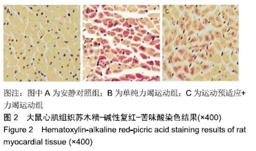
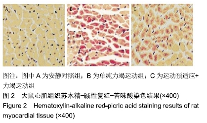
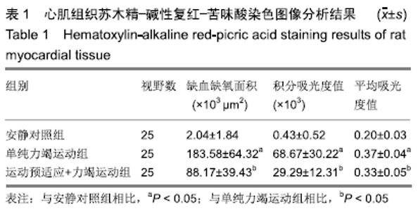
[22] 毕海涛,杨莹,刘霞,等.3种特殊染色在急性心肌梗死死后诊断中的应用比较[J].中国法医学杂志, 2013, 28(4):330-332.
[23] LIEBELT B, PAPAPETROU P, ALI A, et al. Exercise preconditioning reduces neuronal apoptosis in stroke by up-regulating heat shock protein-70 (heat shock protein-72) and extracellular-signal- regulated-kinase 1/2. Neuroscience. 2010;166(4):1091-1100.
[24] WANG Q, ZHANG L, YUAN X, et al. The Relationship between the Bcl-2/Bax Proteins and the Mitochondria-Mediated Apoptosis Pathway in the Differentiation of Adipose-Derived Stromal Cells into Neurons. PLoS One. 2016;11(10):e0163327.
[25] ZHANG Z, LIANG Z, LI H, et al. Perfluorocarbon reduces cell damage from blast injury by inhibiting signal paths of NF-κB, MAPK and Bcl-2/Bax signaling pathway in A549 cells. PLoS One. 2017;12(3):e0173884.
[26] ZHANG B, MONTGOMERY M, CHAMBERLAIN MD, et al. Biodegradable scaffold with built-in vasculature for organ-on-a-chip engineering and direct surgical anastomosis. Nat Mater. 2016;15(6): 669-678.
[27] MAIESE K. Impacting dementia and cognitive loss with innovative strategies: mechanistic target of rapamycin, clock genes, circular non-coding ribonucleic acids, and Rho/Rock. Neural Regen Res. 2019;14(5):773-774.
[28] 杨先金,张新金.RhoA/ROCK 信号通路与心脏重构关系的研究进展[J].心脑血管病防治,2016,16(1):43-45.
[29] BEI Y, HUA-HUY T, NICCO C, et al. RhoA/Rho-kinase activation promotes lung fibrosis in an animal model of systemic sclerosis. Exp Lung Res. 2016;42(1):44-55.
|