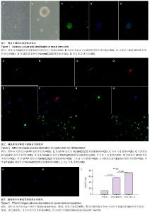
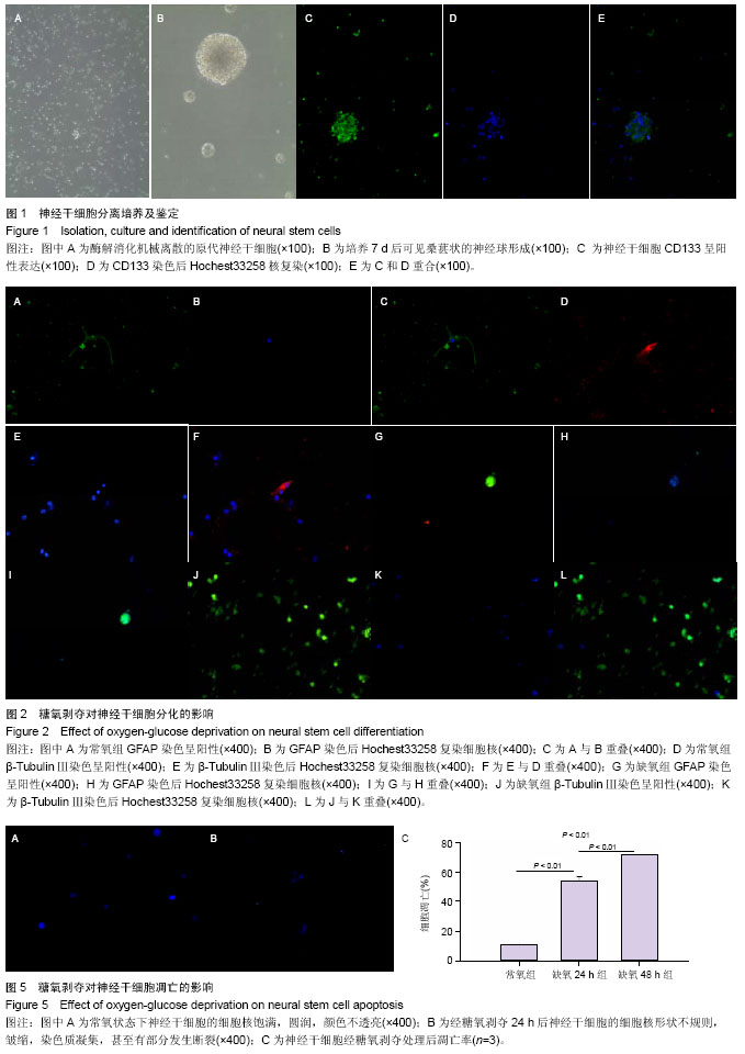
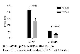
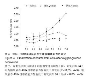
| [1] Brüstle O, Spiro AC, Karram K, et al. In vitro-generated neural precursors participate in mammalian brain development.Proc Natl Acad Sci U S A. 1997;94(26):14809-14814.
[2] Walker MR, Patel KK, Stappenbeck TS.The stem cell niche.J Pathol. 2009;217(2):169-180.
[3] van Strien ME, Sluijs JA, Reynolds BA, et al. Isolation of neural progenitor cells from the human adult subventricular zone based on expression of the cell surface marker CD271. Stem Cells Transl Med. 2014;3(4):470-480.
[4] Mine Y, Tatarishvili J, Oki K, et al. Grafted human neural stem cells enhance several steps of endogenous neurogenesis and improve behavioral recovery after middle cerebral artery occlusion in rats.Neurobiol Dis. 2013;52:191-203.
[5] Jensen MB, Yan H, Krishnaney-Davison R, et al. Survival and differentiation of transplanted neural stem cells derived from human induced pluripotent stem cells in a rat stroke model.J Stroke Cerebrovasc Dis. 2013;22(4):304-308.
[6] Kanno H. Regenerative therapy for neuronal diseases with transplantation of somatic stem cells. World J Stem Cells. 2013;5(4):163-171.
[7] Karussis D, Petrou P, Kassis I. Clinical experience with stem cells and other cell therapies in neurological diseases. J Neurol Sci. 20135;324(1-2):1-9.
[8] Giusto E, Donegà M, Cossetti C, et al. Neuro-immune interactions of neural stem cell transplants: from animal disease models to human trials. Exp Neurol. 2014;260:19-32.
[9] Francis KR, Wei L. Human embryonic stem cell neural differentiation and enhanced cell survival promoted by hypoxic preconditioning. Cell Death Dis. 2010;1:e22.
[10] Grote HE, Hannan AJ. Regulators of adult neurogenesis in the healthy and diseased brain. Clin Exp Pharmacol Physiol. 2007;34(5-6):533-545.
[11] Chen X, Tian Y, Yao L, et al. Hypoxia stimulates proliferation of rat neural stem cells with influence on the expression of cyclin D1 and c-Jun N-terminal protein kinase signaling pathway in vitro. Neuroscience. 2010;165(3):705-714.
[12] Panchision DM. The role of oxygen in regulating neural stem cells in development and disease. J Cell Physiol. 2009;220(3): 562-568.
[13] Urbán N, Guillemot F. Neurogenesis in the embryonic and adult brain: same regulators, different roles. Front Cell Neurosci. 2014;8:396.
[14] Vieira HL, Alves PM, Vercelli A. Modulation of neuronal stem cell differentiation by hypoxia and reactive oxygen species. Prog Neurobiol. 2011;93(3):444-455.
[15] McKay R.Stem cells in the central nervous system.Science. 1997;276(5309):66-71.
[16] Reynolds BA, Weiss S. Generation of neurons and astrocytes from isolated cells of the adult mammalian central nervous system. Science. 1992;255(5052):1707-1710.
[17] Ahmed S.The culture of neural stem cells.J Cell Biochem. 2009;106(1):1-6.
[18] Florek M, Haase M, Marzesco AM, et al. Prominin-1/CD133, a neural and hematopoietic stem cell marker, is expressed in adult human differentiated cells and certain types of kidney cancer. Cell Tissue Res. 2005;319(1):15-26.
[19] Ramasamy S, Narayanan G, Sankaran S, et al. Neural stem cell survival factors. Arch Biochem Biophys. 2013;534(1-2): 71-87.
[20] Erecińska M, Silver IA. Tissue oxygen tension and brain sensitivity to hypoxia. Respir Physiol. 2001;128(3):263-276.
[21] Santilli G, Lamorte G, Carlessi L, et al. Mild hypoxia enhances proliferation and multipotency of human neural stem cells. PLoS One. 2010;5(1):e8575.
[22] Thermet A, Robaczewska M, Rollier C, et al. Identification of antigenic regions of duck hepatitis B virus core protein with antibodies elicited by DNA immunization and chronic infection. J Virol. 2004;78(4):1945-1953.
[23] Bozkaya H, Akarca US, Ayola B, et al. High degree of conservation in the hepatitis B virus core gene during the immune tolerant phase in perinatally acquired chronic hepatitis B virus infection.J Hepatol. 1997;26(3):508-516.
[24] Burns TC, Verfaillie CM, Low WC. Stem cells for ischemic brain injury: a critical review. J Comp Neurol. 2009;515(1): 125-144.
[25] Horie N, So K, Moriya T, et al. Effects of oxygen concentration on the proliferation and differentiation of mouse neural stem cells in vitro.Cell Mol Neurobiol. 2008;28(6):833-845.
[26] Elmore S. Apoptosis: a review of programmed cell death. Toxicol Pathol. 2007;35(4):495-516.
[27] Ziegler U, Groscurth P. Morphological features of cell death. News Physiol Sci. 2004;19:124-128.
[28] Sui Y, Zhao Z, Liu R, et al. Adenosine monophosphate- activated protein kinase activation enhances embryonic neural stem cell apoptosis in a mouse model of amyotrophic lateral sclerosis. Neural Regen Res. 2014;9(19):1770-1778.
[29] Larson TA, Thatra NM, Lee BH, et al. Reactive neurogenesis in response to naturally occurring apoptosis in an adult brain. J Neurosci. 2014;34(39):13066-13076. |