Chinese Journal of Tissue Engineering Research ›› 2014, Vol. 18 ›› Issue (27): 4389-4394.doi: 10.3969/j.issn.2095-4344.2014.27.020
Previous Articles Next Articles
In vitro constructing a of three-dimensional hepatocarcinoma model for drug screening
Liu Jin-song
- Department of General Surgery, the 463 Hospital of Chinese PLA, Shenyang 110000, Liaoning Province, China
-
Online:2014-06-30Published:2014-06-30 -
About author:Liu Jin-song, Attending physician, Department of General Surgery, the 463 Hospital of Chinese PLA, Shenyang 110000, Liaoning Province, China
CLC Number:
Cite this article
Liu Jin-song. In vitro constructing a of three-dimensional hepatocarcinoma model for drug screening[J]. Chinese Journal of Tissue Engineering Research, 2014, 18(27): 4389-4394.
share this article
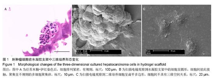
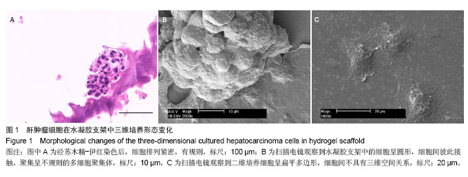
2.1 肝细胞三维培养后的形态变化 人肝癌细胞在水凝胶支架内呈三维立体方式生长,随培养时间的延长聚集成细胞团。将培养14 d的细胞团通过石蜡切片,苏木精-伊红染色结果显示,细胞团内细胞核蓝染,细胞排列紧密,有规则(图1A)。进一步通过扫描电镜观察细胞团的超微结构显示,在水凝胶支架中的细胞呈圆形,大小均一,多层排列,细胞间彼此接触,聚集呈不规则的多细胞聚集体 (图1B)。而二维培养细胞呈扁平多边形(图1C),细胞间不具有三维空间关系。"
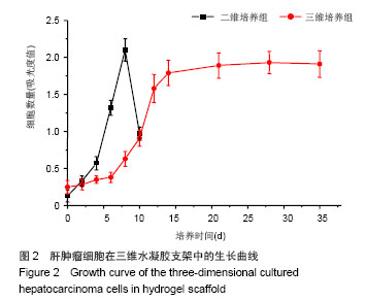
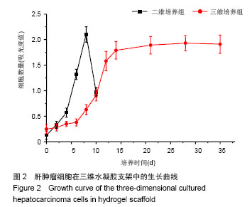
2.2 肝细胞三维培养体系的生长特性 从细胞生长曲线可以看出,与游离的二维培养方式相比,在水凝胶支架中,肝细胞生长曲线的延滞期略长于二维平面培养组。但二维培养的细胞在迅速进入对数生长期后,平台期时间维持很短,很快即从培养瓶中脱落,死亡。相比之下,在水凝胶支架中,细胞在平台期活性维持更持久,更利于细胞活性的长期保持(图2)。"
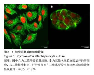
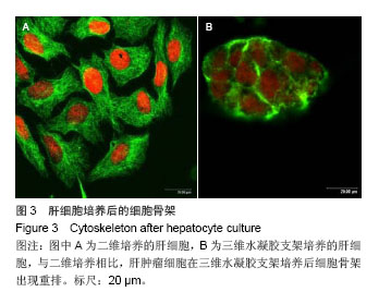
2.3 肝细胞三维培养体系的细胞骨架 细胞骨架是细胞内蛋白质组成的网络系统[42]。与贴壁细胞相比,在水凝胶支架中聚集生长后,细胞形态发生很大变化,由伸展的多边形变成球形。因此,通过标记细胞骨架蛋白,进一步研究在三维培养体系中的细胞骨架排布。借助FITC-鬼笔环肽标记细胞的纤维肌动蛋白,利用激光共聚焦扫描显微镜,观察纤维肌动蛋白在细胞上的分布。结果显示纤维肌动蛋白在水凝胶支架中三维细胞团内,呈现出完全不同于二维贴壁细胞的结构,大量的纤维肌动蛋白趋向于胞周分布,形成纤维肌动蛋白环,极性增强,与小鼠肝组织内的结构更接近(图3)。由此可见,肝细胞在水凝胶支架中聚集生长后,引起细胞骨架的重排和细胞形态的改变,形成更接近在体的结构。"
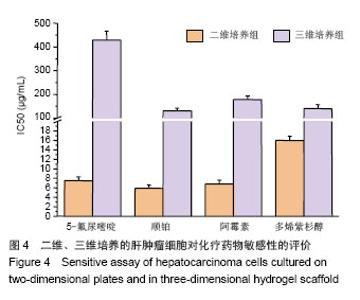
2.4 肝细胞三维培养体系对化疗药物的敏感性 以化疗药物5-氟尿嘧啶、顺铂、阿霉素、多烯紫杉醇为模型,研究上述制备的肝癌细胞三维体系对化疗药物的敏感性。结果显示4种药物对二维平面培养和三维立体培养的细胞均有抑制作用。但在传统的二维平面培养组中所有化疗药物对肝癌细胞抑制作用更显著,而在三维培养体系中,达到半数致死量的药物浓度要较二维平面培养组提高10倍左右。在4种化疗药物中,无论二维培养组、三维培养组,顺铂对肝癌细胞的抑制作用都优于其他药物。但5-氟尿嘧啶在二维培养体系中抑制效果要优于多烯紫杉醇,但在三维培养组中药物的抑制效果相反,5-氟尿嘧啶的化疗效果最差(图4)。"

| [1]Mulhall HJ, Hughes MP, Kazmi B, et al. Epithelial cancer cells exhibit different electrical properties when cultured in 2D and 3D environments. Biochim Biophys Acta. 2013;1830(11): 5136-5141. [2]da Rocha EL, Porto LM, Rambo CR. Nanotechnology meets 3D in vitro models: tissue engineered tumors and cancer therapies. Mater Sci Eng C Mater Biol Appl. 2014;34: 270-279. [3]Ramaiahgari SC, den Braver MW, Herpers B, et al. A 3D in vitro model of differentiated HepG2 cell spheroids with improved liver-like properties for repeated dose high-throughput toxicity studies. Arch Toxicol. 2014;88(5): 1083-1095. [4]Kimlin L, Kassis J, Virador V. 3D in vitro tissue models and their potential for drug screening. Expert Opin Drug Discov. 2013;8(12):1455-1466. [5]Jiguet Jiglaire C, Baeza-Kallee N, Denicolaï E, et al. Ex vivo cultures of glioblastoma in three-dimensional hydrogel maintain the original tumor growth behavior and are suitable for preclinical drug and radiation sensitivity screening. Exp Cell Res. 2014;321(2):99-108. [6]Xu F, Wu J, Wang S, et al. Microengineering methods for cell-based microarrays and high-throughput drug-screening applications. Biofabrication. 2011;3(3):034101. [7]Elliott NT, Yuan F. A review of three-dimensional in vitro tissue models for drug discovery and transport studies. J Pharm Sci. 2011;100(1):59-74. [8]Damania A, Jain E, Kumar A. Advancements in in vitro hepatic models: application for drug screening and therapeutics. Hepatol Int. 2014;8(1):23-28. [9]Moscona A, Moscona H. The dissociation and aggregation of cells from organ rudiments of the early chick embryo. J Anat. 1952;86(3):287-301. [10]Moscona AA. Studies on cell aggregation: demonstration of materials with selective cell-binding activity. Proc Natl Acad Sci U S A. 1963;49(5):742-747. [11]Lilien JE, Moscona AA. Cell aggregation: its enhancement by a supernatant from cultures of homologous cells. Science. 1967;157(3784):70-72. [12]Margolaish E, Schenck JR, Hargie MP, et al. Characterization of specific cell aggregating materials from sponge cells. Biochem Biophys Res Commun. 1965;20(4):383-388. [13]Moscona MH, Moscona AA. Inhibition of adhesiveness and aggregation of dissociated cells by inhibitors of protein and rna synthesis. Science. 1963;142(3595):1070-1071. [14]Moscona A. Rotation-mediated histogenetic aggregation of dissociated cells. A quantifiable approach to cell interactions in vitro. Exp Cell Res. 1961;22:455-475. [15]Moscona A. The development in vitro of chimeric aggregates of dissociated embryonic chick and mouse cells. Proc Natl Acad Sci U S A. 1957;43(1):184-194. [16]Gonzalez-Cordero A, West EL, Pearson RA, et al. Photoreceptor precursors derived from three-dimensional embryonic stem cell cultures integrate and mature within adult degenerate retina. Nat Biotechnol. 2013;31(8): 741-747. [17]Lou YR, Kanninen L, Kuisma T, et al. The use of nanofibrillar cellulose hydrogel as a flexible three-dimensional model to culture human pluripotent stem cells. Stem Cells Dev. 2014; 23(4):380-392. [18]Naito H, Yoshimura M, Mizuno T, et al. The advantages of three-dimensional culture in a collagen hydrogel for stem cell differentiation. J Biomed Mater Res A. 2013;101(10): 2838-2845. [19]Han J, Chen L, Luo G, et al. Three-dimensional culture may promote cell reprogramming. Organogenesis. 2013;9(2): 118-120. [20]Eiraku M, Sasai Y. Self-formation of layered neural structures in three-dimensional culture of ES cells. Curr Opin Neurobiol. 2012;22(5):768-777. [21]Vollmers A, Wallace L, Fullard N, et al. Two- and three-dimensional culture of keratinocyte stem and precursor cells derived from primary murine epidermal cultures. Stem Cell Rev. 2012;8(2):402-413. [22]Pampaloni F, Reynaud EG, Stelzer EH. The third dimension bridges the gap between cell culture and live tissue. Nat Rev Mol Cell Biol. 2007;8(10):839-845. [23]Fang C, Man YG, Cuttitta F, et al. Novel phenotypic fluorescent three-dimensional co-culture platforms for recapitulating tumor in vivo progression and for personalized therapy. J Cancer. 2013;4(9):755-763. [24]Nolte DD, An R, Turek J, et al. Tissue dynamics spectroscopy for phenotypic profiling of drug effects in three-dimensional culture. Biomed Opt Express. 2012;3(11):2825-2841. [25]Shimko VF, Claycomb WC. Effect of mechanical loading on three-dimensional cultures of embryonic stem cell-derived cardiomyocytes. Tissue Eng Part A. 2008;14(1):49-58.. [26]Ajalloueian F, Lim ML, Lemon G, et al. Biomechanical and biocompatibility characteristics of electrospun polymeric tracheal scaffolds. Biomaterials. 2014;35(20):5307-5315. [27]Chen H, Liu Y, Jiang Z, et al. Cell-scaffold interaction within engineered tissue. Exp Cell Res. 2014;323(2):346-351. [28]Kaczmarek M, Jurczyk MU, Rubis B, et al. In vitro biocompatibility of Ti-45S5 bioglass nanocomposites and their scaffolds. J Biomed Mater Res A. 2014;102(5):1316-1324. [29]Rahmanian-Schwarz A, Held M, Knoeller T, et al. In vivo biocompatibility and biodegradation of a novel thin and mechanically stable collagen scaffold. J Biomed Mater Res A. 2014;102(4):1173-1179. [30]Mercuri JJ, Patnaik S, Dion G, et al. Regenerative potential of decellularized porcine nucleus pulposus hydrogel scaffolds: stem cell differentiation, matrix remodeling, and biocompatibility studies. Tissue Eng Part A. 2013;19(7-8): 952-966. [31]García Cruz DM, Salmerón-Sánchez M, Gómez-Ribelles JL. Stirred flow bioreactor modulates chondrocyte growth and extracellular matrix biosynthesis in chitosan scaffolds. J Biomed Mater Res A. 2012;100(9):2330-2341. [32]Oliveira JM, Rodrigues MT, Silva SS, et al. Novel hydroxyapatite/chitosan bilayered scaffold for osteochondral tissue-engineering applications: Scaffold design and its performance when seeded with goat bone marrow stromal cells. Biomaterials. 2006;27(36):6123-6137. [33]Almeida CR, Serra T, Oliveira MI, et al. Impact of 3-D printed PLA- and chitosan-based scaffolds on human monocyte/macrophage responses: unraveling the effect of 3-D structures on inflammation. Acta Biomater. 2014;10(2): 613-622. [34]Ikeda T, Ikeda K, Yamamoto K, et al. Fabrication and characteristics of chitosan sponge as a tissue engineering scaffold. Biomed Res Int. 2014;2014:786892. [35]Inzana JA, Olvera D, Fuller SM, et al. 3D printing of composite calcium phosphate and collagen scaffolds for bone regeneration. Biomaterials. 2014;35(13):4026-4034. [36]Deponti D, Di Giancamillo A, Gervaso F, et al. Collagen scaffold for cartilage tissue engineering: the benefit of fibrin glue and the proper culture time in an infant cartilage model. Tissue Eng Part A. 2014;20(5-6):1113-1126. [37]Fagerholm P, Lagali NS, Ong JA, et al. Stable corneal regeneration four years after implantation of a cell-free recombinant human collagen scaffold. Biomaterials. 2014; 35(8):2420-2427. [38]Lozoya OA, Wauthier E, Turner RA, et al. Regulation of hepatic stem/progenitor phenotype by microenvironment stiffness in hydrogel models of the human liver stem cell niche. Biomaterials. 2011;32(30):7389-7402. [39]Pedron S, Becka E, Harley BA. Regulation of glioma cell phenotype in 3D matrices by hyaluronic acid. Biomaterials. 2013;34(30):7408-7417. [40]Drury JL, Mooney DJ. Hydrogels for tissue engineering: scaffold design variables and applications. Biomaterials. 2003;24(24):4337-4351. [41]Poellmann MJ, Harrell PA, King WP, et al. Geometric microenvironment directs cell morphology on topographically patterned hydrogel substrates. Acta Biomater. 2010;6(9): 3514-3523. [42]Ingber DE, Dike L, Hansen L, et al. Cellular tensegrity: exploring how mechanical changes in the cytoskeleton regulate cell growth, migration, and tissue pattern during morphogenesis. Int Rev Cytol. 1994;150:173-224. [43]Yamada M, Utoh R, Ohashi K, et al. Controlled formation of heterotypic hepatic micro-organoids in anisotropic hydrogel microfibers for long-term preservation of liver-specific functions. Biomaterials. 2012;33(33):8304-8315. [44]You J, Shin DS, Patel D, et al. Multilayered heparin hydrogel microwells for cultivation of primary hepatocytes. Adv Healthc Mater. 2014;3(1):126-132. [45]Du C, Narayanan K, Leong MF, et al. Induced pluripotent stem cell-derived hepatocytes and endothelial cells in multi-component hydrogel fibers for liver tissue engineering. Biomaterials. 2014;35(23):6006-6014. [46]Edelman GM. Cell adhesion molecules in the regulation of animal form and tissue pattern. Annu Rev Cell Biol. 1986;2: 81-116. [47]Takeichi M. Cadherin cell adhesion receptors as a morphogenetic regulator. Science. 1991;251(5000): 1451-1455. [48]Gallin WJ, Sanders EJ. Development of bile canaliculi between chicken embryo liver cells in vivo and in vitro. Exp Cell Res. 1992;200(1):58-69. [49]Kelly HT, Hill E, Reid F, et al. Comparison of RAP-PCR analysis of gene expression in fresh and immortalised rat hepatocyte cell lines. Cytotechnology. 2000;34(1-2): 159-163. [50]Klausner RD. The fabric of cancer cell biology-Weaving together the strands. Cancer Cell. 2002;1(1):3-10. [51]Albini A, Sporn MB. The tumour microenvironment as a target for chemoprevention. Nat Rev Cancer. 2007;7(2):139-147. [52]Liotta LA, Kohn EC. The microenvironment of the tumour-host interface. Nature. 2001;411(6835):375-379. [53]Kunz-Schughart LA, Kreutz M, Knuechel R. Multicellular spheroids: a three-dimensional in vitro culture system to study tumour biology. Int J Exp Pathol. 1998 Feb;79(1):1-23. [54]Yamada KM, Cukierman E. Modeling tissue morphogenesis and cancer in 3D. Cell. 2007;130(4):601-610. [55]Jiguet Jiglaire C, Baeza-Kallee N, Denicolaï E, et al. Ex vivo cultures of glioblastoma in three-dimensional hydrogel maintain the original tumor growth behavior and are suitable for preclinical drug and radiation sensitivity screening. Exp Cell Res. 2014;321(2):99-108. [56]Fischbach C1, Chen R, Matsumoto T, et al. Engineering tumors with 3D scaffolds. Nat Methods. 2007;4(10): 855-860. [57]Teicher BA, Herman TS, Holden SA, et al. Tumor resistance to alkylating agents conferred by mechanisms operative only in vivo. Science. 1990;247(4949 Pt 1):1457-1461. [58]Ng CP, Bonavida B. A new challenge for successful immunotherapy by tumors that are resistant to apoptosis: two complementary signals to overcome cross-resistance. Adv Cancer Res. 2002;85:145-174. [59]Lewis JT, Ketterling RP, Halling KC, et al. Analysis of intratumoral heterogeneity and amplification status in breast carcinomas with equivocal (2+) HER-2 immunostaining. Am J Clin Pathol. 2005;124(2):273-281. [60]Minchinton AI, Tannock IF. Drug penetration in solid tumours. Nat Rev Cancer. 2006;6(8):583-592. [61]Comerford KM, Wallace TJ, Karhausen J, et al. Hypoxia-inducible factor-1-dependent regulation of the multidrug resistance (MDR1) gene. Cancer Res. 2002;62(12): 3387-3394. |
| [1] | Shen Jinbo, Zhang Lin. Micro-injury of the Achilles tendon caused by acute exhaustive exercise in rats: ultrastructural changes and mechanism [J]. Chinese Journal of Tissue Engineering Research, 2021, 25(8): 1190-1195. |
| [2] | Liang Xueqi, Guo Lijiao, Chen Hejie, Wu Jie, Sun Yaqi, Xing Zhikun, Zou Hailiang, Chen Xueling, Wu Xiangwei. Alveolar echinococcosis protoscolices inhibits the differentiation of bone marrow mesenchymal stem cells into fibroblasts [J]. Chinese Journal of Tissue Engineering Research, 2021, 25(7): 996-1001. |
| [3] | Duan Liyun, Cao Xiaocang. Human placenta mesenchymal stem cells-derived extracellular vesicles regulate collagen deposition in intestinal mucosa of mice with colitis [J]. Chinese Journal of Tissue Engineering Research, 2021, 25(7): 1026-1031. |
| [4] | Li Li, Ma Li. Immobilization of lactase on magnetic chitosan microspheres and its effect on enzymatic properties [J]. Chinese Journal of Tissue Engineering Research, 2021, 25(4): 576-581. |
| [5] | Liu Liu, Zhou Qingzhu, Gong Zhuo, Liu Boyan, Yang Bin, Zhao Xian. Characteristics and manufacturing techniques of collagen/inorganic materials for constructing tissue-engineered bone [J]. Chinese Journal of Tissue Engineering Research, 2021, 25(4): 607-613. |
| [6] | Xu Xiaoming, Chen Yan, Song Qian, Yuan Lu, Gu Jiaming, Zhang Lijuan, Geng Jie, Dong Jian. Human placenta derived mesenchymal stem cell gel promotes the healing of radiation skin damage in SD rats [J]. Chinese Journal of Tissue Engineering Research, 2021, 25(25): 3976-3980. |
| [7] | Wang Hao, Chen Mingxue, Li Junkang, Luo Xujiang, Peng Liqing, Li Huo, Huang Bo, Tian Guangzhao, Liu Shuyun, Sui Xiang, Huang Jingxiang, Guo Quanyi, Lu Xiaobo. Decellularized porcine skin matrix for tissue-engineered meniscus scaffold [J]. Chinese Journal of Tissue Engineering Research, 2021, 25(22): 3473-3478. |
| [8] | Chen Lei, Zheng Rui, Jie Yongsheng, Qi Hui, Sun Lei, Shu Xiong. In vitro evaluation of adipose-derived stromal vascular fraction combined with osteochondral integrated scaffold [J]. Chinese Journal of Tissue Engineering Research, 2021, 25(22): 3487-3492. |
| [9] | Li Xinping, Cui Qiuju, Zeng Shuguang, Ran Gaoying, Zhang Zhaoqiang, Liu Xianwen, Fang Wei, Xu Shuaimei. Effect of modification of β-tricalcium phosphate/chitosan hydrogel on growth and mineralization of dental pulp stem cells [J]. Chinese Journal of Tissue Engineering Research, 2021, 25(22): 3493-3499. |
| [10] | Yang Li, Li Xueli, Song Jinghui, Yu Huiqian, Wang Weixia. Effect of cryptotanshinone on hypertrophic scar of rabbit ear and its related mechanism [J]. Chinese Journal of Tissue Engineering Research, 2021, 25(20): 3150-3155. |
| [11] | Liu Zhendong, Wang Rui, Li Xiaolei, Wang Jingcheng. Review of interferon alpha-2b inhibiting scar formation [J]. Chinese Journal of Tissue Engineering Research, 2021, 25(2): 317-321. |
| [12] | Chen Siyu, Li Yannan, Xie Liying, Liu Siqi, Fan Yurong, Fang Changxing, Zhang Xin, Quan Jiayu, Zuo Lin. Thermosensitive chitosan-collagen composite hydrogel loaded with basic fibroblast growth factor retards ventricular remodeling after myocardial infarction in mice [J]. Chinese Journal of Tissue Engineering Research, 2021, 25(16): 2472-2478. |
| [13] | Liu Feng, Zhang Yu, Wang Yanli, Luo Wei, Han Chaoshan, Li Yangxin. Application of temperature-sensitive chitosan hydrogel encapsulated exosomes in ischemic diseases [J]. Chinese Journal of Tissue Engineering Research, 2021, 25(16): 2479-2487. |
| [14] | Chen Liang, Meng Shu, Cheng Guoping, Ding Yi . Effects of fish scale collagen membrane on adhesion, proliferation and osteogenic differentiation of rat bone marrow mesenchymal stem cells [J]. Chinese Journal of Tissue Engineering Research, 2021, 25(16): 2494-2499. |
| [15] | Li Jie, Xu Jianzhen, Hu Ping, Lei Qiqi, Zhang Wenning, Ao Ningjian . Preparation and performance evaluation of carboxymethyl chitosan/oxidized glucomannan/Panax notoginseng compound sponge dressing for chronic wound [J]. Chinese Journal of Tissue Engineering Research, 2021, 25(16): 2528-2534. |
| Viewed | ||||||
|
Full text |
|
|||||
|
Abstract |
|
|||||