Chinese Journal of Tissue Engineering Research ›› 2023, Vol. 27 ›› Issue (16): 2473-2479.doi: 10.12307/2023.464
Previous Articles Next Articles
Preparation and osteoinductive properties of tricalcium phosphate ceramics with submicron topology
Lu Di1, Wan Xinyu1, Yang Jinxin1, Ding Kexin1, Zhang Cheng1, Duan Rongquan1, Liu Zongxiang1, 2
- 1School of Stomatology, Xuzhou Medical University, Xuzhou 221004, Jiangsu Province, China; 2Affiliated Stomatological Hospital of Xuzhou Medical University, Xuzhou 221002, Jiangsu Province, China
-
Received:2022-05-26Accepted:2022-07-21Online:2023-06-08Published:2022-11-10 -
Contact:Liu Zongxiang, Chief physician, Professor, School of Stomatology, Xuzhou Medical University, Xuzhou 221004, Jiangsu Province, China; Affiliated Stomatological Hospital of Xuzhou Medical University, Xuzhou 221002, Jiangsu Province, China Duan Rongquan, MD, Associate professor, School of Stomatology, Xuzhou Medical University, Xuzhou 221004, Jiangsu Province, China -
About author:Lu Di, Master candidate, Physician, School of Stomatology, Xuzhou Medical University, Xuzhou 221004, Jiangsu Province, China -
Supported by:the Transformation Project of Sichuan Provincial Department of Science and Technology, No. 2019ZYZF0081 (to DRQ); Excellent Talents Research Start-up Fund of Xuzhou Medical University, No. D2020005 (to DRQ)
CLC Number:
Cite this article
Lu Di, Wan Xinyu, Yang Jinxin, Ding Kexin, Zhang Cheng, Duan Rongquan, Liu Zongxiang. Preparation and osteoinductive properties of tricalcium phosphate ceramics with submicron topology[J]. Chinese Journal of Tissue Engineering Research, 2023, 27(16): 2473-2479.
share this article
Add to citation manager EndNote|Reference Manager|ProCite|BibTeX|RefWorks
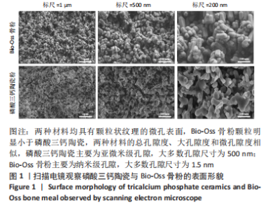
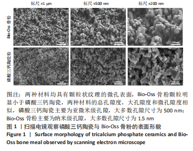
2.1 两种材料的形貌与生物矿化结果 扫描电镜成像显示两种材料均具有颗粒状纹理的微孔表面,其中Bio-Oss骨粉颗粒明显小于磷酸三钙陶瓷,两种材料的总孔隙度、大孔隙度和微孔隙度相似,磷酸三钙陶瓷主要为亚微米级孔隙,大多数孔隙尺寸为500 nm;Bio-Oss骨粉主要为纳米级孔隙,大多数孔隙尺寸为1.5 nm,见图1。"
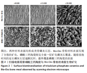
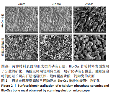
在37 ℃的模拟体液中浸泡7 d后,两种材料表面均形成类骨磷灰石层,但磷酸三钙陶瓷与Bio-Oss骨粉的矿化程度不同,Bio-Oss骨粉材料表面发现了分散的矿化,磷酸三钙陶瓷则完全被一层矿化磷灰石覆盖,随着浸泡时间的延长磷灰石层逐渐沉积,最终覆盖磷酸三钙陶瓷的表面,见图2。表明磷酸三钙陶瓷表面诱导类骨磷灰石沉积的能力明显优于Bio-Oss骨粉。 "

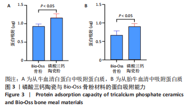
2.2 两种材料的蛋白吸附能力 结果显示,Bio-Oss骨粉相比,磷酸三钙陶瓷能从牛血清白蛋白和胎牛血清溶液中吸附更多的蛋白质(P < 0.05),见图3。"
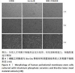
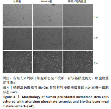
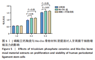
2.3 两种材料浸提液对人牙周膜干细胞增殖的影响 培养后第 1,3,5 天,镜下可见人牙周膜干细胞形态呈长梭形,有较强贴壁能力,细胞数量逐日增加,见图4。CCK-8实验结果显示,培养第1天,3组细胞间增殖能力比较差异无显著性意义(P > 0.05);培养第3,5天,Bio-Oss组、磷酸三钙陶瓷组细胞增殖能力优于对照组(P < 0.05),Bio-Oss组、磷酸三钙陶瓷组细胞增殖能力比较差异无显著性意义(P > 0.05),见图5,结果说明,磷酸三钙陶瓷和 Bio-Oss骨粉浸提液对细胞的增殖有较好的促进作用,均表现出良好的生物相容性,表明两种骨粉材料对人牙周膜干细胞增殖能力的影响相似。"

"
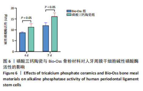
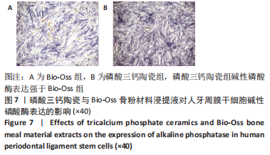
2.4 两种材料对人牙周膜干细胞成骨分化的影响 2.4.1 碱性磷酸酶染色和活性检测结果 培养第4,7天,磷酸三钙陶瓷组的碱性磷酸酶活性均高于Bio-Oss组(P < 0.05),见图6。培养第7天的碱性磷酸酶染色显示,磷酸三钙陶瓷组碱性磷酸酶表达强于Bio-Oss组,与碱性磷酸酶活性测定结果一致,见图7。"


"
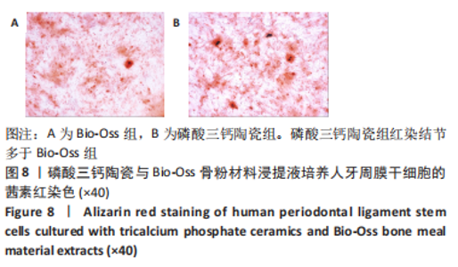
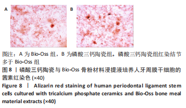
2.4.2 茜素红染色结果 Bio-Oss组和磷酸三钙陶瓷组均有红染矿化结节,且磷酸三钙陶瓷组红染结节多于Bio-Oss组,见图8,表明磷酸三钙陶瓷对成骨晚期矿化有促进作用。"
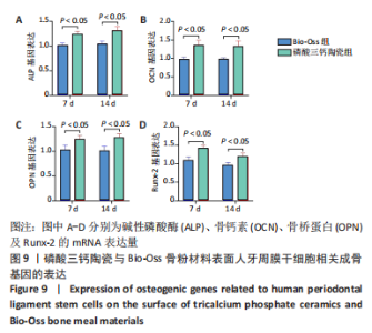
2.4.3 qRT-PCR检测结果 培养第7,14天,与Bio-Oss组相比,磷酸三钙陶瓷组人牙周膜干细胞中成骨相关因子骨桥蛋白、骨钙素、Runx-2、碱性磷酸酶的mRNA表达量升高(P < 0.05),见图9。也就是说明磷酸三钙陶瓷可能要比 Bio-Oss 骨粉更加有利于人牙周膜干细胞的成骨分化。"


2.5 两种材料的生物相容性 由CCK-8实验结果可知,磷酸三钙陶瓷与Bio-Oss骨粉材料具有良好的生物相容性。"

| [1] TANG Z, LI X, TAN Y, et al. The material and biological characteristics of osteoinductive calcium phosphate ceramics. Regen Biomater. 2018; 5(1):43-59. [2] DUAN R, VAN DIJK LA, BARBIERI D, et al. Accelerated bone formation by biphasic calcium phosphate with a novel sub-micron surface topography. Eur Cell Mater. 2019;37:60-73. [3] 肖莎,高承志,周冬平.三种骨替代材料修复即刻种植下颌后牙区周围骨缺损的比较[J].中国组织工程研究,2021,25(34):5495-5500. [4] 刘一.新型骨修复材料和Bio-oss骨粉用于种植牙骨缺损的填充修复效果观察[J].当代医学,2020,26(1):17-19. [5] 路佳佳,李伟琼,许敏,等. BoneCeramic和Bio-Oss骨粉对狗骨髓间充质干细胞体外成骨分化能力影响的比较研究[J].中国药理学通报,2021,37(4):511-516. [6] KIM SE, PARK K. Recent Advances of Biphasic Calcium Phosphate Bioceramics for Bone Tissue Regeneration. Adv Exp Med Biol. 2020; 1250:177-188. [7] GUO T, KANG W, XIAO D, et al. Molecular docking characterization of a four-domain segment of human fibronectin encompassing the RGD loop with hydroxyapatite. Molecules. 2013;19(1):149-158. [8] DUAN R, BARBIERI D, LUO X, et al. Variation of the bone forming ability with the physicochemical properties of calcium phosphate bone substitutes. Biomater Sci. 2017;6(1):136-145. [9] ELIAZ N, METOKI N. Calcium Phosphate Bioceramics: A Review of Their History, Structure, Properties, Coating Technologies and Biomedical Applications. Materials (Basel). 2017;10(4):334. [10] WANG P, WANG W, GENG T, et al. EphrinB2 regulates osteogenic differentiation of periodontal ligament stem cells and alveolar bone defect regeneration in beagles. J Tissue Eng. 2019;10:1543343527. [11] WANG W, YUAN C, LIU Z, et al. Characteristic comparison between canine and human dental mesenchymal stem cells for periodontal regeneration research in preclinical animal studies. Tissue Cell. 2020; 67:101405. [12] AN Y, ZHANG H, WANG C, et al. Activation of ROS/MAPKs/NF-κB/NLRP3 and inhibition of efferocytosis in osteoclast-mediated diabetic osteoporosis. FASEB J. 2019;33(11):12515-12527. [13] LIU Q, DOUGLAS T, ZAMPONI C, et al. Comparison of in vitro biocompatibility of NanoBone(®) and BioOss(®) for human osteoblasts. Clin Oral Implants Res. 2011;22(11):1259-1264. [14] DUAN R, ZHANG Y, VAN DIJK L, et al. Coupling between macrophage phenotype, angiogenesis and bone formation by calcium phosphates. Mater Sci Eng C Mater Biol Appl. 2021;122:111948. [15] BEREBICHEZ-FRIDMAN R, MONTERO-OLVERA PR. Sources and Clinical Applications of Mesenchymal Stem Cells: State-of-the-art review. Sultan Qaboos Univ Med J. 2018;18(3):e264-e277. [16] ABE T, SUMI K, KUNIMATSU R, et al. Bone Regeneration in a Canine Model of Artificial Jaw Cleft Using Bone Marrow-Derived Mesenchymal Stem Cells and Carbonate Hydroxyapatite Carrier. Cleft Palate Craniofac J. 2020;57(2):208-217. [17] CHEN Z, BACHHUKA A, HAN S, et al. Tuning Chemistry and Topography of Nanoengineered Surfaces to Manipulate Immune Response for Bone Regeneration Applications. ACS Nano. 2017;11(5):4494-4506. [18] SAFRONOVA TV, SELEZNEVA II, TIKHONOVA SA, et al. Biocompatibility of biphasic α,β-tricalcium phosphate ceramics in vitro. Bioact Mater. 2020;5(2):423-427. [19] AYDIN S, ŞAHIN F. Stem Cells Derived from Dental Tissues. Adv Exp Med Biol. 2019;1144:123-132. [20] WU Y, YANG Y, YANG P, et al. The osteogenic differentiation of PDLSCs is mediated through MEK/ERK and p38 MAPK signalling under hypoxia. Arch Oral Biol. 2013;58(10):1357-1368. [21] SUN YY, HU WP, LIU ZX, et al. [Effects of Wnt3a on osteogenic differentiation of dental pulp stem cells]. Zhonghua Kou Qiang Yi Xue Za Zhi. 2017;52(7):427-431. [22] LEE YC, CHAN YH, HSIEH SC, et al. Comparing the Osteogenic Potentials and Bone Regeneration Capacities of Bone Marrow and Dental Pulp Mesenchymal Stem Cells in a Rabbit Calvarial Bone Defect Model. Int J Mol Sci. 2019;20(20):5015. [23] LIU Y, LIU C, ZHANG A, et al. Down-regulation of long non-coding RNA MEG3 suppresses osteogenic differentiation of periodontal ligament stem cells (PDLSCs) through miR-27a-3p/IGF1 axis in periodontitis. Aging (Albany NY). 2019;11(15):5334-5350. [24] VAN DIJK LA, DUAN R, LUO X, et al. Biphasic calcium phosphate with submicron surface topography in an Ovine model of instrumented posterolateral spinal fusion. JOR Spine. 2018;1(4):e1039. [25] FU X, LIU G, HALIM A, et al. Mesenchymal Stem Cell Migration and Tissue Repair. Cells. 2019;8(8):784. [26] DENG C, SHEN X, YANG W, et al. Construction of zinc-incorporated nano-network structures on a biomedical titanium surface to enhance bioactivity. Appl Surf Sci. 2018;453;263-270. [27] DUAN R, BARBIERI D, DE GROOT F, et al. Modulating Bone Regeneration in Rabbit Condyle Defects with Three Surface-Structured Tricalcium Phosphate Ceramics. ACS Biomater Sci Eng. 2018;4(9):3347-3355. [28] WANG H, ZHI W, LU X, et al. Comparative studies on ectopic bone formation in porous hydroxyapatite scaffolds with complementary pore structures. Acta Biomater. 2013;9(9):8413-8421. [29] LUO X, BARBIERI D, DUAN R, et al. Strontium-containing apatite/polylactide composites enhance bone formation in osteopenic rabbits. Acta Biomater. 2015;26:331-337. [30] DUAN R, BARBIERI D, LUO X, et al. Submicron-surface structured tricalcium phosphate ceramic enhances the bone regeneration in canine spine environment. J Orthop Res. 2016;34(11):1865-1873. |
| [1] | Wen Xinghua, Ding Huanwen, Cheng Kai, Yan Xiaonan, Peng Yuanhao, Wang Yuning, Liu Kang, Zhang Huiwu. Three-dimensional finite element model analysis of intramedullary nailing fixation design for large femoral defects in Beagle dogs [J]. Chinese Journal of Tissue Engineering Research, 2023, 27(9): 1371-1376. |
| [2] | Du Xueting, Zhang Xiaodong, Chen Yanjun, Wang Mei, Chen Wubiao, Huang Wenhua. Application of compressed sensing technology in two-dimensional magnetic resonance imaging of the ankle joint [J]. Chinese Journal of Tissue Engineering Research, 2023, 27(9): 1396-1402. |
| [3] | Long Guiyue, Li Dongdong, Liao Hongbing. Calcium phosphate cement/poly(lactic-co-glycolic acid) degradation products promote osteoclast differentiation of mouse monocytes [J]. Chinese Journal of Tissue Engineering Research, 2023, 27(8): 1193-1198. |
| [4] | Yang Zhishan, Tang Zhenglong. YAP/TAZ, a core factor of the Hippo signaling pathway, is involved in bone formation [J]. Chinese Journal of Tissue Engineering Research, 2023, 27(8): 1264-1271. |
| [5] | Xu Cong, Zhao He, Sun Yan. Regeneration of facial nerve injury repaired by biomaterial nerve conduits [J]. Chinese Journal of Tissue Engineering Research, 2023, 27(7): 1089-1095. |
| [6] | Lu Di, Zhang Cheng, Duan Rongquan, Liu Zongxiang. Osteoinductive properties of calcium phosphate ceramic bone repair materials [J]. Chinese Journal of Tissue Engineering Research, 2023, 27(7): 1103-1109. |
| [7] | Tang Haotian, Liao Rongdong, Tian Jing. Application and design of piezoelectric materials for bone defect repair [J]. Chinese Journal of Tissue Engineering Research, 2023, 27(7): 1117-1125. |
| [8] | Xu Yan, Li Ping, Lai Chunhua, Zhu Peijun, Yang Shuo, Xu Shulan. Piezoelectric materials for vascularized bone regeneration [J]. Chinese Journal of Tissue Engineering Research, 2023, 27(7): 1126-1132. |
| [9] | Li Wenjie, You Aijia, Zhou Junli, Fang Sujuan, Li Chun. Effects of different dressings in the treatment of burn wounds: a network meta-analysis [J]. Chinese Journal of Tissue Engineering Research, 2023, 27(7): 1141-1148. |
| [10] | Ke Weiqiang, Chen Xianghui, Chen Xiaoling, Meng Jie, Ma Yanlin. Rituximab combined with autologous peripheral blood stem cell transplantation in the treatment of diffuse large B-cell lymphoma and the expression of related factors [J]. Chinese Journal of Tissue Engineering Research, 2023, 27(6): 915-920. |
| [11] | Xu Qijing, Yang Yichun, Lei Wei, Yang Ying, Yu Jiang, Xia Tingting, Zhang Meng, Zhang Tao, Zhang Qian. Advances and problems in cell-free treatment of diabetic skin chronic wounds [J]. Chinese Journal of Tissue Engineering Research, 2023, 27(6): 962-969. |
| [12] | Shen Lianwei, Zhu Hongliu, Wang Wei. Risk factor analysis of metabolic syndrome and construction of a nomogram prediction model in middle-aged and elderly people [J]. Chinese Journal of Tissue Engineering Research, 2023, 27(5): 657-662. |
| [13] | Li Long, Li Guangdi, Shi Hao, Deng Keqi. Circular RNA as a competing endogenous RNA is involved in the regulation of osteoarthritis [J]. Chinese Journal of Tissue Engineering Research, 2023, 27(5): 751-757. |
| [14] | Zhang Min, Zhang Xiaoming, Liu Tongbin. Application potential of naringin in bone tissue regeneration [J]. Chinese Journal of Tissue Engineering Research, 2023, 27(5): 787-792. |
| [15] | Liu Yuan. Effect of hypoxic training on the oxygen sensing pathway [J]. Chinese Journal of Tissue Engineering Research, 2023, 27(5): 793-798. |
| Viewed | ||||||
|
Full text |
|
|||||
|
Abstract |
|
|||||