Chinese Journal of Tissue Engineering Research ›› 2023, Vol. 27 ›› Issue (9): 1396-1402.doi: 10.12307/2023.205
Previous Articles Next Articles
Application of compressed sensing technology in two-dimensional magnetic resonance imaging of the ankle joint
Du Xueting1, Zhang Xiaodong2, Chen Yanjun2, Wang Mei3, Chen Wubiao1, Huang Wenhua4, 5
- 1Department of Radiology, 4Department of Orthopedics, Affiliated Hospital of Guangdong Medical University, Zhanjiang 524001; 2Department of Radiology, Third Affiliated Hospital of Southern Medical University, Guangzhou 510000; 3Department of Radiology, Nansha Hosptial, Guangzhou First People’s Hosptial, Guangzhou 511458; 5Guangdong Engineering Research Center for Translation of Medical 3D Printing Application, Guangzhou 510515
-
Received:2021-12-23Accepted:2022-01-22Online:2023-03-28Published:2022-07-01 -
Contact:Huang Wenhua, MD, Doctoral supervisor, Professor, Department of Orthopedics, Affiliated Hospital of Guangdong Medical University, Zhanjiang 524001, Guangdong Province, China; Guangdong Engineering Research Center for Translation of Medical 3D Printing Application, Guangdong Provincial Key Laboratory of Medical Biomechanics, National Key Discipline of Human Anatomy, School of Basic Medical Sciences, Southern Medical University, Guangzhou 510515, Guangdong Province, China Chen Wubiao, Chief physician, Master’s supervisor, Department of Radiology, Affiliated Hospital of Guangdong Medical University, Zhanjiang 524001, Guangdong Province, China -
About author:Du Xueting, Master, Technician, Department of Radiology, Affiliated Hospital of Guangdong Medical University, Zhanjiang 524001, Guangdong Province, China -
Supported by:Science and Technology Planning Project of Guangdong Province, No. 2016B090917001 (to HWH), No. 2017B090912006 (to ZXD); “Sanming Project” of Medicine in Shenzhen, No. SZSM201612019 (to HWH); Fundamental and Applied Basic Research Fund of Guangdong Province, No. 2020B1515120001 (to HWH)
CLC Number:
Cite this article
Du Xueting, Zhang Xiaodong, Chen Yanjun, Wang Mei, Chen Wubiao, Huang Wenhua. Application of compressed sensing technology in two-dimensional magnetic resonance imaging of the ankle joint[J]. Chinese Journal of Tissue Engineering Research, 2023, 27(9): 1396-1402.
share this article
Add to citation manager EndNote|Reference Manager|ProCite|BibTeX|RefWorks
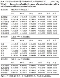
2.1 定性和定量资料的一致性分析 结果显示,2位观察者对38例踝关节图像质量主观评分的一致性较好,加权Kappa系数为0.778-0.971。重复2次踝关节图像背景噪声标准差和各结构信号强度的测量结果一致性较好,组内相关系数ICC值为0.689-0.999。 2.2 图像定性评价结果 不同加速因子的踝关节图像主观评分统计结果见表3。"
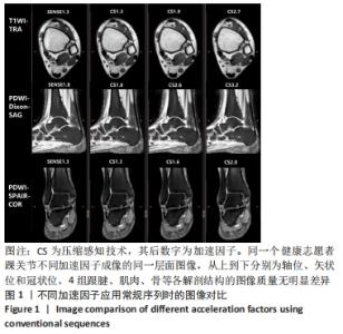
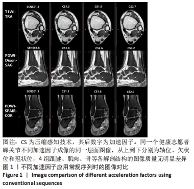
结果如下:①总体上CS组各解剖结构主观评分均随加速因子的升高而下降,但均≥3分;②在轴位T1WI序列上,4组加速因子间胫后肌腱主观评分比较差异无显著性意义(P > 0.05),而不同加速因子间肌肉、韧带及除胫后肌腱外的其他肌腱的主观评分比较差异均有显著性意义(P < 0.05);③在矢状位PDWI序列上,4组加速因子间软骨、跟腱和骨的主观评分比较差异有显著性意义(P < 0.05);④在冠状位PDWI序列上,4组加速因子间软骨和韧带的主观评分比较差异有显著性意义(P < 0.05)。常规序列不同加速因子的图像对比见图1。 "
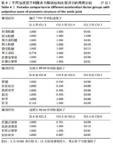
对各组踝关节解剖结构的主观评分进行两两比较,见表4。 "

结果如下:①在同一方位序列下,当S组和CS组的加速因子相同时,肌腱、肌肉、软骨和韧带的主观评分比较差异均无显著性意义(P > 0.05);②在轴位上,CS2.7组胫前肌腱、腓肠肌腱、腓短肌腱、肌肉和韧带的主观评分均低于CS1.3组(P < 0.05);③在矢状位上,CS3.2组跟腱、软骨和骨的主观评分均低于CS1.8组(P < 0.05);④在冠状位上,CS2.0组的韧带和软骨均低于S1.3组(P < 0.05)。 2.3 图像定量评价结果 在测量各解剖结构信号强度和背景噪声标准差时发现,CS组对运动伪影显示更为敏感,以高信号半圆环形显示,多在图像背景噪声中显示。 不同加速因子的踝关节解剖结构信噪比和对比噪声比统计结果,见表5。 "

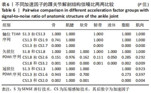
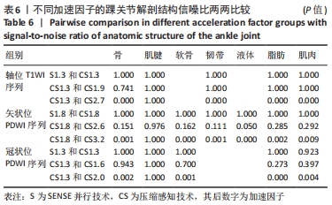
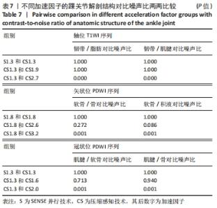
结果如下:①在轴位序列中,不同加速因子间肌腱信噪比的比较差异无显著性意义(P > 0.05),不同加速因子间骨、肌肉、脂肪和韧带信噪比的比较差异有显著性意义(P < 0.05),不同加速因子间韧带/脂肪和韧带/肌腱对比噪声比的比较差异有显著性意义(P < 0.05);②在矢状位序列中,不同加速因子间肌腱信噪比的比较差异无显著性意义(P > 0.05),而骨、软骨、液体、肌肉、脂肪和韧带信噪比的比较差异有显著性意义(P < 0.05),软骨/骨和软骨/积液对比噪声比的比较差异有显著性意义(P < 0.05);③在冠状位4组中,不同加速因子间肌腱信噪比的比较差异无显著性意义(P > 0.05),不同加速因子间骨、脂肪、肌肉和软骨信噪比的比较差异有显著性意义(P < 0.05),不同加速因子间肌腱/软骨和肌腱/骨对比噪声比的比较差异有显著性意义(P < 0.05)。 对各组的踝关节解剖结构信噪比和对比噪声比进行两两比较,见表6,7。 "
"

结果如下:①在相同方位序列下,当S组和CS组的加速因子相同时,两组间骨、肌腱、软骨、韧带、液体、脂肪、肌肉信噪比的比较差异无显著性意义(P > 0.05),且总体上来说,CS组的信噪比和对比噪声比大于S组;②在轴位T1WI序列上,当加速因子从CS1.3增加到CS2.7时,两组间肌腱信噪比的比较差异无显著性意义(P > 0.05),骨、肌肉、脂肪和韧带的信噪比逐渐降低(P < 0.05),韧带/脂肪和韧带/肌腱的对比噪声比逐渐降低(P < 0.05);③在矢状位PDWI序列上,当加速因子从CS1.8增加到CS3.2时,两组间肌腱信噪比的比较差异无显著性意义(P > 0.05),而骨、软骨、韧带、液体、脂肪、肌肉的信噪比逐渐降低(P < 0.05),软骨/骨和软骨/积液的对比噪声比逐渐降低(P < 0.05);④在冠状位PDWI序列上,当加速因子从CS1.3增加到CS2.0时,两组间肌腱信噪比的比较差异无显著性意义(P > 0.05),而骨、软骨、脂肪和肌肉的信噪比逐渐降低(P < 0.05),肌腱/软骨和肌腱/骨的对比噪声比逐渐降低(P < 0.05)。 2.4 扫描时间 随着加速因子的增大,踝关节常规序列扫描时间减少,以SENSE并行技术的扫描时间为基准,CS扫描时间减少量计算如公式(3): 扫描时间减少量=(S组扫描时间-CS组扫描时间)/S组扫描时间×100% (3) 踝关节轴位T1WI序列采用S1.3、CS1.3、CS1.9、CS2.7的扫描时间及减少量分别是2 min 13 s、2 min 13 s、1 min 32 s(减少31%)、1 min 5 s(减少51%);矢状位PDWI序列采用S1.8、CS1.8、CS2.6、CS3.2的扫描时间及减少量分别是2 min 30 s、2 min 30 s、1 min 42 s(减少32%)、1 min 18 s(减少48%);冠状位PDWI序列采用S1.3、CS1.3、CS1.6、CS2.0的扫描时间及减少量分别是2 min 12 s、2 min 12 s、1 min 48 s(减少18%)、1 min 24 s(减少36%)。 "

| [1] CREMA MD, KRIVOKAPIC B, GUERMAZI A, et al. MRI of ankle sprain: the association between joint effusion and structural injury severity in a large cohort of athletes. Eur Radiol. 2019;29(11):6336-6344. [2] SIRIWANARANGSUN P, BAE WC, STATUM S, et al. Advanced MRI Techniques for the Ankle. AJR Am J Roentgenol. 2017;209(3):511-524. [3] FRITZ B, FRITZ J, SUTTER R. 3D MRI of the Ankle: A Concise State-of-the-Art Review. Semin Musculoskelet Radiol. 2021;25(3):514-526. [4] GERSING AS, BODDEN J, NEUMANN J, et al. Accelerating anatomical 2D turbo spin echo imaging of the ankle using compressed sensing. Eur J Radiol. 2019;118(9):277-284. [5] JASPAN ON, FLEYSHER R, LIPTON ML. Compressed sensing MRI: a review of the clinical literature. Br J Radiol. 2015;88(1056):20150487. [6] HOLLINGSWORTH KG. Reducing acquisition time in clinical MRI by data undersampling and compressed sensing reconstruction. Phys Med Biol. 2015;60(21):297-322. [7] KLAAS PP, MARKUS W, MARKUS BS, et al. SENSE: Sensitivity Encoding for Fast MRI. Magn Reson Med. 1999;42(5):952-962. [8] LUSTIG M, DONOHO D, PAULY JM. Sparse MRI: The application of compressed sensing for rapid MR imaging. Magn Reson Med. 2007;58(6):1182-1195. [9] DONOHO DL. Compressed sensing. IEEE Trans Inf Theory. 2006;52(4): 1289-1306. [10] DESHMANE A, GULANI V, GRISWOLD MA, et al. Parallel MR imaging. J Magn Reson Imaging. 2012;36(1):55-72. [11] NOTOHAMIPRODJO M, KUSCHEL B, HORNG A, et al. 3D-MRI of the ankle with optimized 3D-SPACE. Invest Radiol. 2012;47(4):231-239. [12] KIM HS, YOON YC, KWON JW, et al. Qualitative and quantitative assessment of isotropic ankle magnetic resonance imaging: three-dimensional isotropic intermediate-weighted turbo spin echo versus three-dimensional isotropic fast field echo sequences. Korean J Radiol. 2012;13(4):443-449. [13] DIETRICH O, RAYA JG, REEDER SB, et al. Measurement of signal-to-noise ratios in MR images: influence of multichannel coils, parallel imaging, and reconstruction filters. J Magn Reson Imaging. 2007;26(2):375-385. [14] GOERNER FL, CLARKE GD. Measuring signal-to-noise ratio in partially parallel imaging MRI. Med Phys. 2011;38(9):5049-5057. [15] LI X, HUANG W, ROONEY WD. Signal-to-noise ratio, contrast-to-noise ratio and pharmacokinetic modeling considerations in dynamic contrast-enhanced magnetic resonance imaging. Magn Reson Imaging. 2012;30(9):1313-1322. [16] FONG DT, HONG Y, CHAN LK, et al. A systematic review on ankle injury and ankle sprain in sports. Sports Med. 200;37(1):73-94. [17] TAKATSU Y, KYOTANI K, UEYAMA T, et al. Assessment of the Quality of Breast MR Imaging Using the Modified Dixon Method and Frequency-Selective Fat Suppression: A Phantom Study. Magn Reson Med Sci. 2018;17(4):350-355. [18] CANDES EJ, ROMBERG JK, TAO T. Stable signal recovery from incomplete and inaccurate measurements. Commun Pure Appl Math. 2006;59(8):1207-1223. [19] CRAVEN D, MCGINLEY B, KILMARTIN L, et al. Compressed sensing for bioelectric signals: a review. IEEE J Biomed Health Inform. 2015;19(2): 529-540. [20] VRANIC JE, CROSS NM, WANG Y, et al. Compressed Sensing-Sensitivity Encoding (CS-SENSE) Accelerated Brain Imaging: Reduced Scan Time without Reduced Image Quality. AJNR Am J Neuroradiol. 2019;40(1): 92-98. [21] MOLNAR U, NIKOLOV J, NIKOLIĆ O, et al. Diagnostic quality assessment of compressed SENSE accelerated magnetic resonance images in standard neuroimaging protocol: Choosing the right acceleration. Phys Medica. 2021;88:158-166. [22] LI B, LI H, KONG H, et al. Compressed sensing based simultaneous black- and gray-blood carotid vessel wall MR imaging. Magn Reson Imaging. 2017;38(4):214-223. [23] PARK CJ, CHA J, AHN SS, et al. Contrast-Enhanced High-Resolution Intracranial Vessel Wall MRI with Compressed Sensing: Comparison with Conventional T1 Volumetric Isotropic Turbo Spin Echo Acquisition Sequence. Korean J Radiol. 2020;21(12):1334-1344. [24] YOON JK, KIM MJ, LEE S. Compressed Sensing and Parallel Imaging for Double Hepatic Arterial Phase Acquisition in Gadoxetate-Enhanced Dynamic Liver Magnetic Resonance Imaging. Invest Radiol. 2019;54(6): 374-382. [25] ZIBETTI MVW, SHARAFI A, OTAZO R, et al. Compressed sensing acceleration of biexponential 3D-T(1ρ) relaxation mapping of knee cartilage. Magn Reson Med. 2019;81(2):863-880. [26] ZIBETTI MVW, JOHNSON PM, SHARAFI A, et al. Rapid mono and biexponential 3D-T(1ρ) mapping of knee cartilage using variational networks. Sci Rep. 2020;10(1):19144. [27] ZIBETTI MVW, SHARAFI A, OTAZO R, et al. Accelerated mono- and biexponential 3D-T1ρ relaxation mapping of knee cartilage using golden angle radial acquisitions and compressed sensing. Magn Reson Med. 2020;83(4):1291-1309. [28] ZEILINGER MG, WIESMüLLER M, FORMAN C, et al. 3D Dixon water-fat LGE imaging with image navigator and compressed sensing in cardiac MRI. Eur Radiol. 2021;31(6):3951-3961. [29] 魏强,王家正,伊东娜,等.结合压缩感知技术与敏感度编码的3D mDixon序列以及3D Vane序列对肝脏成像的影响的对比研究[J].磁共振成像,2020,11(9):781-785. [30] 李爽,陆敏杰,赵世华.压缩感知技术及其在心脏磁共振中的应用进展[J].磁共振成像,2018,9(4):299-302. [31] WORTERS PW, SUNG K, STEVENS KJ, et al. Compressed-sensing multispectral imaging of the postoperative spine. J Magn Reson Imaging. 2013;37(1):243-248. [32] TAKATO Y, HATA H, INOUE Y, et al. Evaluation of a novel reconstruction method based on the compressed sensing technique: Application to cervical spine MR imaging. Clin Imaging. 2019;56:140-145. [33] MORITA K, NAKAURA T, MARUYAMA N, et al. Hybrid of Compressed Sensing and Parallel Imaging Applied to Three-dimensional Isotropic T(2)-weighted Turbo Spin-echo MR Imaging of the Lumbar Spine . Magn Reson Med Sci. 2020;19(1):48-55. [34] VASANAWALA SS, ALLEY MT, HARGREAVES BA, et al. Improved pediatric MR imaging with compressed sensing. Radiology. 2010;256(2):607-616. [35] IUGA AI, ABDULLAYEV N, WEISS K, et al. Accelerated MRI of the knee. Quality and efficiency of compressed sensing. Eur J Radiol. 2020;132: 109273. [36] KIJOWSKI R, ROSAS H, SAMSONOV A, et al. Knee imaging: Rapid three-dimensional fast spin-echo using compressed sensing. J Magn Reson Imaging. 2017;45(6):1712-1722. [37] BAUR OL, DEN HARDER JM, HEMKE R, et al. The road to optimal acceleration of Dixon imaging and quantitative T2-mapping in the ankle using compressed sensing and parallel imaging. Eur J Radiol. 2020;132: 109295. [38] MATCUK GR, GROSS JS, FIELDS BKK, et al. Compressed Sensing MR Imaging (CS-MRI) of the Knee: Assessment of Quality, Inter-reader Agreement, and Acquisition Time. Magn Reson Med Sci. 2020;19(3):254-258. [39] DELATTRE BMA, BOUDABBOUS S, HANSEN C, et al. Compressed sensing MRI of different organs: ready for clinical daily practice? Eur Radiol. 2020;30(1):308-319. [40] SARTORETTI T, REISCHAUER C, SARTORETTI E, et al. Common artefacts encountered on images acquired with combined compressed sensing and SENSE. Insights Imaging. 2018;9(6):1107-1115. |
| [1] | Guo Yingqi, Gong Xianxu, Zhang Yan, Xiao Han, Wang Ye, Gu Wenguang. Meniscus extrusion and patellofemoral joint cartilage injury and bone marrow lesions: MRI semi-quantitative score [J]. Chinese Journal of Tissue Engineering Research, 2023, 27(4): 600-605. |
| [2] | Liu Hao, Yang Hongsheng, Zeng Zhimou, Wang Liping, Yang Kunhai, Hu Yongrong, Qu Bo. Lumbar MRI vertebral bone quality score to evaluate the severity of osteoporosis in postmenopausal women [J]. Chinese Journal of Tissue Engineering Research, 2023, 27(4): 606-611. |
| [3] | Wu Tong, Yin Caiyun, Zhao Mingzhe, Zhu Yishen. Application of functional peptides for biomedical diagnosis [J]. Chinese Journal of Tissue Engineering Research, 2023, 27(3): 478-485. |
| [4] | Bai Zixing, Cao Xuhan, Sun Chengyi, Yang Yanjun, Chen Si, Wen Jianmin, Lin Xinxiao, Sun Weidong. Construction and biomechanical analysis of ankle joint finite element model in gait cycle [J]. Chinese Journal of Tissue Engineering Research, 2022, 26(9): 1362-1366. |
| [5] | Xu Kuishuai, Zhang Liang, Chen Jinli, Ren Zhongkai, Zhao Xia, Li Tianyu, Yu Tengbo. Effect of force line changes on lower limb joints after medial open wedge high tibial osteotomy [J]. Chinese Journal of Tissue Engineering Research, 2022, 26(6): 821-826. |
| [6] | Shao Yangyang, Zhang Junxia, Jiang Meijiao, Liu Zelong, Gao Kun, Yu Shuhan. Kinematics characteristics of lower limb joints of young men running wearing knee pads [J]. Chinese Journal of Tissue Engineering Research, 2022, 26(6): 832-837. |
| [7] | Ma Jiang, Zhang Di, Zhao Tianyu, Liu Xiaoxiao, Wang Ju, Lu Li, Wang Ying, Jin Song. Mechanism and application prospects of motor imagery in spinal cord injury [J]. Chinese Journal of Tissue Engineering Research, 2022, 26(36): 5897-5904. |
| [8] | Ding Junwen, Mi Tao, Li Zeqing, Ren Rong, Tang Baoming, Luo Wei, Li Zhaowei. Biomechanical finite element analysis of lower tibiofibular ligament [J]. Chinese Journal of Tissue Engineering Research, 2022, 26(33): 5259-5264. |
| [9] | Jia Caixia, He Si, Ha Xiaoqin. In vitro isolation, culture and identification of chondrocytes from the ankle joint of aged mice [J]. Chinese Journal of Tissue Engineering Research, 2022, 26(32): 5097-5101. |
| [10] | Zhang Jian, Lin Jianping, Zhou Gang, Fang Yehan, Wang Benchao, Wu Yongchang. Semi-quantitative MRI evaluation of cartilage degeneration in early knee osteoarthritis [J]. Chinese Journal of Tissue Engineering Research, 2022, 26(3): 425-429. |
| [11] | Liu Yubo, Zhang Huizeng, Zhang Tongrun, Sui Gengyi, Ma Nan, Cheng Xu, Gao Xupeng, Xu Jing, Wang Chaoliang. Correlation analysis between the morphological changes of ankle acupoints and the ankle function after ankle fracture surgery [J]. Chinese Journal of Tissue Engineering Research, 2022, 26(3): 440-445. |
| [12] | Min Youjiang, Yao Haihua, Sun Jie, Zhou Xuan, Yu Hang, Sun Qianpu, Hong Ensi. Effect of “three-tong acupuncture” on brain function of patients with spinal cord injury based on magnetic resonance technology [J]. Chinese Journal of Tissue Engineering Research, 2021, 25(在线): 1-8. |
| [13] | Min Youjiang, , Yao Haihua, Sun Jie, Zhou Xuan, Yu Hang, Sun Qianpu, Hong Ensi. Effect of “three-tong acupuncture” on brain function of patients with spinal cord injury based on magnetic resonance technology [J]. Chinese Journal of Tissue Engineering Research, 2021, 25(29): 4600-4607. |
| [14] | Yi Meizhi, Luo Guanghua, Xiao Yawen, Hu Rong, Chen Xiaolong, Zhao Heng. MRI findings of anatomical variations of the talus [J]. Chinese Journal of Tissue Engineering Research, 2021, 25(24): 3888-3893. |
| [15] | Chen Xiaolong, Zhao Heng, Hu Rong, Luo Guanghua, Liu Jincai . Correlation of infrapatellar fat pad edema with trochlear and patellofemoral joint morphology: MRI evaluation [J]. Chinese Journal of Tissue Engineering Research, 2021, 25(15): 2410-2415. |
| Viewed | ||||||
|
Full text |
|
|||||
|
Abstract |
|
|||||