Chinese Journal of Tissue Engineering Research ›› 2023, Vol. 27 ›› Issue (21): 3325-3331.doi: 10.12307/2023.417
Previous Articles Next Articles
Preparation and biological evaluation of decellularized dermal matrix hydrogel
Xu Xin, Liu Yaowei, Mu Yunping, Wang Jianying, Li Fanghong, Zhao Zijian
- School of Biomedical and Pharmaceutical Sciences, Guangdong University of Technology, Guangzhou 510006, Guangdong Province, China
-
Received:2022-03-01Accepted:2022-06-02Online:2023-07-28Published:2022-11-24 -
Contact:Li Fanghong, PhD, Professor, School of Biomedical and Pharmaceutical Sciences, Guangdong University of Technology, Guangzhou 510006, Guangdong Province, China Zhao Zijian, PhD, Professor, School of Biomedical and Pharmaceutical Sciences, Guangdong University of Technology, Guangzhou 510006, Guangdong Province, China -
About author:Xu Xin, Master candidate, School of Biomedical and Pharmaceutical Sciences, Guangdong University of Technology, Guangzhou 510006, Guangdong Province, China -
Supported by:The National Key Research & Development Program of China, No. 2018YFA0800603 (to ZZJ); The Key Research and Development Program of Guangdong Province for “Innovative Drug Creation”, No. 2019B020201015 (to LFH); The Guangdong “Pearl River Talent Plan” Program, No. 2016ZT06Y432 (to ZZJ); The Startup Scientific Research Funding of Guangdong University of Technology, No. 50010102 (to ZZJ and LFH)
CLC Number:
Cite this article
Xu Xin, Liu Yaowei, Mu Yunping, Wang Jianying, Li Fanghong, Zhao Zijian. Preparation and biological evaluation of decellularized dermal matrix hydrogel[J]. Chinese Journal of Tissue Engineering Research, 2023, 27(21): 3325-3331.
share this article
Add to citation manager EndNote|Reference Manager|ProCite|BibTeX|RefWorks
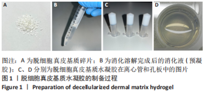
2.1 脱细胞真皮基质水凝胶的大体外观 脱细胞真皮基质水凝胶由猪真皮脱细胞、冻干、碎片化后,经胃蛋白酶和HCl溶液溶解消化,加入NaOH和PBS调节pH值至中性后得到预凝胶,在37 ℃条件下,预凝胶可自组装形成白色近无色的固态凝胶,见图1。"
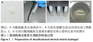
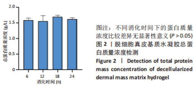
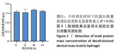
2.2 脱细胞真皮基质水凝胶的蛋白质量浓度 定量分析结果显示,消化时间无论是6,12,18 h还是24 h,10 g/L脱细胞真皮基质预凝胶蛋白质量浓度均在1.6 g/L左右,无统计学差异,见图2。但消化时间越长大体可见的真皮碎片越少,表明延长消化时间(18-24 h)是必要的。"

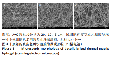
2.3 脱细胞真皮基质水凝胶的超微结构 扫描电镜结果显示,脱细胞真皮基质水凝胶呈现一种不规则随机走向的多孔纤维结构,孔径大小不一,见图3,表明脱细胞真皮基质水凝胶是一种具有多孔纤维状结构的三维立体支架。"
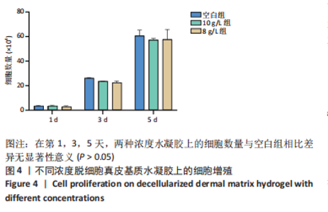
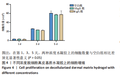
2.4 脱细胞真皮基质水凝胶的细胞毒性 细胞增殖实验结果显示,两种浓度水凝胶上的细胞呈正常生长趋势。在第1,3,5天,两种浓度水凝胶上的细胞数量与空白组相比差异无显著性意义(P > 0.05),见图4,表明制备的脱细胞真皮基质水凝胶无明显细胞毒性。"
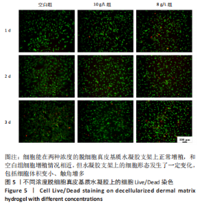
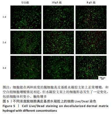
2.5 脱细胞真皮基质水凝胶的细胞相容性 荧光染色结果显示,细胞能在两种浓度的脱细胞真皮基质水凝胶支架上正常增殖,和空白组细胞增殖情况相近,但水凝胶支架上的细胞形态发生了一定变化,包括细胞体积变小、触角增多,见图5,具体原因有待进一步研究。"
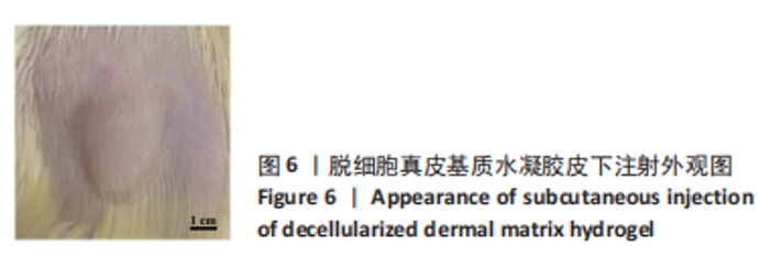
2.6 脱细胞真皮基质水凝胶的组织相容性 在大鼠皮下注射水凝胶后,其背部有明显突起,后慢慢消失,见图6。蛋白质量浓度0.25 g/L的水凝胶在2周后降解,0.5 g/L水凝胶在4周后降解。"

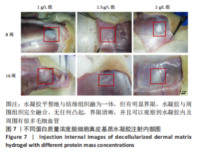
蛋白质量浓度1,1.5,2 g/L的水凝胶在体内16周均未降解,甚至可能保留更长时间。当取出水凝胶时,水凝胶平整地与结缔组织融为一体,但有明显界限,水凝胶与周围组织完全融合,无任何凸起,界限清晰,并且可以观察到水凝胶内及周围有很多毛细血管,见图7。"

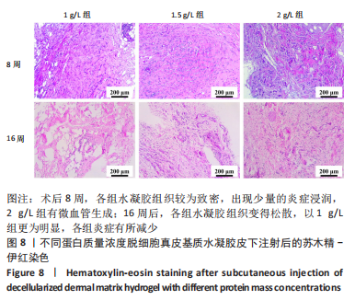
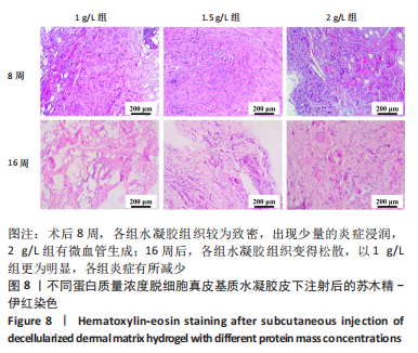
苏木精-伊红染色结果显示,术后8周,各组水凝胶组织较为致密,出现少量的炎症浸润,2 g/L组有微血管生成;16周后,各组水凝胶组织较8周变得松散,以1 g/L组更为明显,各组炎症有所减少,大鼠体内有新生肉芽组织,1.5 g/L组和2 g/L组出现明显的微血管,见图8。"
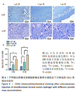
CD31免疫组化染色结果显示,术后8周,CD31在3种蛋白质量浓度的水凝胶中均出现阳性表达,半定量统计数据表明,水凝胶的蛋白质量浓度越高,新生微血管数量越多;16周后,与第8周类似,各组微血管数量也与水凝胶蛋白质量浓度有关,并且同一浓度水凝胶中微血管数量比8周时明显增多,见图9。"


蛋白质量浓度1 g/L水凝胶中的微血管数量在8周时和16周时无差异,这可能与其在体内慢慢降解有关,在苏木精-伊红染色中也能观察到该水凝胶在16周时结构变得松散。总的来说,脱细胞真皮基质水凝胶支持新生血管生成,具有良好的生物相容性。"

| [1] BAYER IS. Hyaluronic Acid and Controlled Release: A Review. Molecules. 2020;25(11):2649. [2] CHOW KV, URMAN DS, CABRAL ES, et al. Hyaluronic Acid Filler Incidentally Found During Mohs Micrographic Surgery: Observations in 36 Patients Regarding Skin Depth, Degradation Size, and Estimated Persistence Time. Dermatol Surg. 2022;48(4):401-405. [3] 杨彪,郭学平,臧恒昌,等.交联透明质酸凝胶修饰度的测定方法研究进展[J].药物生物技术,2015,22(2):177-180. [4] TANG S, CHI K, XU H, et al. A covalently cross-linked hyaluronic acid/bacterial cellulose composite hydrogel for potential biological applications. Carbohydr Polym. 2021;252:117123. [5] ZERBINATI N, SOMMATIS S, MACCARIO C, et al. Toward Physicochemical and Rheological Characterization of Different Injectable Hyaluronic Acid Dermal Fillers Cross-Linked with Polyethylene Glycol Diglycidyl Ether. Polymers (Basel). 2021;13(6):948. [6] KENNE L, GOHIL S, NILSSON EM, et al. Modification and cross-linking parameters in hyaluronic acid hydrogels--definitions and analytical methods. Carbohydr Polym. 2013;91(1):410-418. [7] RZANY B, CONVERSET-VIETHEL S, HARTMANN M, et al. Efficacy and Safety of 3 New Resilient Hyaluronic Acid Fillers, Crosslinked With Decreased BDDE, for the Treatment of Dynamic Wrinkles: Results of an 18-Month, Randomized Controlled Trial Versus Already Available Comparators. Dermatol Surg. 2019;45(10):1304-1314. [8] CECCHI R, SPOTA A, FRATI P, et al. Migrating granulomatous chronic reaction from hyaluronic acid skin filler (Restylane): review and histopathological study with histochemical stainings. Dermatology. 2014;228(1):14-17. [9] SHOME D, DOSHI K, VADERA S, et al. Delayed hypersensitivity reaction to hyaluronic acid dermal filler post-COVID-19 viral infection. J Cosmet Dermatol. 2021;20(5):1549-1550. [10] SUN W, GREGORY DA, TOMEH MA, et al. Silk Fibroin as a Functional Biomaterial for Tissue Engineering. Int J Mol Sci. 2021;22(3):1499. [11] CHEN D, ZHOU X, CHANG L, et al. Hemostatic Self-Healing Hydrogel with Excellent Biocompatibility Composed of Polyphosphate-Conjugated Functional PNIPAM-Bearing Acylhydrazide. Biomacromolecules. 2021; 22(5):2272-2283. [12] HINDERER S, LAYLAND SL, SCHENKE-LAYLAND K. ECM and ECM-like materials - Biomaterials for applications in regenerative medicine and cancer therapy. Adv Drug Deliv Rev. 2016;97:260-269. [13] KOU L, SUN R, BHUTIA YD, et al. Emerging advances in P-glycoprotein inhibitory nanomaterials for drug delivery. Expert Opin Drug Deliv. 2018;15(9):869-879. [14] HOGANSON DM, OWENS GE, MEPPELINK AM, et al. Decellularized extracellular matrix microparticles as a vehicle for cellular delivery in a model of anastomosis healing. J Biomed Mater Res A. 2016;104(7): 1728-1735. [15] HWANG J, SULLIVAN MO, KIICK KL. Targeted Drug Delivery via the Use of ECM-Mimetic Materials. Front Bioeng Biotechnol. 2020;8:69. [16] PISHAVAR E, KHOSRAVI F, NASERIFAR M, et al. Multifunctional and Self-Healable Intelligent Hydrogels for Cancer Drug Delivery and Promoting Tissue Regeneration In Vivo. Polymers (Basel). 2021;13(16):2680. [17] FREYTES DO, MARTIN J, VELANKAR SS, et al. Preparation and rheological characterization of a gel form of the porcine urinary bladder matrix. Biomaterials. 2008;29(11):1630-1637. [18] WASEEQ UR REHMAN, ASIM M, HUSSAIN S, et al. Hydrogel: A Promising Material in Pharmaceutics. Curr Pharm Des. 2020;26(45):5892-5908. [19] CASCONE S, LAMBERTI G. Hydrogel-based commercial products for biomedical applications: A review. Int J Pharm. 2020;573:118803. [20] ALI F, KHAN I, CHEN J, et al. Emerging Fabrication Strategies of Hydrogels and Its Applications. Gels. 2022;8(4):205. [21] KAMOUN EA, LOUTFY SA, HUSSEIN Y, et al. Recent advances in PVA-polysaccharide based hydrogels and electrospun nanofibers in biomedical applications: A review. Int J Biol Macromol. 2021;187: 755-768. [22] ARKABAN H, BARANI M, AKBARIZADEH MR, et al. Polyacrylic Acid Nanoplatforms: Antimicrobial, Tissue Engineering, and Cancer Theranostic Applications. Polymers (Basel). 2022;14(6):1259. [23] STEPANOVSKA J, SUPOVA M, HANZALEK K, et al. Collagen Bioinks for Bioprinting: A Systematic Review of Hydrogel Properties, Bioprinting Parameters, Protocols, and Bioprinted Structure Characteristics. Biomedicines. 2021;9(9):1137. [24] GOODARZI K, RAO SS. Hyaluronic acid-based hydrogels to study cancer cell behaviors. J Mater Chem B. 2021;9(31):6103-6115. [25] ZOU Z, ZHANG B, NIE X, et al. A sodium alginate-based sustained-release IPN hydrogel and its applications. RSC Adv. 2020;10(65): 39722-39730. [26] RAO F, WANG Y, ZHANG D, et al. Aligned chitosan nanofiber hydrogel grafted with peptides mimicking bioactive brain-derived neurotrophic factor and vascular endothelial growth factor repair long-distance sciatic nerve defects in rats. Theranostics. 2020;10(4):1590-1603. [27] ZHONG G, YAO J, HUANG X, et al. Injectable ECM hydrogel for delivery of BMSCs enabled full-thickness meniscus repair in an orthotopic rat model. Bioact Mater. 2020;5(4):871-879. [28] VOYTIK-HARBIN SL, BRIGHTMAN AO. Small intestinal submucosa: a tissue derived extracellular matrix that promotes tissue-specifific growth and differentiation of cells in vitro. Tissue Eng. 1998;4(2): 157-174. [29] GIOBBE GG, CROWLEY C, LUNI C, et al. Extracellular matrix hydrogel derived from decellularized tissues enables endodermal organoid culture. Nat Commun. 2019;10(1):5658. [30] XU Y, ZHOU J, LIU C, et al. Understanding the role of tissue-specific decellularized spinal cord matrix hydrogel for neural stem/progenitor cell microenvironment reconstruction and spinal cord injury. Biomaterials. 2021;268:120596. [31] CHEN Z, ZHANG B, SHU J, et al. Human decellularized adipose matrix derived hydrogel assists mesenchymal stem cells delivery and accelerates chronic wound healing. J Biomed Mater Res A. 2021; 109(8):1418-1428. [32] BORDBAR S, LOTFI BAKHSHAIESH N, KHANMOHAMMADI M, et al. Production and evaluation of decellularized extracellular matrix hydrogel for cartilage regeneration derived from knee cartilage. J Biomed Mater Res A. 2020;108(4):938-946. [33] YAZDANPANAH G, SHAH R, RAGHURAMA R, et al. In-situ porcine corneal matrix hydrogel as ocular surface bandage. Ocul Surf. 2021;21:27-36. [34] GOLDFRACHT I, EFRAIM Y, SHINNAWI R, et al. Engineered heart tissue models from hiPSC-derived cardiomyocytes and cardiac ECM for disease modeling and drug testing applications. Acta Biomater. 2019;92:145-159. [35] FARNEBO S, WOON CY, SCHMITT T, et al. Design and characterization of an injectable tendon hydrogel: a novel scaffold for guided tissue regeneration in the musculoskeletal system. Tissue Eng Part A. 2014; 20(9-10):1550-1561. [36] FU W, XU P, FENG B, et al. A hydrogel derived from acellular blood vessel extracellular matrix to promote angiogenesis. J Biomater Appl. 2019;33(10):1301-1313. [37] LEE JS, SHIN J, PARK HM, et al. Liver extracellular matrix providing dual functions of two-dimensional substrate coating and three-dimensional injectable hydrogel platform for liver tissue engineering. Biomacromolecules. 2014;15(1):206-218. [38] BANKOTI K, RAMESHBABU AP, DATTA S, et al. Dual Functionalized Injectable Hybrid Extracellular Matrix Hydrogel for Burn Wounds. Biomacromolecules. 2021;22(2):514-533. [39] 梁成宵,古瑞,李婷,等.肝脏基质水凝胶的制备及其细胞相容性研究[J].解放军医学杂志,2018,43(5):409-413. [40] WOLF MT, DALY KA, BRENNAN-PIERCE EP, et al. A hydrogel derived from decellularized dermal extracellular matrix. Biomaterials. 2012; 33(29):7028-7038. [41] KADLER KE, HILL A, CANTY-LAIRD EG. Collagen fibrillogenesis: fibronectin, integrins, and minor collagens as organizers and nucleators. Curr Opin Cell Biol. 2008;20(5):495-501. [42] OZUDOGRU E, ISIK M, EYLEM CC, et al. Decellularized spinal cord meninges extracellular matrix hydrogel that supports neurogenic differentiation and vascular structure formation. J Tissue Eng Regen Med. 2021;15(11):948-963. |
| [1] | Sun Kexin, Zeng Jinshi, Li Jia, Jiang Haiyue, Liu Xia. Mechanical stimulation enhances matrix formation of three-dimensional bioprinted cartilage constructs [J]. Chinese Journal of Tissue Engineering Research, 2023, 27(在线): 1-7. |
| [2] | Lian Shilin, Zhang Yan, Jiang Qiang, Zhang Hanshuo, Li Tusheng, Ding Yu. Interventional effects of whole blood and platelet-rich plasma with different preparation methods on nucleus pulposus cells [J]. Chinese Journal of Tissue Engineering Research, 2023, 27(8): 1199-1204. |
| [3] | Liu Xiaolin, Mu Xinyue, Ma Ziyu, Liu Shutai, Wang Wenlong, Han Xiaoqian, Dong Zhiheng. Effect of hydrogel-loaded simvastatin microspheres on osteoblast proliferation and differentiation [J]. Chinese Journal of Tissue Engineering Research, 2023, 27(7): 998-1003. |
| [4] | Xu Xingxing, Wen Chaoju, Meng Maohua, Wang Qinying, Chen Jingqiao, Dong Qiang. Carbon nanomaterials in oral implant [J]. Chinese Journal of Tissue Engineering Research, 2023, 27(7): 1062-1070. |
| [5] | Yang Yitian, Wang Lu, Yao Wei, Zhao Bin. Application of the interaction between biological scaffolds and macrophages in bone regeneration [J]. Chinese Journal of Tissue Engineering Research, 2023, 27(7): 1071-1079. |
| [6] | Li Cheng, Zheng Guoshuang, Kuai Xiandong, Yu Weiting. Alginate scaffold in articular cartilage repair [J]. Chinese Journal of Tissue Engineering Research, 2023, 27(7): 1080-1088. |
| [7] | Chen Shisong, Liu Xiaohong, Xu Zhiyun. Current status and prospects of bioprosthetic heart valves [J]. Chinese Journal of Tissue Engineering Research, 2023, 27(7): 1096-1102. |
| [8] | Shi Yehong, Wang Cheng, Chen Shijiu. Early thrombosis and prevention of small-diameter blood vessel prosthesis [J]. Chinese Journal of Tissue Engineering Research, 2023, 27(7): 1110-1116. |
| [9] | Tang Haotian, Liao Rongdong, Tian Jing. Application and design of piezoelectric materials for bone defect repair [J]. Chinese Journal of Tissue Engineering Research, 2023, 27(7): 1117-1125. |
| [10] | Xu Yan, Li Ping, Lai Chunhua, Zhu Peijun, Yang Shuo, Xu Shulan. Piezoelectric materials for vascularized bone regeneration [J]. Chinese Journal of Tissue Engineering Research, 2023, 27(7): 1126-1132. |
| [11] | Li Xinyue, Li Xiheng, Mao Tianjiao, Tang Liang, Li Jiang. Three-dimensional culture affects morphology, activity and osteogenic differentiation of human periodontal ligament stem cells [J]. Chinese Journal of Tissue Engineering Research, 2023, 27(6): 846-852. |
| [12] | Li Xiaoyin, Yang Xiaoqing, Chen Shulian, Li Zhengchao, Wang Ziqi, Song Zhen, Zhu Daren, Chen Xuyi. Collagen/silk fibroin scaffold combined with neural stem cells in the treatment of traumatic spinal cord injury [J]. Chinese Journal of Tissue Engineering Research, 2023, 27(6): 890-896. |
| [13] | Yuan Bo, Xie Lide, Fu Xiumei. Schwann cell-derived exosomes promote the repair and regeneration of injured peripheral nerves [J]. Chinese Journal of Tissue Engineering Research, 2023, 27(6): 935-940. |
| [14] | Qin Yuxing, Ren Qiangui, Li Zilong, Quan Jiaxing, Shen Peifeng, Sun Tao, Wang Haoyu. Action mechanism and prospect of bone microvascular endothelial cells for treating femoral head necrosis [J]. Chinese Journal of Tissue Engineering Research, 2023, 27(6): 955-961. |
| [15] | Li Long, Li Guangdi, Shi Hao, Deng Keqi. Circular RNA as a competing endogenous RNA is involved in the regulation of osteoarthritis [J]. Chinese Journal of Tissue Engineering Research, 2023, 27(5): 751-757. |
| Viewed | ||||||
|
Full text |
|
|||||
|
Abstract |
|
|||||