Chinese Journal of Tissue Engineering Research ›› 2023, Vol. 27 ›› Issue (21): 3293-3299.doi: 10.12307/2023.162
Previous Articles Next Articles
Mechanical stimulation enhances matrix formation of three-dimensional bioprinted cartilage constructs
Sun Kexin, Zeng Jinshi, Li Jia, Jiang Haiyue, Liu Xia
- Research Center of Plastic Surgery Hospital, Chinese Academy of Medical Science & Peking Union Medical College, Beijing 100144, China
-
Online:2023-07-28Published:2022-11-23 -
Contact:Liu Xia, MD, Researcher, Research Center of Plastic Surgery Hospital, Chinese Academy of Medical Science & Peking Union Medical College, Beijing 100144, China -
About author:Sun Kexin, Master, Research Center of Plastic Surgery Hospital, Chinese Academy of Medical Science & Peking Union Medical College, Beijing 100144, China -
Supported by:General Program of the National Natural Science Foundation of China, No. 81871575 (to LX); The Chinese Academy of Medical Sciences Innovation Fund for Medical Sciences, No. 2021-I2M-1-052 (to JHY)
CLC Number:
Cite this article
Sun Kexin, Zeng Jinshi, Li Jia, Jiang Haiyue, Liu Xia. Mechanical stimulation enhances matrix formation of three-dimensional bioprinted cartilage constructs[J]. Chinese Journal of Tissue Engineering Research, 2023, 27(21): 3293-3299.
share this article
Add to citation manager EndNote|Reference Manager|ProCite|BibTeX|RefWorks

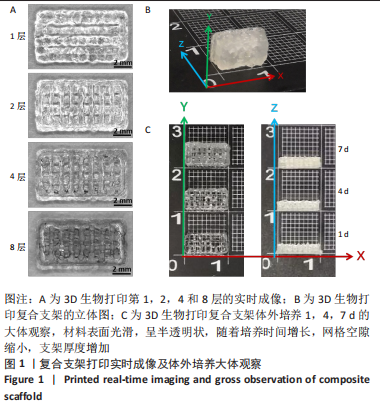
2.1 复合支架的大体观及细胞活/死检测 2.1.1 实时成像 3D生物打印软骨细胞-GelMA复合支架第1,2,4和8层的打印实时成像,图中清晰可见直线线条形状,支架材料表面光滑,呈半透明状,随着层数的增多,其形态越来越清晰,呈网格状,见图1A。 2.1.2 大体观察 3D生物打印完成后的支架大小约为10 mm×5 mm×2 mm,结构清晰,呈网格状,质地较软;置于37 ℃、体积分数5%CO2培养箱中,加入完全培养基培养1,4,7 d,可见支架形态稳定,随着培养时间的增长,至第7天网格状结构仍清晰可见,但网格空隙缩小,支架厚度略有增加,见图1B、C。"

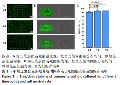
2.1.3 复合支架中细胞密度、分布及细胞存活率 对复合支架进行活死细胞染色观察拍照得到二维与三维成像图,如图2A、B所示,复合支架内细胞分布均匀,以绿色活细胞为主,中间分布有坏死的红色细胞,第1天时,红色细胞数量相对较多。统计学分析显示,培养1,4,7天支架内细胞存活率分别为(90.57%±1.84)%,(93.41±1.84)%,(94.53±1.62)%,第1天与第4,7天的细胞存活率比较差异有显著性意义 (P < 0.05),第4,7天的细胞存活率比较差异无显著性意义(P > 0.05),见图2C,这可能与第1天时,细胞经历打印过程后坏死较多有关。"
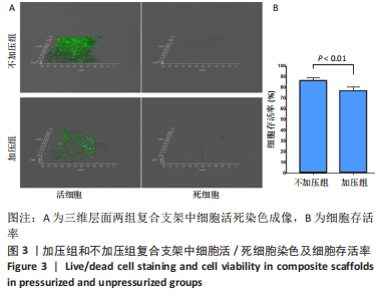
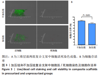
2.1.4 加压组和不加压组复合支架细胞存活率检测 培养2周后,对两组复合支架活死细胞染色观察与拍照得到三维成像图,并进行细胞计数,见图3。加压组与不加压组复合支架中的细胞存活率分别为(76.61±3.78)%,(86.42±2.51)%,加压组细胞存活率低于不加压组(P < 0.05),说明加压培养过程中会导致细胞死亡增加。"

2.2 复合支架体外成软骨检测 2.2.1 大体观察 加压组与不加压组复合支架在大体外观上无明显区别,网格状结构仍清晰可见,见图4A。 2.2.2 组织学染色结果 苏木精-伊红染色结果显示两组均有明显的软骨陷窝结构,细胞在材料中分布较均匀,加压组复合支架的材料间空隙区域有更多新生软骨组织形成,见图4B;番红O染色显示两组组织均可见红色软骨基质形成,加压组细胞周基质染色更深,见图4C,提示经加压培养后,细胞分泌糖胺多糖基质成分的能力增加;Ⅰ型胶原免疫组织化学染色也显示加压组着色更为明显,见图4D,提示加压刺激能够促进软骨细胞的胶原分泌。"

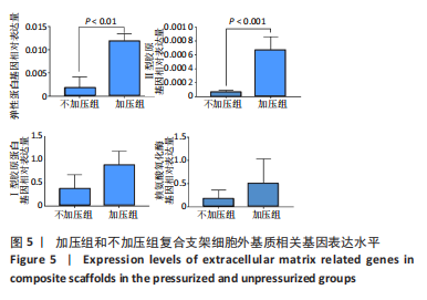
2.2.3 qRT-PCR 检测成软骨相关基因表达 见图5。"

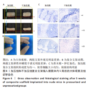
加压组复合支架中成软骨相关基因Ⅱ型胶原和弹性蛋白的mRNA表达水平明显高于不加压组(P < 0.05),进一步说明加压培养能够促进软骨细胞的胶原蛋白合成能力;Ⅰ型胶原蛋白和赖氨酸氧化酶mRNA表达量也有升高趋势,但组间差异无显著性意义(P > 0.05)。 2.3 复合支架裸鼠皮下植入后的体内成软骨检测 植入裸鼠体内5周后取材,大体观察显示两组外观无明显差异,动图显示两组复合支架弹性和硬度手感无明显差异,见图6A、B;苏木精-伊红染色结果显示加压组复合支架组织形成更为均一,软骨细胞大小均匀,陷窝结构明显,见图6C。"


2.4 复合支架的生物相容性 由活死染色裸鼠皮下植入实验可知,3D生物打印软骨细胞-GelMA复合支架具有良好的生物相容性。"

| [1] ZHOU G, JIANG H, YIN Z, et al. In Vitro Regeneration of Patient-specific Ear-shaped Cartilage and Its First Clinical Application for Auricular Reconstruction. EBioMedicine. 2018;28:287-302. [2] NABZDYK C, PRADHAN L, MOLINA J, et al. Review: auricular chondrocytes - from benchwork to clinical applications. In Vivo. 2009;23(3):369-380. [3] 董可欣.弹性纤维相关组分在工程化软骨组织中的表达及作用研究[D].北京:北京协和医学院,2020. [4] MOHAGHEGH S, HOSSEINI SF, RAD MR, et al. 3D Printed Composite Scaffolds in Bone Tissue Engineering: A systematic review. Curr Stem Cell Res Ther. 2021.doi: 10.2174/1574888X16666210810111754. [5] MANDRYCKY C, WANG Z, KIM K, et al. 3D bioprinting for engineering complex tissues. Biotechnol Adv. 2016;34(4):422-434. [6] CHIMENE D, KAUNAS R, GAHARWAR AK. Hydrogel Bioink Reinforcement for Additive Manufacturing: A Focused Review of Emerging Strategies. Adv Mater. 2020;32(1):e1902026. [8] YANG J, ZHANG YS, YUE K, et al. Cell-laden hydrogels for osteochondral and cartilage tissue engineering. Acta Biomater. 2017;57:1-25. [9] GUNGOR-OZKERIM PS, INCI I, ZHANG YS, et al. Bioinks for 3D bioprinting: an overview. Biomater Sci. 2018;6(5):915-946. [7] YUE K, TRUJILLO-DE SANTIAGO G, ALVAREZ MM, et al. Synthesis, properties, and biomedical applications of gelatin methacryloyl (GelMA) hydrogels. Biomaterials. 2015;73:254-271. [10] BYAMBAA B, ANNABI N, YUE K, et al. Bioprinted Osteogenic and Vasculogenic Patterns for Engineering 3D Bone Tissue. Adv Healthc Mater. 2017;6(16). doi: 10.1002/adhm.201700015. [11] HU H, DONG L, BU Z, et al. miR-23a-3p-abundant small extracellular vesicles released from Gelma/nanoclay hydrogel for cartilage regeneration. J Extracell Vesicles. 2020;9(1):1778883. [12] KAUPP JA, WEBER JF, WALDMAN SD. Mechanical stimulation of chondrocyte-agarose hydrogels. J Vis Exp. 2012;(68):e4229. [13] BOOS MA, LAMANDé SR, STOK KS. Multiscale Strain Transfer in Cartilage. Front Cell Dev Biol. 2022;10:795522. [14] APPELMAN TP, MIZRAHI J, ELISSEEFF JH, et al. The differential effect of scaffold composition and architecture on chondrocyte response to mechanical stimulation. Biomaterials. 2009;30(4):518-525. [15] 周丽斌,徐冰心,丁瑞英,等.应用微纤维胶原支架构建组织工程软骨[J].中国组织工程研究,2017,21(22):3483-3487. [16] YU K, ZHANG X, SUN Y, et al. Printability during projection-based 3D bioprinting. Bioact Mater. 2022;11:254-267. [17] CHOE R, DEVOY E, KUZEMCHAK B, et al. Computational investigation of interface printing patterns within 3D printed multilayered scaffolds for osteochondral tissue engineering. Biofabrication. 2022; 14(2).10.1088/1758-5090/ac5220. doi: 10.1088/1758-5090/ac5220. [18] WANG Y, CAO X, MA M, et al. A GelMA-PEGDA-nHA Composite Hydrogel for Bone Tissue Engineering. Materials (Basel). 2020;13(17):3735. [19] KILIC BEKTAS C, HASIRCI V. Cell Loaded GelMA:HEMA IPN hydrogels for corneal stroma engineering. J Mater Sci Mater Med. 2019;31(1):2. [20] KOSIK-KOZIOŁ A, COSTANTINI M, MRóZ A, et al. 3D bioprinted hydrogel model incorporating β-tricalcium phosphate for calcified cartilage tissue engineering. Biofabrication. 2019;11(3):035016. [21] JORGENSEN I, RAYAMAJHI M, MIAO EA. Programmed cell death as a defence against infection. Nat Rev Immunol. 2017;17(3):151-164. [22] JØRGENSEN AEM, KJÆR M, HEINEMEIER KM. The Effect of Aging and Mechanical Loading on the Metabolism of Articular Cartilage. J Rheumatol. 2017;44(4):410-417. [23] ZHAO DL, LI HT, LIU SH. TIMP3/TGF-β1 axis regulates mechanical loading‑induced chondrocyte degeneration and angiogenesis. Mol Med Rep. 2020;22(4):2637-2644. [24] MAUCK RL, NICOLL SB, SEYHAN SL, et al. Synergistic action of growth factors and dynamic loading for articular cartilage tissue engineering . Tissue Eng. 2003;9(4):597-611. [25] VAN SUSANTE JLC, PIEPER J, BUMA P, et al. Linkage of chondroitin-sulfate to type I collagen scaffolds stimulates the bioactivity of seeded chondrocytes in vitro. Biomaterials. 2001;22(17):2359-2369. [26] SALINAS EY, HU JC, ATHANASIOU K. A Guide for Using Mechanical Stimulation to Enhance Tissue-Engineered Articular Cartilage Properties. Tissue Eng Part B Rev. 2018;24(5):345-358. [27] QIN L, LIU W, CAO H, et al. Molecular mechanosensors in osteocytes. Bone Res. 2020;8:23. [28] 吕晓杰,周广东,李宏,等.灌流-液压式反应器体外构建组织工程化软骨[J].上海交通大学学报(医学版),2006,26(9):1006-1010. [29] PATTAPPA G, ZELLNER J, JOHNSTONE B, et al. Cells under pressure - the relationship between hydrostatic pressure and mesenchymal stem cell chondrogenesis. Eur Cell Mater. 2019;37:360-381. [30] YOUNG IC, CHUANG ST, GEFEN A, et al. A novel compressive stress-based osteoarthritis-like chondrocyte system. Exp Biol Med (Maywood). 2017;242(10):1062-1071. [31] ANDERSON DE, JOHNSTONE B. Dynamic Mechanical Compression of Chondrocytes for Tissue Engineering: A Critical Review. Front Bioeng Biotechnol. 2017;5:76. [32] RICHARDSON BM, WALKER CJ, MAPLES MM, et al. Mechanobiological Interactions between Dynamic Compressive Loading and Viscoelasticity on Chondrocytes in Hydrazone Covalent Adaptable Networks for Cartilage Tissue Engineering. Adv Healthc Mater. 2021;10(9):e2002030. [33] SHIN SJ, YANAGISAWA H. Recent updates on the molecular network of elastic fiber formation. Essays Biochem. 2019;63(3):365-376. [34] KOZEL BA, MECHAM RP. Elastic fiber ultrastructure and assembly. Matrix Biol. 2019;84:31-40. |
| [1] | Sun Kexin, Zeng Jinshi, Li Jia, Jiang Haiyue, Liu Xia. Mechanical stimulation enhances matrix formation of three-dimensional bioprinted cartilage constructs [J]. Chinese Journal of Tissue Engineering Research, 2023, 27(在线): 1-7. |
| [2] | Zhang Hui, Wang Jiayang, Wang Qian, Gan Hongquan, Wang Zhiqiang. Effects of hyaluronic acid combined with domestic porous tantalum on chondrocyte function under the dynamic environment [J]. Chinese Journal of Tissue Engineering Research, 2023, 27(3): 339-345. |
| [3] | Wang Buyu, Zhang Yong, Li Feifei, Dong Xiaoyu, Deng Jiang, Ruan Shiqiang. Role and application of bone morphogenetic protein 2 in the repair of osteochondral defects [J]. Chinese Journal of Tissue Engineering Research, 2023, 27(20): 3259-3265. |
| [4] | Zhang Weiye, Zhan Jiawen, Zhu Liguo, Wang Shangquan, Chen Ming, Wei Xu, Feng Minshan, Yu Jie, Han Tao, Cai Chuhao, Zhou Shuaiqi, Shao Chenchen. Effect of nucleus pulposus cells-derived exosomes under cyclic mechanical tension on endplate chondrocytes [J]. Chinese Journal of Tissue Engineering Research, 2023, 27(2): 223-229. |
| [5] | Wang Huanhuan, Wang Qing, Tang Peng, Zhang Rui, Min Hongwei. Effects of extracorporeal shock wave on the proliferation and autophagy of chondrocytes from osteoarthritis rats [J]. Chinese Journal of Tissue Engineering Research, 2023, 27(2): 252-257. |
| [6] | Li Zhe, Yuan Changshen, Guan Yanbing, Xu Wenfei, Liao Shuning, Rong Weiming, Mei Qijie, Duan Kan. Bioinformatic analysis and experimental validation of ferroptosis in osteoarthritis [J]. Chinese Journal of Tissue Engineering Research, 2023, 27(17): 2637-2643. |
| [7] | Huang Haoran, Wei Yangwenxiang, Zhang Jiahao, Mo Liang, Liu Yuhao, Chen Zhenqiu, Wang Haibin, Zhou Chi. Piezo1-mediated mechanical stress stimulation in anti-osteoporosis treatment [J]. Chinese Journal of Tissue Engineering Research, 2023, 27(17): 2716-2722. |
| [8] | Yang Jun, Li Peng. Differentiation of bone marrow mesenchymal stem cells into meniscus fibrochondrocytes induced by transforming growth factor beta [J]. Chinese Journal of Tissue Engineering Research, 2023, 27(15): 2412-2419. |
| [9] | Liao Jianzhao, Yang Nan, Zhou Yi, Xu Hang, Xia Tian, Song Shilei, Zeng Qi, Chen Yueping. Protective effect of astragaloside IV against inflammatory injury in chondrocytes [J]. Chinese Journal of Tissue Engineering Research, 2023, 27(14): 2158-2163. |
| [10] | Ye Xuwen, Gu Yong, Chen Liang. Curcumin loaded injectable microspheres retard progression of intervertebral disc degeneration [J]. Chinese Journal of Tissue Engineering Research, 2023, 27(12): 1884-1891. |
| [11] | Feng Junming, Xiong Xianmei, Ma Liqiong, Zhang Yan, Chen Zijie, Li Shijie, Chen Baixing, Jiang Ziwei, Zeng Zhanpeng, Gao Yijia . Comparison of platelet-rich plasma, concentrated growth factor and 3D micro-nanostructure composite scaffolds in repair of rabbit radius defects [J]. Chinese Journal of Tissue Engineering Research, 2023, 27(12): 1906-1913. |
| [12] | Wu Cong, Jia Quanzhong, Liu Lun. Relationship between transforming growth factor beta1 expression and chondrocyte migration in adult articular cartilage after fragmentation [J]. Chinese Journal of Tissue Engineering Research, 2022, 26(8): 1167-1172. |
| [13] | Lin Xuchen, Zhu Hainian, Wang Zengshun, Qi Tengmin, Liu Limin, Suonan Angxiu. Effect of xanthohumol on inflammatory factors and articular cartilage in a mouse mode of osteoarthritis [J]. Chinese Journal of Tissue Engineering Research, 2022, 26(5): 676-681. |
| [14] | Xu Lei, Han Xiaoqiang, Zhang Jintao, Sun Haibiao. Hyaluronic acid around articular chondrocytes: production, transformation and function characteristics [J]. Chinese Journal of Tissue Engineering Research, 2022, 26(5): 768-773. |
| [15] | Wu Yongli, Liu Di, Wang Duo, Liu Junwei, Ma Yuyuan. Effect of warm acupuncture on PI3K/Akt signaling pathway in articular cartilage of a rabbit knee osteoarthritis model [J]. Chinese Journal of Tissue Engineering Research, 2022, 26(35): 5596-5601. |
| Viewed | ||||||
|
Full text |
|
|||||
|
Abstract |
|
|||||