Chinese Journal of Tissue Engineering Research ›› 2021, Vol. 25 ›› Issue (25): 4065-4069.doi: 10.12307/2021.021
Previous Articles Next Articles
Mesenchymal stem cells for the treatment of ulcerative colitis: possibility and feasibility
Liang Yuan1, Jiang Xiaoke2, Bai Yangqiu2, Luo Xiaoying2, Zhang Bingyong2
- 1Zhengzhou University People’s Hospital, Zhengzhou 450000, Henan Province, China; 2Department of Gastroenterology, Zhengzhou University People’s Hospital, Zhengzhou 450000, Henan Province, China
-
Received:2020-06-08Revised:2020-06-11Accepted:2020-07-20Online:2021-09-08Published:2021-03-30 -
Contact:Zhang Bingyong, MD, Chief physician, Master’s supervisor, Department of Gastroenterology, Zhengzhou University People’s Hospital, Zhengzhou 450000, Henan Province, China -
About author:Liang Yuan, Master candidate, Zhengzhou University People’s Hospital, Zhengzhou 450000, Henan Province, China -
Supported by:the Joint Provincial and Ministry Project of Henan Provincial Health and Family Planning Commission Medical Science and Technology, No. 201701024 (to ZBY)
CLC Number:
Cite this article
Liang Yuan, Jiang Xiaoke, Bai Yangqiu, Luo Xiaoying, Zhang Bingyong. Mesenchymal stem cells for the treatment of ulcerative colitis: possibility and feasibility[J]. Chinese Journal of Tissue Engineering Research, 2021, 25(25): 4065-4069.
share this article
Add to citation manager EndNote|Reference Manager|ProCite|BibTeX|RefWorks
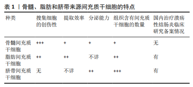
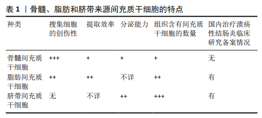
2.1 干细胞在溃疡性结肠炎中的应用 干细胞是一类具有高度繁殖、自我更新和分化潜能的细胞,分为全能、多能和单能干细胞。1991年CAPLAN在前人研究的基础上[7],首次将可分化为间质/基质细胞谱系并为造血干细胞提供造血微环境的基质干细胞命名为间充质干细胞。间充质干细胞是一类起源于中胚层的成体干细胞,属于多能干细胞,来源广泛,免疫原性低,具有自我更新、多项分化、分泌和免疫调节功能,可在不同诱导条件下分化为成骨细胞、脂肪细胞、软骨细胞和其他类型的细胞,直接参与受损器官的修复与再生[8]。目前已有13项有关间充质干细胞的项目被批准使用,如急性心肌梗死、膝关节软骨缺损、移植物抗宿主病、克罗恩病、血栓闭塞性动脉炎等疾病,表现出了强大的治疗潜力。大量文献报道显示,用于研究溃疡性结肠炎的间充质干细胞主要包括骨髓间充质干细胞、脂肪间充质干细胞、脐带间充质干细胞。 2.1.1 骨髓间充质干细胞 骨髓是第一个被报道含有间充质干细胞的组织,骨髓间充质干细胞既往称为骨髓基质成纤维细胞,不仅对骨髓中的造血干细胞有机械支持作用,分泌各种生长因子来支持造血,还具有间充质干细胞的共同特性。既往文献报道中,多采用骨髓间充质干细胞治疗溃疡性结肠炎,但抽取骨髓是有创性操作,并且细胞的寿命和分化潜能随着年龄的增长而减弱[9],限制了其在临床方面的研究进展。 2.1.2 脂肪间充质干细胞 主要来源于皮下脂肪组织,取材方便,可从减脂术中获得的脂肪组织中提取,其提取效率是骨髓间充质干细胞的40倍,且增殖速率快[10]。与骨髓间充质干细胞不同,脂肪干细胞的附着力与增殖能力与年龄无关,而与供者的体质有关,不会随着供者年龄的增加而降低。目前已有关于脂肪间充质干细胞治疗溃疡性结肠炎的Ⅰ/Ⅱ期随机对照临床研究的项目备案。 2.1.3 脐带间充质干细胞 以往认为骨髓和脂肪组织来源的间充质干细胞最有希望应用于临床,相关的研究报道也较多,但随着对间充质干细胞研究的进一步深入,脐带来源间充质干细胞逐渐引起大家的关注。脐带间充质干细胞无成脂性,分离成功率虽然较骨髓间充质干细胞、脂肪间充质干细胞低,但脐带组织中含有的间充质干细胞数量明显高于骨髓和脂肪组织(每厘米脐带5×10⁴个,每克脂肪组织5×10³个,0.01%骨髓单核细胞),分泌能力强于骨髓间充质干细胞,在大多数情况下,脐带间充质干细胞分泌生物活性因子的浓度比骨髓间充质干细胞分泌的生物活性因子浓度高出10-100倍[11],并且脐带来源间充质干细胞具有取材方便、免疫原性低、分化潜能高、增殖能力强[9]、伦理学争议少、制备周期短等特点,有可能成为最具有临床研究和应用前景的多功能干细胞。3种间充质干细胞的特点见表1。 2.2 间充质干细胞治疗溃疡性结肠炎的安全性和有效性 2.2.1 间充质干细胞治疗溃疡性结肠炎的安全性 间充质干细胞的生物安全性一直是备受关注的问题。何小文等[12]在葡聚糖硫酸钠诱导的结肠炎小鼠模型中发现,间充质干细胞不仅能明显改善小鼠的结肠病变,还能抑制结肠炎向恶性肿瘤转化。HU等[13]对接受脐带间充质干细胞治疗的34例溃疡性结肠炎患者进行了长达2年的随访,在随访期间未发现恶性肿瘤、免疫排斥反应等远期并发症。KNYAZEV等[14]对140例溃疡性结肠炎患者(56例接受间充质干细胞治疗和84例进行标准治疗)进行了5年的随访,比较两组患者的不良反应发生率,研究发现:与标准治疗组相比,接受间充质干细胞治疗的患者除了偶有短暂发热外,在急性输血后反应、严重感染、恶性肿瘤等致命性的不良反应方面,两组无显著性差异。 2.2.2 间充质干细胞治疗溃疡性结肠炎的疗效 一项Meta分析共纳入了7项间充质干细胞治疗溃疡性结肠炎的临床研究(n=312)[15],其中包括4项单臂试验[16-19](n=117)和3项对照试验[13,20-21](n=195)。4项单臂试验显示,间充质干细胞治疗溃疡性结肠炎有效率的比例明显高于无效率的比例;3项对照试验显示,与标准治疗组相比,间充质干细胞能够显著提高溃疡性结肠炎的缓解率(RR=3.08,95%CI:2.15-4.41)。 由此可见,间充质干细胞治疗溃疡性结肠炎安全有效,无移植物排斥、致瘤、严重感染等并发症。但由于目前纳入研究的样本数量较少,尚需要大量的随机双盲对照试验来进一步评价间充质干细胞治疗溃疡性结肠炎的安全性和疗效。 2.3 间充质干细胞治疗溃疡性结肠炎的移植剂量和方式 目前间充质干细胞治疗溃疡性结肠炎的最佳剂量和输注方式尚不明确。明确间充质干细胞治疗溃疡性结肠炎的最佳移植剂量和移植方式,可为临床应用提供个体化治疗。 2.3.1 移植剂量 明确间充质干细胞治疗溃疡性结肠炎的最佳剂量,不仅可以减少取材,降低生产成本,还可优化治疗方案。熊轩轩等[22]比较了输注不同剂量间充质干细胞(1×105,1×106,1×107)的溃疡性结肠炎小鼠在疾病活动指数、大体形态、组织学评分等方面的差异,结果显示:当输注剂量为1×106,1×107时,与剂量1×10⁵相比,疾病活动指数、大体形态、组织学评分均明显下降,三者差异有显著性意义(P < 0.05);输注剂量为1×10⁶与1×10⁷相比,三者改变无显著性差异(P > 0.05),故提示最佳治疗剂量为1×10⁶。ROBINSON等[23]研究表明,在葡聚糖硫酸钠诱导的结肠炎动物模型中,当间充质干细胞输注剂量为1×10⁵时,间充质干细胞迁移到结肠后主要集中在黏膜固有层;剂量为1×10⁶时,间充质干细胞可跨壁迁移,并移植入肌间神经节,且结肠病变处白细胞数量明显下降;与剂量为1×10⁵相比,剂量为1×106,3×106治疗的结肠黏膜层和黏膜肌层白细胞数量明显减少(均P < 0.01);与3×10⁶相比,治疗剂量为1×10⁶的黏膜层白细胞数量减少 (P < 0.01)。由此推测,间充质干细胞治疗溃疡性结肠炎的最佳剂量为1×10⁶。但临床研究方面,尚无统一的治疗剂量。 2.3.2 移植方式 目前了解到间充质干细胞的输注方式包括静脉注射、腹腔注射、灌肠、内镜下局部黏膜注射、肠系膜下动脉注射[19],尚未发现关于最佳移植方式的临床研究。在动物研究层面上注射方式使用最多的是腹腔注射和静脉注射。GONCALVES等[24]和葛翠翠[25]通过对照研究间充质干细胞治疗结肠炎小鼠模型,发现与腹腔注射相比,受损肠黏膜在静脉移植治疗后明显改善;而WANG等[26]从体质量变化、大便潜血情况、小鼠生存率及间充质干细胞迁移到结肠组织的数量等方面比较了静脉移植治疗组和腹腔注射组的差异,其认为腹腔注射可能是最佳的移植方式;PAN等[27]在间充质干细胞治疗结肠炎小鼠模型中,从体质量变化、生存率、疾病活动指数方面也证实腹腔注射比静脉注射有效。目前尚未得出统一的结论,需要更多的研究进一步探索最佳的移植方式,以指导临床试验方案的进行。但是静脉注射简单易行,创伤性小,且静脉可为间充质干细胞的迁移、归巢、分化创造适宜的微环境,修复受损肠黏膜。因此在临床研究中,采用静脉注射移植的方式比较多。 2.4 间充质干细胞治疗溃疡性结肠炎的作用机制 2.4.1 定植、分化 干细胞定向迁移的数量与组织的损伤程度有关。随着损伤的加重,间充质干细胞迁移率增加,恢复期数量则明显减少[28-29]。当肠道出现炎症时,间充质干细胞可在体内迁移并定居于肠黏膜表面,增殖并分化为新的结肠黏膜上皮细胞从而修复损伤部位[19,30]。BRITTAN等[31]在诱导的溃疡性结肠炎动物模型中发现,骨髓间充质干细胞移植后可在肠道定植并分化成肠上皮下肌成纤维细胞,通过改善肠道微环境促进肠黏膜修复和新生血管形成。在人体内,间充质干细胞是直接分化为肠黏膜上皮细胞还是分化为肌成纤维细胞,通过改善肠道微环境来促进肠上皮细胞修复和新生血管形成,尚需要进一步研究证实。 2.4.2 促进微循环重建 研究提示,血管内皮生长因子可增加血管通透性,促进炎症细胞浸润,在溃疡性结肠炎的发病机制中起着重要作用[32]。结肠微血管功能障碍和新生血管异常生成所致组织持续低灌注和缺血,最终导致溃疡性结肠炎反复发作,溃疡迁延不愈。动物实验表明,间充质干细胞可迁移至结肠分化为血管内皮细胞进而促进受损部位新生血管的形成[33-35],促进微循环的重建,从而有利于结肠黏膜炎症的修复。间充质干细胞是否通过调节血管内皮生长因子促进肠黏膜血运重建,需要进一步探讨。 2.4.3 免疫调节作用 免疫功能紊乱被认为在溃疡性结肠炎的发生发展中发挥关键作用。研究提示间充质干细胞可能通过抑制不适当的免疫反应并提供各种细胞因子而不是直接恢复受损细胞来协助组织再生[36]。 溃疡性结肠炎发病的机制尚不明确。研究发现,在活动性溃疡性结肠炎患者的肠黏膜中,存在细胞因子风暴,特别是白细胞介素17水平明显增高[37]。调节性T细胞(Regulatory T cells,Tregs)/辅助性T细胞17(T help cell 17,Th17)比例失衡可能与溃疡性结肠炎的发生发展有关。仅有Foxp3表达的CD4+CD25+调节性T细胞才有免疫调节作用,Foxp3与核受体结合可明显抑制白细胞介素17的转录,从而影响Th17细胞的分化[38]。研究发现,Rab27A和Rab27B是与外泌体有关的GTP酶,与外泌体的分泌及其在各种细胞中质膜的对接有关。与健康对照组相比,在活动性溃疡性结肠炎组的结肠黏膜中可观察到Rab27A+或Rab27B+肠道免疫细胞数量明显增加,这表明外泌体介导的免疫反应在溃疡性结肠炎的发病机制中起着重要的作用[39]。间充质干细胞可通过分泌外泌体,诱导T淋巴细胞凋亡,刺激单核细胞分泌白细胞介素10和转化生长因子β,促使CD4+CD25+ Foxp3+调节性T细胞上调,降低炎性因子白细胞介素4水平,增加抗炎因子白细胞介素10水平来调节免疫应答。转化生长因子β、白细胞介素10等抗炎因子又能够刺激体外间充质干细胞更有效地分泌外泌体,反过来促进调节性T细胞上调,减轻肠道炎症反应,促进受损组织的修复与再生[40-41]。 "
| [1] LI X, SONG P, LI J, et al. The Disease Burden and Clinical Characteristics of Inflammatory Bowel Disease in the Chinese Population: A Systematic Review and Meta-Analysis. Int J Environ Res Public Health. 2017;14(3):238. [2] YANG H, LI Y, WU W, et al. The incidence of inflammatory bowel disease in Northern China: a prospective population-based study. PLoS One. 2014; 9(7):e101296. [3] ROGLER G. Chronic ulcerative colitis and colorectal cancer. Cancer Lett. 2014;345(2):235-241. [4] 王晓蕾,欧阳春晖,高翔,等.溃疡性结肠炎降阶梯治疗对象的选择[J].中华炎性肠病杂志,2020,4(1):67-70. [5] GEARRY RB, BARCLAY ML. Azathioprine and 6-mercaptopurine pharmacogenetics and metabolite monitoring in inflammatory bowel disease. J Gastroenterol Hepatol. 2005;20(8):1149-1157. [6] PANÉS J, GARCÍA-OLMO D, VAN ASSCHE G, et al. Expanded allogeneic adipose-derived mesenchymal stem cells (Cx601) for complex perianal fistulas in Crohn’s disease: a phase 3 randomised, double-blind controlled trial. Lancet. 2016;388(10051):1281-1290. [7] HORWITZ EM, LE BLANC K, DOMINICI M, et al. Clarification of the nomenclature for MSC: The International Society for Cellular Therapy position statement. Cytotherapy. 2005;7(5):393-395. [8] UDER C, BRÜCKNER S, WINKLER S, et al. Mammalian MSC from selected species: Features and applications. Cytometry A. 2018;93(1):32-49. [9] YU YB, SONG Y, CHEN Y, et al. Differentiation of umbilical cord mesenchymal stem cells into hepatocytes in comparison with bone marrow mesenchymal stem cells. Mol Med Rep. 2018;18(2):2009-2016. [10] 刘美辰,刘霞,肖苒,等.脂肪间充质干细胞治疗皮肤老化的研究进展[J].组织工程与重建外科杂志,2014,10(4):236-239. [11] ROMANOV YA, VOLGINA NE, VTORUSHINA VV, et al. Comparative Analysis of Secretome of Human Umbilical Cord- and Bone Marrow-Derived Multipotent Mesenchymal Stromal Cells. Bull Exp Biol Med. 2019;166(4): 535-540. [12] 何小文,陈泽贤,张珑涓,等.骨髓间充质干细胞移植治疗炎症性肠病小鼠结肠炎的有效性及肿瘤学安全性[J].中国组织工程研究,2014, 18(23):3696-3701. [13] HU J, ZHAO G, ZHANG L, et al. Safety and therapeutic effect of mesenchymal stem cell infusion on moderate to severe ulcerative colitis. Exp Ther Med. 2016;12(5):2983-2989. [14] KNYAZEV OV, PARFENOV AI, KONOPLYANNIKOV AG, et al. Safety of mesenchymal stromal cell therapy for inflammatory bowel diseases: results of a 5-year follow-up. Ter Arkh. 2015;87(2):39-44. [15] SHI X, CHEN Q, WANG F. Mesenchymal stem cells for the treatment of ulcerative colitis: a systematic review and meta-analysis of experimental and clinical studies. Stem Cell Res Ther. 2019;10(1):266. [16] LIANG J, ZHANG H, WANG D, et al. Allogeneic mesenchymal stem cell transplantation in seven patients with refractory inflammatory bowel disease. Gut. 2012;61(3):468-469. [17] LAZEBNIK LB, KONOPLIANNIKOV AG, KNIAZEV OV, et al. Use of allogeneic mesenchymal stem cells in the treatment of intestinal inflammatory diseases. Ter Arkh. 2010;82(2):38-43. [18] LAZEBNIK LB, KNIAZEV OV, KONOPLIANNIKOV AG, et al. Allogeneic mesenchymal stromal cells in patients with ulcerative colitis: two years of observation. Eksp Klin Gastroenterol. 2010;(11):3-15. [19] KNYAZEV O, KAGRAMANOVA A, FADEEVA N, et al. Relative frequency of relapses in patients with ulcerative colitis and Crohn’s disease treated with mesenchymal stromal cells-5 years of follow-up. United European Gastroenterol J. 2017;5(5):A291. [20] 杨波,赵振林,樊强.自体骨髓间充质干细胞治疗溃疡性结肠炎疗效分析[J].临床医药实践,2015,24(7):493-497. [21] KNYAZEV OV, PARFENOV AI, KONOPLYANNIKOV AG, et al. Use of mesenchymal stem cells in the combination therapy of ulcerative colitis. Ter Arkh. 2016;88(2):44-48. [22] 熊轩轩,吴克俭,费素娟,等.骨髓间充质干细胞治疗大鼠溃疡性结肠炎的作用及机制研究[J]. 中华临床医师杂志(电子版),2013,7(7):3029-3035. [23] ROBINSON AM, RAHMAN AA, MILLER S, et al. The neuroprotective effects of human bone marrow mesenchymal stem cells are dose-dependent in TNBS colitis. Stem Cell Res Ther. 2017;8(1):87. [24] GONÇALVES FDA C, SCHNEIDER N, PINTO FO, et al. Intravenous vs intraperitoneal mesenchymal stem cells administration: what is the best route for treating experimental colitis? World J Gastroenterol. 2014;20(48): 18228-18239. [25] 葛翠翠.人脐带间充质干细胞对炎症性肠病小鼠模型的治疗作用及移植相关问题探讨[D].合肥:安徽医科大学,2013. [26] WANG M, LIANG C, HU H, et al. Intraperitoneal injection (IP), Intravenous injection (IV) or anal injection (AI)? Best way for mesenchymal stem cells transplantation for colitis. Sci Rep. 2016;6:30696. [27] PAN XH, LI QQ, ZHU XQ, et al. Mechanism and therapeutic effect of umbilical cord mesenchymal stem cells in inflammatory bowel disease. Sci Rep. 2019;9(1):17646. [28] 林艳,郑长青.干细胞移植治疗炎症性肠病:造血干细胞和间充质干细胞的应用[J].华西医学,2015,30(3):565-568. [29] 段征,徐艳华,陈小云,等.间充质干细胞移植后在溃疡性结肠炎大鼠体内的迁移[J].重庆医科大学学报,2010,35(8):1152-1155. [30] 翟俊山,李方,孙亚梅,等.人脐带间充质干细胞对小鼠实验性溃疡性结肠炎的疗效[J].中华细胞与干细胞杂志(电子版),2017,7(2):87-92. [31] BRITTAN M, HUNT T, JEFFERY R, et al. Bone marrow derivation of pericryptal myofibroblasts in the mouse and human small intestine and colon. Gut. 2002;50(6):752-757. [32] TOLSTANOVA G, KHOMENKO T, DENG X, et al. New molecular mechanisms of the unexpectedly complex role of VEGF in ulcerative colitis. Biochem Biophys Res Commun. 2010;399(4):613-616. [33] DENG X, SZABO S, CHEN L, et al. New cell therapy using bone marrow-derived stem cells/endothelial progenitor cells to accelerate neovascularization in healing of experimental ulcerative colitis. Curr Pharm Des. 2011;17(16):1643-1651. [34] HAYASHI Y, TSUJI S, TSUJII M, et al. Topical implantation of mesenchymal stem cells has beneficial effects on healing of experimental colitis in rats. J Pharmacol Exp Ther. 2008;326(2):523-531. [35] 张夏梦,寿折星,石月萍,等.骨髓间充质干细胞对溃疡性结肠炎大鼠结肠组织血管内皮的修复作用[J].世界华人消化杂志,2013,21(28):2908-2914. [36] GIRDLESTONE J. Mesenchymal stromal cells with enhanced therapeutic properties. Immunotherapy. 2016;8(12):1405-1416. [37] CHRISTOPHI GP, RONG R, HOLTZAPPLE PG, et al. Immune markers and differential signaling networks in ulcerative colitis and Crohn’s disease. Inflamm Bowel Dis. 2012;18(12):2342-2356. [38] ZHANG F, MENG G, STROBER W. Interactions among the transcription factors Runx1, RORgammat and Foxp3 regulate the differentiation of interleukin 17-producing T cells. Nat Immunol. 2008;9(11):1297-1306. [39] XU AT, LU JT, RAN ZH, et al. Exosome in intestinal mucosal immunity. Journal of Gastroenterology and Hepatology. 2016;31(10):1694-1699. [40] 刘与进,范恒,刘星星.炎症性肠病与干细胞源性微囊泡的研究进展[J].华中科技大学学报(医学版),2017,46(6):709-714. [41] 朱立伟,朱达坚.外泌体在炎症性肠病中的研究进展[J].广东医科大学学报,2018,36(6):609-612. [42] 张丹,肖凤军,聂文博,等.间充质干细胞治疗溃疡性结肠炎的研究现状与展望[J].军事医学,2019,43(10):797-801. |
| [1] | Pu Rui, Chen Ziyang, Yuan Lingyan. Characteristics and effects of exosomes from different cell sources in cardioprotection [J]. Chinese Journal of Tissue Engineering Research, 2021, 25(在线): 1-. |
| [2] | Lin Qingfan, Xie Yixin, Chen Wanqing, Ye Zhenzhong, Chen Youfang. Human placenta-derived mesenchymal stem cell conditioned medium can upregulate BeWo cell viability and zonula occludens expression under hypoxia [J]. Chinese Journal of Tissue Engineering Research, 2021, 25(在线): 4970-4975. |
| [3] | Zhang Chao, Lü Xin. Heterotopic ossification after acetabular fracture fixation: risk factors, prevention and treatment progress [J]. Chinese Journal of Tissue Engineering Research, 2021, 25(9): 1434-1439. |
| [4] | Zhou Jihui, Li Xinzhi, Zhou You, Huang Wei, Chen Wenyao. Multiple problems in the selection of implants for patellar fracture [J]. Chinese Journal of Tissue Engineering Research, 2021, 25(9): 1440-1445. |
| [5] | Wang Debin, Bi Zhenggang. Related problems in anatomy mechanics, injury characteristics, fixed repair and three-dimensional technology application for olecranon fracture-dislocations [J]. Chinese Journal of Tissue Engineering Research, 2021, 25(9): 1446-1451. |
| [6] | Ji Zhixiang, Lan Changgong. Polymorphism of urate transporter in gout and its correlation with gout treatment [J]. Chinese Journal of Tissue Engineering Research, 2021, 25(8): 1290-1298. |
| [7] | Yuan Mei, Zhang Xinxin, Guo Yisha, Bi Xia. Diagnostic potential of circulating microRNA in vascular cognitive impairment [J]. Chinese Journal of Tissue Engineering Research, 2021, 25(8): 1299-1304. |
| [8] | Zhang Xiumei, Zhai Yunkai, Zhao Jie, Zhao Meng. Research hotspots of organoid models in recent 10 years: a search in domestic and foreign databases [J]. Chinese Journal of Tissue Engineering Research, 2021, 25(8): 1249-1255. |
| [9] | Wang Xianyao, Guan Yalin, Liu Zhongshan. Strategies for improving the therapeutic efficacy of mesenchymal stem cells in the treatment of nonhealing wounds [J]. Chinese Journal of Tissue Engineering Research, 2021, 25(7): 1081-1087. |
| [10] | Wan Ran, Shi Xu, Liu Jingsong, Wang Yansong. Research progress in the treatment of spinal cord injury with mesenchymal stem cell secretome [J]. Chinese Journal of Tissue Engineering Research, 2021, 25(7): 1088-1095. |
| [11] | Liao Chengcheng, An Jiaxing, Tan Zhangxue, Wang Qian, Liu Jianguo. Therapeutic target and application prospects of oral squamous cell carcinoma stem cells [J]. Chinese Journal of Tissue Engineering Research, 2021, 25(7): 1096-1103. |
| [12] | Zhao Min, Feng Liuxiang, Chen Yao, Gu Xia, Wang Pingyi, Li Yimei, Li Wenhua. Exosomes as a disease marker under hypoxic conditions [J]. Chinese Journal of Tissue Engineering Research, 2021, 25(7): 1104-1108. |
| [13] | Xie Wenjia, Xia Tianjiao, Zhou Qingyun, Liu Yujia, Gu Xiaoping. Role of microglia-mediated neuronal injury in neurodegenerative diseases [J]. Chinese Journal of Tissue Engineering Research, 2021, 25(7): 1109-1115. |
| [14] | Li Shanshan, Guo Xiaoxiao, You Ran, Yang Xiufen, Zhao Lu, Chen Xi, Wang Yanling. Photoreceptor cell replacement therapy for retinal degeneration diseases [J]. Chinese Journal of Tissue Engineering Research, 2021, 25(7): 1116-1121. |
| [15] | Jiao Hui, Zhang Yining, Song Yuqing, Lin Yu, Wang Xiuli. Advances in research and application of breast cancer organoids [J]. Chinese Journal of Tissue Engineering Research, 2021, 25(7): 1122-1128. |
| Viewed | ||||||
|
Full text |
|
|||||
|
Abstract |
|
|||||