| [1]Feldmann M.Pathogenesis of arthritis: recent research progress.Nat Immunol. 2001;2:771-773.
[2]Vanwanseele B,Lucchinetti E,St¨ussi E.The effects of immobilization on the characteristics of articular cartilage: current concepts and future directions. Osteorarthr. Carti. 2002;10:408-419.
[3]Hayashi M, Ninomiya Y, Parsons T,et al.Differential localization of mRNAs of collagen types I and II in chick fibroblasts, chondrocytes, and corneal cells by in situ hybridization using cDNA probes.J Cell Biol.1986; 102: 2302-2309.
[4]Albert B,Johnson A,Lewis J,et al,Molecular Biology of the Cell. 4th ed.New York: Garland Science,2007.
[5]Miosge N, Waletzko K, Bode C, et al.Light and electron microscopic in-situ hybridization of collagen type I and type II mRNA in the fibrocartilaginous tissue of late-stage osteoarthritis. Osteoarthritis Cartilage. 1998;6(4):278-285.
[6]Lorenz H, Wenz W, Ivancic M, et al.Early and stable upregulation of collagen type II, collagen type I and YKL40 expression levels in cartilage during early experimental osteoarthritis occurs independent of joint location and histological grading. Arthritis Res Ther. Arthritis Res Ther. 2005;7(1):R156-165.
[7]Eyre DR, Wu JJ. Collagen structure and cartilage matrix integrity. Journal of Rheumatology. Supplement.1995;43: 82-85.
[8]Henrotina Y, Addisonb S, Krausb V,et al. Type II collagen markers in osteoarthritis: what do they indicate? Curr. Opin. Rheumatol.2007;19,444-450.
[9]Goldwasser M, Astley T, van der Rest M, et al. Analysis of the type of collagen present in osteoarthritic human cartilage. Clin Orthop.1982;167:296-302.
[10]Ronziere MC, Richard-Blum S.Comparative analysis of collagens solubilized from human foetal, and normal and osteoarthritic adult articular cartilage, with emphasis on type VI collagen. Biochim Biophy Acta.1990; 1038:2226230.
[11]Miosge N, Hartmann M, Maelicke C, et al.Expression of collagen type I and type II in consecutive stages of human osteoarthritis.Histochem Cell Biol.2004; 122:229–236.
[12]Eyre DR, McDevitt CA, Billingham ME, et al.Biosynthesis of collagen and other matrix proteins by articular cartilage in experiment osteoarthritis.biochem J.1980;188:823-837.
[13]Marlovits S, Hombauer M, Truppe M,et al.Changes in the ratio of type-I and type-II collagen expression J Bone Joint Surg Br.2004;86:286-295.
[14]Roos EM.Joint injury causes knee osteoarthritis in young adults.Current Opinion in Rheumatology.2005;(2):195-200.
[15]Buckwalter JA. Articular cartilage injuries. Clin Orthop Relat Res.2002;402:21-37.
[16]Fitzgerald J, Rich C, Burkhardt D,et al.Evidence for articular cartilage regeneration in MRL/MpJ mice.OsteoarthritisCartilage2008;16:1319e26.
[17]Hayami T, Pickarski M, Wesolowski GA,et al.The role of subchondral bone remodeling in osteoarthritis: reduction of cartilage degeneration and prevention of osteophyte formation by alendronate in the rat anterior cruciate ligament transaction model. Arthritis Rheum.2004; 50: 1193-1206.
[18]Oestergaard S,Rasmussen KE, Doyle N,et al. Evaluation of cartilage and bone degradation in a murine collagen antibodyinduced arthritis model. Scand J Immunol.2008;67: 304-312.
[19]Marijnissen AC, van Roermund PM, TeKoppele JM, et al.The canine ‘groove’ model, compared with the ACLT model of osteoarthritis. Osteoarthritis Cartilage. 2002; 10(2): 145-55. 1
[20]Panula HE, Hyttinen MM, Arokoski J,et al.Articular cartilage superWcial zone collagen Wbrillation in experimental osteoarthritis. Ann Rheum Dis. 1988;57:237-245.
[21]Stoop R, Buma P, Peter M,et al.DiVerences in type II collagen degradation between peripheral and central cartilage of rat stiXe joints after cruciate ligament transection. Arthritis Rheum.2000;43:2121-2131.
[22]Thibault M,Poole AR,Buschmann MD.Cyclic compression of cartilage/bone explants in vitro leads to physical weakening, mechanical breakdoan of collagen and release of matrix fragments. J Orthop Res.2002;20:1265-1273.
[23]Bevill SL,Thambyah A,Broom ND.New insights into the role of the superficial tangential zone in influencing the microstructural response of articular cartilage to compression. Osteoarthr Cartilage.2010;18:1310-1318.
[24]Aigner T,Kurz B,Fukui N,et al.Roles of chondrocytes in the pathogenesis of osteoarthritis. Curr Opin Rheumato. 2002; 114:578-584.
[25]Goldring MB.Update on the biology of the chondrocyte and new approaches to treating cartilage diseases. Best Pract Res Clin Rheumatol.2006;20:1003-1025.
[26]Fitzgerald J, Rich C, Burkhardt D,et al.Evidence for articular cartilage regeneration in MRL/MpJ mice. Osteoarthritis Cartilage.2008;16:1319e26.
[27]任红革,崔逢德.细胞因子在骨性关节炎中的表达与应用[J].中国组织工程研究,2012,16(52):9828-9835.
[28]Valcourt U, Gouttenoire J, Aubert-Foucher E,et al. Alternative splicing of type II procollagen pre-mRNA in chondrocytes is oppositely regulated by BMP-2 and TGF-[beta]1. FEBS Lett. 2003;545:115-119.
[29]Pfander D, Swoboda B, Kirsch T.Expression of early and late differentiation markers (proliferating cell nuclear antigen, Syndecan-3, Annexin VI, and Alkaline Phosphatase) by human osteoarthritic chondrocytes. Am J Pathol. 2001;159:1777-1783.
[30]Yamaguchi Y,Mann DM,Ruoslathi E.Negative regulation of transforming growth facyor-β by the proteoglycan decorin. Nature.1990;346:281.
[31]Hay ED.Extracellular matrix.J Cell Biol.1981;91:205.
[32]Lee JW, Qi WN, Scully SP.The involvement of beta1 integrin in the modulation by collagen of chondrocyte response to transforming growth factor-beta1.J Orthop Res.2002;20(1):66-75.
[33]Yang X, Chen L, Xu X,et al.TGF-β/Smad3 signals repress chondrocyte hypertrophic differentiation and are required for maintaining articular cartilage.J Cell Biol.2001;153: 35-46.
[34]van der Kraan PM, Blaney Davidson EN, van den Berg WB.A role for age-related changes in TGFβ signaling in aberrant chondrocyte differentiation and osteoarthritis. Arthritis Res Ther. Arthritis Res Ther.2010;12(1):201.
[35]袁绍辉,刘伟,吴滨奇,等.骨质疏松性骨折愈合过程中Ⅰ,Ⅱ型胶原蛋白表达与其力学性能[J].中国组织工程研究与临床康复, 2011,15(2):208-212.
[36]张述卿,张春秋,高丽兰,等.组织工程修复关节软骨缺损的力学状态研究[J].中国组织工程研究与临床康复,2011, 15(20): 3629-3632. |
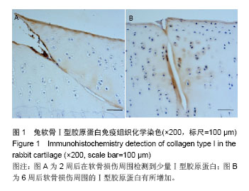
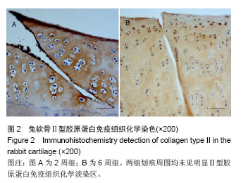
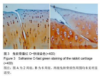
.jpg)