中国组织工程研究 ›› 2013, Vol. 17 ›› Issue (7): 1174-1179.doi: 10.3969/j.issn.2095-4344.2013.07.007

• 皮肤粘膜组织构建 skin and mucosal tissue construction • 上一篇    下一篇

原代培养人增生性瘢痕成纤维细胞的生物学行为

杜启翠1,肖文林2,孙桂兰2,马秀兴1,姚如永3   

  1. 1青岛大学医学院,山东省青岛市 266000
    2青岛大学医学院附属医院口腔科,山东省青岛市 266000
    3青岛大学医学院附属医院中心实验室,山东省青岛市 266000
  • 收稿日期:2012-07-25 修回日期:2012-09-15 出版日期:2013-02-12 发布日期:2013-02-12
  • 通讯作者: 肖文林,博士,副主任医师,副教授,青岛大学医学院附属医院口腔科,山东省青岛市 266000 daybreaker@163.com
  • 作者简介:杜启翠★,女,1985年生,山东省莒县人,汉族,青岛大学在读硕士,主要从事口腔修复学研究。 duqicui@126.com

Primary culture of human hypertrophic scar fibroblasts and its biological behavior

Du Qi-cui1, Xiao Wen-lin2, Sun Gui-lan2, Ma Xiu-xing1, Yao Ru-yong3   

  1. 1 Qingdao University Medical College, Qingdao 266000, Shandong Province, China
    2 Department of Stomatology, Affiliated Hospital of Qingdao University Medical College, Qingdao 266000, Shandong Province, China
    3 Central Laboratory, Affiliated Hospital of Qingdao University Medical College, Qingdao 266000, Shandong Province, China
  • Received:2012-07-25 Revised:2012-09-15 Online:2013-02-12 Published:2013-02-12
  • Contact: Xiao Wen-lin, Doctor, Associate professor, Associate chief physician, Department of Stomatology, Affiliated Hospital of Qingdao University Medical College, Qingdao 266000, Shandong Province, China daybreaker@163.com
  • About author:Du Qi-cui★, Studying for master’s degree, Qingdao University Medical College, Qingdao 266000, Shandong Province, China duqicui@126.com

摘要:

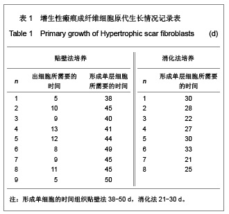
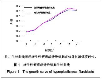
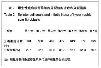
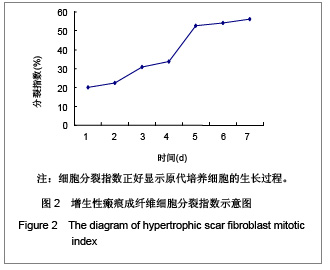
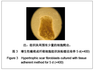
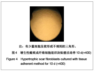
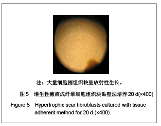
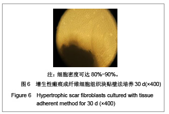
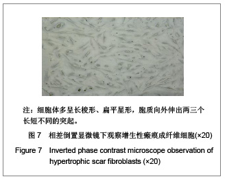
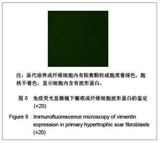
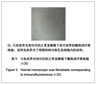
背景:增生性瘢痕组织的形成是创面愈合过程中不可避免的,但是增生性瘢痕组织的形成产生很多不良的影响。
目的:建立可靠的人增生性瘢痕成纤维细胞的原代培养方法。
方法:采用组织贴壁法和消化法分别进行人皮肤增生性瘢痕成纤维细胞的原代培养,使用含体积分数10%胎牛血清的DMEM培养基,37 ℃,体积分数5%CO2,饱和湿度下培养,分别描述其生长曲线、形态及波形蛋白的表达。
结果与结论:组织贴壁法人皮肤增生性瘢痕成纤维细胞培养成功,20-40 d可传第1代,以后每7-10 d可传1代,细胞为长梭形,波形蛋白表达阳性;消化法培养人皮肤增生性瘢痕成纤维细胞成功,15-20 d细胞可融合成片,细胞为长梭形,波形蛋白表达阳性。进一步证实两种方法进行人增生性瘢痕成纤维细胞的原代培养均培养成功。

关键词: 组织构建, 皮肤组织构建, 人增生性瘢痕, 成纤维细胞, 原代培养, 细胞培养, 贴壁法, 消化法, 波形蛋白, 生物学行为, 细胞分裂, 分裂指数, 组织构建图片文章

Abstract:

BACKGROUND: Hypertrophic scar is inevitable in the process of wound healing, but the hypertrophic scar tissue can result in many adverse effects.
OBJECTIVE: To establish a reliable method to culture human hypertrophic scar fibroblasts in vitro.
METHODS: Human hypertrophic scar fibroblasts were cultured primarily in Dulbecco’s modified Eagle’s medium containing 10% fetal bovine serum using tissue adherent method and digestion method. Fibroblasts were incubated at 37℃in a humidified atmosphere of 5% carbon dioxide. Cellular morphologies were observed, the growth curve was drawn, and vimentin expression wass detected.
RESULTS AND CONCLUSION: (1) After cultured with tissue adherent method, human hypertrophic scar fibroblasts were cultured successfully, first passaged within 20-40 days and then continued to passage every 7-10 days. The cells were long spindle-shaped, and vimentin was positive. (2) Human hypertrophic scar fibroblasts, cultured using digestion method, were cultured successfully and confluent at 15-20 days. The cells were long spindle-shaped, and vimentin was positive. These data have confirmed that both tissue adherent method and digestion method are successful in the primary culture of human hypertrophic scar fibroblasts.

Key words: tissue construction, skin tissue construction, human hypertrophic scar, fibroblasts, primary culture, cell culture, adherent method, digestion, vimentin, biological behavior, cell division, mitotic index, tissue construction photographs-containing paper

中图分类号: