[1] AWAD H, RAMADAN ME, EL SAYED HF, et al. Spinal Cord Injury after Thoracic Endovascular Aortic Aneurysm Repair. Can J Anaesth. 2017; 64(12):1218-1235.
[2] ALZGHARI T, AN KR, HARIK L, et al. Spinal Cord Injury after Open and Endovascular Repair of Descending Thoracic Aneurysm and Thoracoabdominal Aortic Aneurysm: An Updated Systematic Review and Meta-Analysis. Ann Cardiothorac Sur. 2023;12(5):409-417.
[3] MALINOVIC M, WALKER J, LEE F. Diffuse and Persistent Blood-Spinal Cord Barrier Disruption after Contusive Spinal Cord Injury Rapidly Recovers Following Intravenous Infusion of Bone Marrow Mesenchymal Stem Cells. Cureus. 2021;13(9):e18298.
[4] 高煜,韩佳慧,葛新.脊髓缺血再灌注损伤后的免疫炎性微环境[J].中国组织工程研究,2023,8(27):1300-1305.
[5] 艾琪勇,罗越,刘辉.脊髓缺血再灌注损伤的发病机制与治疗进展[J].实用医学杂志,2014,16(30):2678-2680.
[6] RONG Y, FAN J, JI C, et al. USP11 Regulates Autophagy-Dependent Ferroptosis after Spinal Cord Ischemia-Reperfusion Injury by Deubiquitinating Beclin 1. Cell Death Differ. 2022;29(6):1164-1175.
[7] LUAN X, CHEN P, MIAO L, et al. Ferroptosis in Organ Ischemia-Reperfusion Injuries: Recent Advancements and Strategies. Mol Cell Biochem. 2024;480(1):19-41.
[8] DIXON SJ, LEMBERG KM, LAMPRECHT MR, et al. Ferroptosis: An Iron-Dependent Form of Nonapoptotic Cell Death. Cell. 2012;149(5):1060-1072.
[9] YU QJ, YANG Y. Function of SOD1, SOD2, and PI3K/AKT Signaling Pathways in the Protection of Propofol on Spinal Cord Ischemic Reperfusion Injury in a Rabbit Model. Life Sci. 2016;148:86-92.
[10] DONG Y, AI C, CHEN Y, et al. Eph Receptor A4 Regulates Motor Neuron Ferroptosis in Spinal Cord Ischemia/Reperfusion Injury in Rats. Neural Regen Res. 2023;18(10):2219-2228.
[11] GUO L, ZHANG D, REN X, et al. SYVN1 Attenuates Ferroptosis and Alleviates Spinal Cord Ischemia-Reperfusion Injury in Rats by Regulating the HMGB1/NRF2/HO-1 Axis. Int Immunopharmacol. 2023;123:110802.
[12] LIU S, CHEN F, HAN J, et al. Ferrostatin-1 Improves Neurological Impairment Induced by Ischemia/Reperfusion Injury in the Spinal Cord through ERK1/2/SP1/GPX4. Exp Neurol. 2024;373:114659.
[13] WANG P, REN Q, SHI M, et al. Overexpression of Mitochondrial Ferritin Enhances Blood–Brain Barrier Integrity Following Ischemic Stroke in Mice by Maintaining Iron Homeostasis in Endothelial Cells. Antioxidants. 2022;11(7):1257.
[14] ZHOU R, LI J, WANG R, et al. The Neurovascular Unit in Healthy and Injured Spinal Cord. J Cereb Blood Flow Metab. 2023;43(9):1437-1455.
[15] YOU Z, GAO X, KANG X, et al. Microvascular Endothelial Cells Derived from Spinal Cord Promote Spinal Cord Injury Repair. Bioact Mater. 2023;29:36-49.
[16] LIU L, ZHANG H, SHI Y, et al. Prostaglandin E1 Improves Cerebral Microcirculation Through Activation of Endothelial NOS and GRPCH1. J Mol Neurosci. 2020;70(12):2041-2048.
[17] XIE X, LU W, CHEN Y, et al. Prostaglandin E1 Alleviates Cognitive Dysfunction in Chronic Cerebral Hypoperfusion Rats by Improving Hemodynamics. Front Neurosci. 2019;13:549.
[18] SHEN J, CAO MS, ZHOU T, et al. PGE1 Triggers Nrf2/HO-1 Signal Pathway to Resist Hemin-Induced Toxicity in Mouse Cortical Neurons. Ann Transl Med. 2021;9(8):634.
[19] QIN Z, KONG B, ZHENG J, et al. Alprostadil Injection Attenuates Coronary Microembolization-Induced Myocardial Injury Through GSK-3β/Nrf2/HO-1 Signaling-Mediated Apoptosis Inhibition. Drug Des Devel Ther. 2020;14:4407-4422.
[20] DODSON M, CASTRO-PORTUGUEZ R, ZHANG DD. NRF2 Plays a Critical Role in Mitigating Lipid Peroxidation and Ferroptosis. Redox Biol. 2019;23:101107.
[21] LIU J, ZHANG S, FAN X, et al. Dexmedetomidine Preconditioning Ameliorates Inflammation and Blood–Spinal Cord Barrier Damage After Spinal Cord Ischemia-Reperfusion Injury by Down-Regulation High Mobility Group Box 1-Toll-Like Receptor 4-Nuclear Factor κB Signaling Pathway. Spine. 2019;44(2):E74-E81.
[22] YE J, LI F, HUANG S, et al. Effects of Ginsenoside Rb1 on Spinal Cord Ischemia-Reperfusion Injury in Rats. J Orthop Surg Res. 2019;14(1):259.
[23] SMITH PD, PUSKAS F, MENG X, et al. The Evolution of Chemokine Release Supports a Bimodal Mechanism of Spinal Cord Ischemia and Reperfusion Injury. Circulation. 2012;126(11 Suppl 1):S110-117.
[24] COHEN J. Statistical Power Analysis for the Behavioral Sciences. New York: Routledge, 2013:567.
[25] YANG H, MAJNO P, MOREL P, et al. Prostaglandin E(1) Protects Human Liver Sinusoidal Endothelial Cell from Apoptosis Induced by Hypoxia Reoxygenation. Microvasc Res. 2002;64(1):94-103.
[26] YANG M, FAN Z, ZHANG Z, et al. MitoQ Protects against High Glucose-Induced Brain Microvascular Endothelial Cells Injury via the Nrf2/HO-1 Pathway. J Pharmacol Sci. 2021;145(1):105-114.
[27] LI W, ZHAO X, ZHANG R, et al. Ferroptosis Inhibition Protects Vascular Endothelial Cells and Maintains Integrity of the Blood-Spinal Cord Barrier after Spinal Cord Injury. Neural Regen Res. 2023;18(11):2474-2481.
[28] 潘玉,赵刃峰,李兴平.铁过载诱导成骨前体细胞铁死亡并抑制成骨分化[J].中国组织工程研究,2025,29(30):6381-6390.
[29] CEMIL B, GOKCE EC, KAHVECI R, et al. Aged Garlic Extract Attenuates Neuronal Injury in a Rat Model of Spinal Cord Ischemia/Reperfusion Injury. J Med Food. 2016;19(6):601-606.
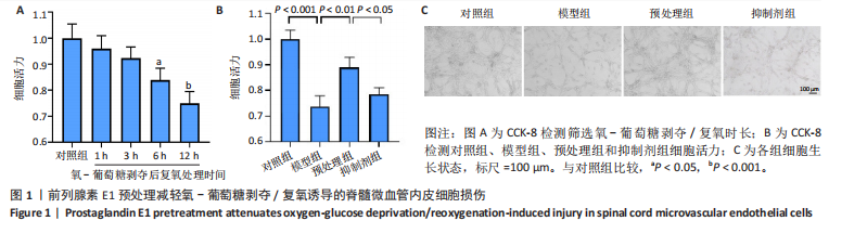
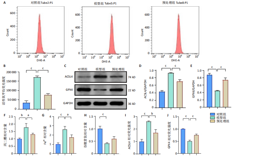
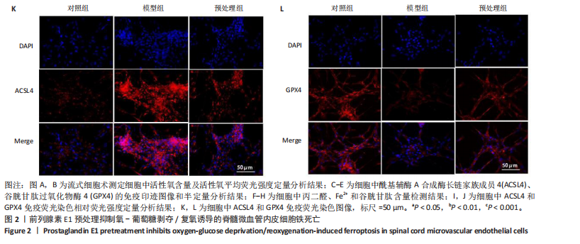
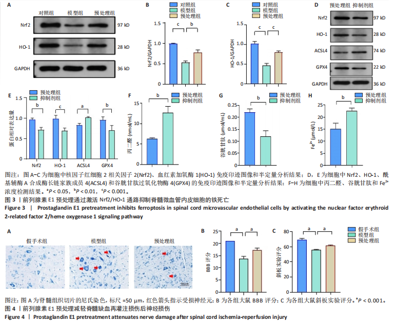
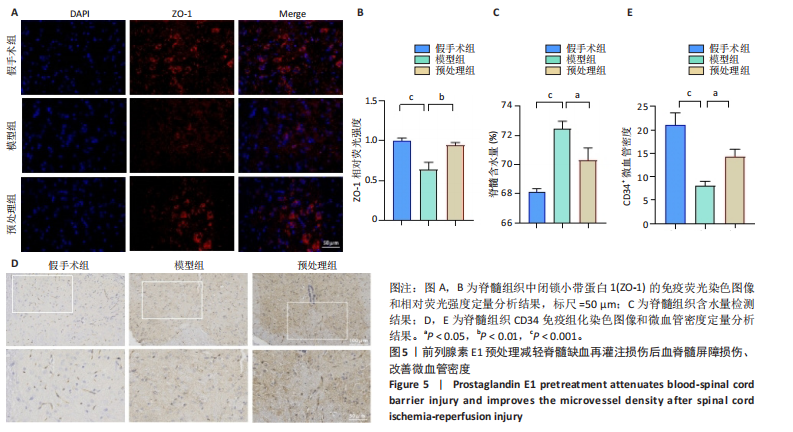
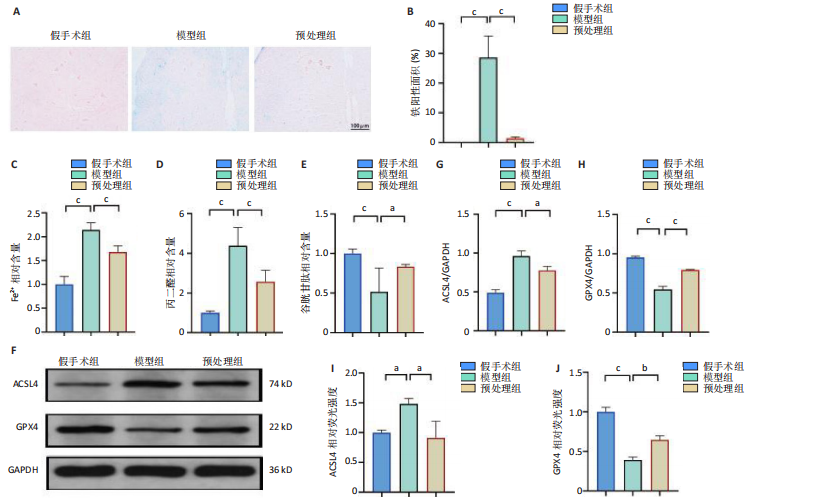
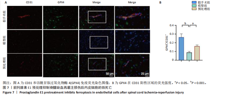
[30] 王荣荣,黄玉珊,李湘淼,等.创伤性脊髓损伤急性期前列腺素E1对血管相关因子的调节和微循环功能的保护[J].中国组织工程研究,2025,29(5):958-967.
[31] BAI T, LI M, LIU Y, et al. Inhibition of Ferroptosis Alleviates Atherosclerosis through Attenuating Lipid Peroxidation and Endothelial Dysfunction in Mouse Aortic Endothelial Cell. Free Radical Biol Med. 2020;160:92-102.
[32] HU J, YU Q, XIE L, et al. Targeting the Blood-Spinal Cord Barrier: A Therapeutic Approach to Spinal Cord Protection against Ischemia-Reperfusion Injury. Life Sci. 2016;158:1-6.
[33] YING X, XIE Q, YU X, et al. Water Treadmill Training Protects the Integrity of the Blood-Spinal Cord Barrier Following SCI via the BDNF/TrkB-CREB Signalling Pathway. Neurochem Int. 2021;143:104945.
[34] SHEN N, WANG L, WU Y, et al. Adeno-Associated Virus Packaged TRPC5 Gene Therapy Alleviated Spinal Cord Ischemic Reperfusion Injury in Rats. Neuroreport. 2020;31(1):29.
[35] REN ZX, XU JH, CHENG X, et al. Pathophysiological Mechanisms of Chronic Compressive Spinal Cord Injury Due to Vascular Events. Neural Regen Res. 2023;18(4):790-796.
[36] MATSUSHITA T, LANKFORD KL, ARROYO EJ, et al. Diffuse and Persistent Blood-Spinal Cord Barrier Disruption after Contusive Spinal Cord Injury Rapidly Recovers Following Intravenous Infusion of Bone Marrow Mesenchymal Stem Cells. Exp Neurol. 2015;267:152-164.
[37] XU J, LI P, LU F, et al. Domino Reaction of Neurovascular Unit in Neuropathic Pain after Spinal Cord Injury. Exp Neurol. 2023;359: 114273.
[38] LUO Y, YAO F, HU X, et al. M1 Macrophages Impair Tight Junctions between Endothelial Cells after Spinal Cord Injury. Brain Res Bull. 2022;180:59-72.
[39] LIU Q, SONG T, CHEN B, et al. Ferroptosis of Brain Microvascular Endothelial Cells Contributes to Hypoxia-Induced Blood-Brain Barrier Injury. FASEB J. 2023;37(5):e22874.
[40] LI C, CHEN X, DU Z, et al. Inhibiting Ferroptosis in Brain Microvascular Endothelial Cells: A Potential Strategy to Mitigate Polystyrene Nanoplastics‒induced Blood‒brain Barrier Dysfunction. Environ Res. 2024;250:118506.
[41] JIA B, LI J, SONG Y, et al. ACSL4-Mediated Ferroptosis and Its Potential Role in Central Nervous System Diseases and Injuries. Int J Mol Sci. 2023;24(12):10021.
[42] SEIBT TM, PRONETH B, CONRAD M. Role of GPX4 in Ferroptosis and Its Pharmacological Implication. Free Radical Bio Med. 2019; 133:144-152.
[43] WANG D, FANG B, WANG Z, et al. Sevoflurane Pretreatment Regulates Abnormal Expression of MicroRNAs Associated with Spinal Cord Ischemia/Reperfusion Injury in Rats. Ann Transl Med. 2021;9(9):752-752.
[44] LI X, CAO X, WANG J, et al. Sevoflurane Preconditioning Ameliorates Neuronal Deficits by Inhibiting Microglial MMP-9 Expression after Spinal Cord Ischemia/Reperfusion in Rats. Mol Brain. 2014;7(1):69.
[45] GÜR FM, BILGIÇ S. A Synthetic Prostaglandin E1 Analogue, Misoprostol, Ameliorates Paclitaxel-Induced Oxidative Damage in Rat Brain. Prostag Oth Lipid M. 2022;162:106663.
[46] AYALA A, MUÑOZ MF, ARGÜELLES S. Lipid Peroxidation: Production, Metabolism, and Signaling Mechanisms of Malondialdehyde and 4-Hydroxy-2-Nonenal. Oxid Med Cell Longev. 2014; 2014:360438.
[47] XU Y, LI Y, LI J, et al. Ethyl Carbamate Triggers Ferroptosis in Liver through Inhibiting GSH Synthesis and Suppressing Nrf2 Activation. Redox Biol. 2022;53:102349.
[48] CHEN L, HUANG J, YAO ZM, et al. Procyanidins Alleviated Cerebral Ischemia/Reperfusion Injury by Inhibiting Ferroptosis via the Nrf2/HO-1 Signaling Pathway. Molecules. 2023;28(8):3582.
[49] LI R, ZHANG J, ZHOU Y, et al. Transcriptome Investigation and In Vitro Verification of Curcumin-Induced HO-1 as a Feature of Ferroptosis in Breast Cancer Cells. Oxid Med Cell Longev. 2020;2020:e3469840.
[50] GE H, XUE X, XIAN J, et al. Ferrostatin-1 Alleviates White Matter Injury Via Decreasing Ferroptosis Following Spinal Cord Injury. Mol Neurobiol. 2022;59(1):161-176.
[51] SHI J, XUE X, YUAN L, et al. Amelioration of White Matter Injury Through Mitigating Ferroptosis Following Hepcidin Treatment After Spinal Cord Injury. Mol Neurobiol. 2023;60(6):3365-3378. |