中国组织工程研究 ›› 2023, Vol. 27 ›› Issue (31): 5035-5039.doi: 10.12307/2023.440

• 骨科植入物相关临床实践 Clinical practice of orthopedic implant • 上一篇    下一篇

绝经后膝骨关节炎患者不同部位的骨量分布特征

陈建超,宋会平   

  1. 华北理工大学附属医院骨科,河北省唐山市  063000
  • 收稿日期:2022-05-25 接受日期:2022-07-14 出版日期:2023-11-08 发布日期:2023-01-31
  • 通讯作者: 宋会平,博士,教授,华北理工大学附属医院骨科,河北省唐山市 063000
  • 作者简介:陈建超,男,1994年生,河北省唐山市人,汉族,2021年华北理工大学毕业,硕士,医师,主要从事骨质疏松症与膝骨关节炎方向研究工作。
  • 基金资助:
    2018年河北省政府资助专科带头人项目(361036),项目负责人:宋会平;2019年度河北省医学科学研究课题计划(20191136),项目负责人:宋会平

Distribution characteristics of bone mass in different parts of postmenopausal women with knee osteoarthritis

Chen Jianchao, Song Huiping   

  1. Department of Orthopedics, Affiliated Hospital of North China University of Science and Technology, Tangshan 063000, Hebei Province, China
  • Received:2022-05-25 Accepted:2022-07-14 Online:2023-11-08 Published:2023-01-31
  • Contact: Song Huiping, MD, Professor, Department of Orthopedics, Affiliated Hospital of North China University of Science and Technology, Tangshan 063000, Hebei Province, China
  • About author:Chen Jianchao, Master, Physician, Department of Orthopedics, Affiliated Hospital of North China University of Science and Technology, Tangshan 063000, Hebei Province, China
  • Supported by:
    Specialist Leader Project Funded by Hebei Provincial Government in 2018, No. 361036 (to SHP); Hebei Medical Science Research Project Plan in 2019, No. 20191136 (to SHP)

摘要:


文题释义:

骨质疏松症(osteoporosis,OP):是一种系统性骨病,其特征是骨量下降和骨的微细结构破坏,表现为骨的脆性增加,因而骨折的危险性大为增加,即使是轻微的创伤或无外伤的情况下也容易发生骨折。
膝骨关节炎(knee osteoarthritis,KOA):亦称退行性膝骨关节病,是一种以关节软骨退行性改变为核心,累及骨质并包括滑膜、关节囊及关节其它结构的全方位、多层次、不同程度的膝关节疾病。
感兴趣区(Region of Interest,ROIs):是图像的一部分,它通过在图像上选择或使用诸如设定阈值(thresholding) 或者从其他文件(如矢量> 转换获得等方法生成。感兴趣区可以是点、线、面或不规则的形状,通常用来作为图像分类的样本、掩膜、裁剪区或其他操作。

背景:膝骨关节炎与骨质疏松症两种疾病的相互影响关系仍存在较大争议,膝骨关节炎患者通常伴有骨密度的改变,目前对于膝关节骨密度与腰椎和髋部骨密度的关系尚不清楚。
目的:探讨膝骨关节炎患者腰椎、股骨颈和膝关节局部骨密度分布情况及其相关性。
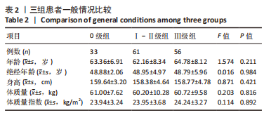
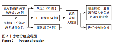
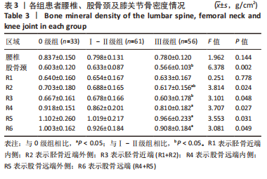
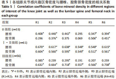
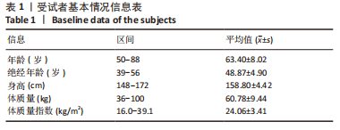
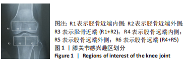
方法:选取膝骨关节炎患者150例,均为绝经后女性,测量腰椎、股骨颈和同侧膝关节骨密度,然后将膝关节划分为6个感兴趣区(ROI),分别标记为R1-R6(R1:胫骨近端内侧,R2:胫骨近端外侧,R3=R1+R2,R4:股骨远端内侧,R5:股骨远端外侧,R6=R5+R4)。根据K-L分级将患者分为0级组(33例)、Ⅰ-Ⅱ级组(61例)和Ⅲ级组(56例),比较3组患者腰椎、股骨颈和膝关节各感兴趣区骨密度情况,再分别将各感兴趣区骨密度值进行两两比较,并分析各感兴趣区骨密度与腰椎和股骨颈骨密度的相关性。
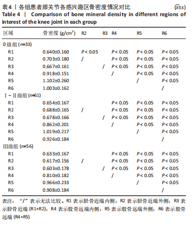
结果与结论:①Ⅲ级组患者R2,R4,R5和R6骨密度低于0级组患者(P < 0.05);Ⅲ级组患者股骨颈、R2和R3骨密度低于Ⅰ-Ⅱ级组患者

(P < 0.05);②3组患者膝关节各感兴趣区骨密度R5 > R6 > R4> (R1,R2,R3);0级组患者R1骨密度低于R2(P < 0.05);③在0级组患者中,R2,R5和R6骨密度与腰椎和股骨颈骨密度均呈正相关(P < 0.05),而R1与R3骨密度仅与腰椎骨密度呈正相关(P < 0.05);在Ⅰ-Ⅱ级组患者中,各感兴趣区骨密度与腰椎和股骨颈骨密度均呈正相关(P < 0.05);在Ⅲ级组患者中,R1和R3骨密度与腰椎和股骨颈骨密度呈正相关(P < 0.05),而R2,R4,R5和R6骨密度只与股骨颈骨密度呈正相关(P < 0.05);④结果表明,膝关节不同区域骨密度存在差异,股骨远端外侧骨密度最高;随着膝骨关节炎的进展,股骨颈骨密度和除胫骨近端内侧以外的膝关节局部骨密度均呈减低趋势,膝关节不同部位骨密度与腰椎和同侧股骨颈骨密度具有相关性。

https://orcid.org/0000-0002-2820-7666(陈建超)

中国组织工程研究杂志出版内容重点:人工关节;骨植入物;脊柱;骨折;内固定;数字化骨科;组织工程

关键词: 膝关节, 骨关节炎, 骨质疏松症, 骨密度, 相关性, 绝经, 腰椎, 股骨颈

Abstract: BACKGROUND: There is still a big controversy about the interaction between knee osteoarthritis and osteoporosis. Patients with knee osteoarthritis often present with changes in bone mineral density (BMD). The relationship between the BMD of the knee joint and the BMD of the lumbar spine and hip is unclear.
OBJECTIVE: To investigate the distribution and correlation of BMD in the lumbar spine, femoral neck, and knee joint of patients with knee osteoarthritis.
METHODS: 150 postmenopausal women with primary knee osteoarthritis were selected to measure the BMD of the lumbar spine, femoral neck, and ipsilateral knee joint. Then the knee joint was divided into six regions of interest (ROI), which were marked as R1-R6, with R1 representing the medial side of proximal tibia, R2 representing the lateral side of proximal tibia, R3 representing the proximal tibia (R3=R1+R2), R4 representing the medial side of distal femur, R5 representing the lateral side of distal femur and R6 representing the distal femur (R6=R5+R4). According to the K-L classification, all the patients were divided into grade 0 group (33 cases), grade I-II group (61 cases), and grade III group (56 cases). The BMD of each ROI was compared with the BMD of the lumbar spine and femoral neck, and then the BMD of different ROIs was compared in pairs. The correlation between the BMD of each ROI and the BMD of the lumbar spine and femoral neck was analyzed.
RESULTS AND CONCLUSION: (1) The BMD of R2, R4, R5, and R6 in the grade III group was lower than that in the grade 0 group (P < 0.05). The BMD of the femoral neck, R2, and R3 in the grade III group was lower than that in the grade I-II group (P < 0.05). (2) The BMD of each ROI of the knee joint in the three groups was ranked as R5 > R6 > R4 > (R1, R2, and R3). The BMD of R1 was lower than that of R2 in the grade 0 group (P < 0.05). (3) In grade 0 patients, the BMD of R2, R5, and R6 was positively correlated with the BMD of the lumbar spine and femoral neck (P < 0.05), while the BMD of R1 and R3 was only positively correlated with the BMD of the lumbar spine (P < 0.05). In grade I-II patients, the BMD of each ROI of the knee joint was positively correlated with the BMD of the lumbar spine and femoral neck (P < 0.05). In grade III patients, the BMD of R1 and R3 was positively correlated with the BMD of the lumbar spine and femoral neck (P < 0.05), while the BMD of R2, R4, R5, and R6 was only positively correlated with the BMD of the femoral neck (P < 0.05). (4) All these findings suggest that there are differences in the BMD of different ROIs of the knee joint, and the BMD of the lateral side of distal femur is the highest. With the progression of knee osteoarthritis, the BMD of the femoral neck and the BMD of the knee joint except the medial side of proximal tibia are on the decline, and the BMD of different parts of the knee joint is correlated with the BMD of the lumbar spine and ipsilateral femoral neck.

Key words: knee joint, osteoarthritis, osteoporosis, bone mineral density, correlation, menopause, lumbar spine, femoral neck

中图分类号: