[1] ARMIENTO AR, STODDART MJ, ALINI M, et al. Biomaterials for articular cartilage tissue engineering: Learning from biology. Acta Biomater. 2018;65:1-20.
[2] 张雪梅,马征,吴宓勋.纳米羟基磷灰石前体与胶原自组装成类骨质复合材料的表征[J].中国组织工程研究,2020,24(10):1534-1539.
[3] BRETSCHNEIDER H, QUADE M, LODE A, et al. Chemotactic and Angiogenic Potential of Mineralized Collagen Scaffolds Functionalized with Naturally Occurring Bioactive Factor Mixtures to Stimulate Bone Regeneration. Int J Mol Sci. 2021;22(11):5836.
[4] DE TULLIO I, CAPUTI S, PERFETTI G, et al. A Human Clinical and Histomorphometrical Study on Different Resorbable and Non-Resorbable Bone Substitutes Used in Post-Extractive Sites. Materials (Basel). 2019;12(15):2408.
[5] LI Z, DU T, RUAN C, et al. Bioinspired mineralized collagen scaffolds for bone tissue engineering. Bioactive Materials. 2021;6(5):1491-1511.
[6] 陈琬雯.基于硒活性的壳聚糖复合物的制备研究[D].无锡:江南大学,2020.
[7] 刘婉霞.壳聚糖基复合材料的制备及其吸附钴离子的研究[D].北京:北京建筑大学,2020.
[8] DREIFKE MB, EBRAHEIM NA, JAYASURIYA AC. Investigation of potential injectable polymeric biomaterials for bone regeneration. J Biomed Mater Res A. 2013;101:2436-2447.
[9] GAIHRE B, USWATTA S, JAYASURIYA AC. Nano-scale characterization of nano-hydroxyapatite incorporated chitosan particles for bone repair. Colloids Surf B Biointerfaces. 2018;165:158-164.
[10] RUIXIN L, CHENG X, YINGJIE L, et al. Degradation behavior and compatibility of micro, nanoHA/chitosan scaffolds with interconnected spherical macropores. Int J Biol Macromol. 2017;103:385-394.
[11] SHAKIR M, ZIA I, REHMAN A, et al. Fabrication and characterization of nanoengineered biocompatible n-HA/chitosan-tamarind seed polysaccharide: Bio-inspired nanocomposites for bone tissue engineering. Int J Biol Macromol. 2018;111:903-916.
[12] CHEN GG, QI XM, GUAN Y, et al. High strength hemicellulose-based nanocomposite film for food packaging applications. ACS Sustain Chem Eng. 2016;4(4):1985-1993.
[13] YANG C, GAO XT, YOUNIS MR, et al. Non-invasive monitoring of in vivo bone regeneration based on alkaline phosphatase-responsive scaffolds. Chem Eng J. 2021;408:127959.
[14] 庄均珺.生物功能化胶原涂层的可控制备及其评价[D].杭州:浙江大学,2018.
[15] AMINI AR, LAURENCIN CT, NUKAVARAPU SP. Bone tissue engineering: recent advances and challenges. Crit Rev Biomed Eng. 2012;40(5):363-408.
[16] MAJI K, DASGUPTA S, PRAMANIK K, et al. Preparation and characterization of gelatin-chitosan-nano beta-TCP based scaffold for orthopaedic application. Mater Sci Eng C Mater Biol Appl. 2018;86:83-94.
[17] 刘来俊,张宇,李超婧,等.用于骨组织再生的仿生骨膜的研究进展[J].中国生物医学工程学报,2020,39(4):493-503.
[18] LING T, LIN J, TU JJ, et al. Mineralized collagen coatings formed by electrochemical deposition. J Mater Sci Mater Med. 2013;24(12):2709-2718.
[19] 李博,王硕,赵勇刚,等.仿生矿化胶原骨材料用于儿童颅骨再生修复的最新研究进展[J].中国修复重建外科杂志,2021,17(2):49-54.
[20] TW SUN, WL YU, YJ ZHU, et al. Porous Nanocomposite Comprising Ultralong Hydroxyapatite Nanowires Decorated with Zinc‐Containing Nanoparticles and Chitosan: Synthesis and Application in Bone Defect Repair. Chem Eur J. 2018;24:8809.
[21] LIAO J, LI Y, LI H, et al. Preparation, bioactivity and mechanism of nano-hydroxyapatite/sodium alginate/chitosan bone repair material. J Appl Biomater Funct Mater. 2018;16(1):28-35.
[22] KONG ZQ, YU MF, CHENG K, et al. Incorporation of chitosan nanospheres into thin mineralized collagen coatings for improving the antibacterial effect. Colloids Surf B Biointerfaces. 2013;111:536-541.
[23] KONG M, CHEN XG, XING K, et al. Antimicrobial properties of chitosan and mode of action: a state of the art review. Int J Food Microbiol. 2010;144(1):51-63.
[24] DAI C, LI Y, PAN W, et al. Three-Dimensional High-Porosity Chitosan/Honeycomb Porous Carbon/Hydroxyapatite Scaffold with Enhanced Osteoinductivity for Bone Regeneration. ACS Biomater Sci Eng. 2020; 6(1):575-586.
[25] LI M, JIA WB, ZHANG XL, et al. Hyaluronic acid oligosaccharides modified mineralized collagen and chitosan with enhanced osteoinductive properties for bone tissue engineering. Carbohydr Polym. 2021;260:117780.
[26] BERNHARDT A, LODE A, MIETRACH C, et al. In vitroosteogenic potential of human bone marrow stromal cells cultivated in porous scaffolds from mineralized collagen. J Biomed Mater Res. 2009;90(3):852-862.
[27] WANG S, YANG YD, KOONS GL, et al. Tuning pore features of mineralized collagen/PCL scaffolds for cranial bone regeneration in a rat model. Mater Sci Eng C Mater Biol Appl. 2020;106:110186.
[28] JIANG L, LI Y, XIONG C, et al. Preparation and properties of bamboo fiber/nano-hydroxyapatite/poly(lactic-co-glycolic) composite scaffold for bone tissue engineering. ACS Appl Mater Interfaces. 2017;9(5): 4890-4897.
[29] 李锐.羟基磷灰石/壳聚糖复合二甲双胍用于大鼠骨缺损的生物学特点[J].中国组织工程研究,2021,25(28):4460-4464.
[30] SADEGHINIA A, SOLTANI S, AGHAZADEH M, et al. Design and fabrication of clinoptilolite-nanohydroxyapatite/chitosan-gelatin composite scaffold and evaluation of its effects on bone tissue engineering. J Biomed Mater Res A. 2020; 108(2):221-233.
[31] 李扬,刘怡卓,沈超,等.生物玻璃/壳聚糖三维多孔支架的抗感染性能和生物相容性研究[J].生物骨科材料与临床研究,2020, 17(2):49-54.
[32] 陈黎,黄蕾,余梦瑶,等.用于生物人工肝反应器构建三维支架材料的选择与优化[J].中国组织工程研究,2021,25(28):4429-4434.
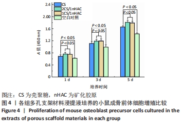
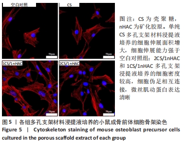
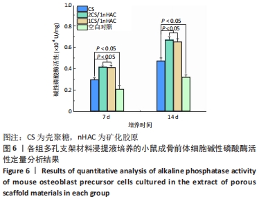
|