中国组织工程研究 ›› 2019, Vol. 23 ›› Issue (26): 4194-4199.doi: 10.3969/j.issn.2095-4344.1359

• 纳米生物材料 nanobiomaterials • 上一篇    下一篇

载阿霉素壳聚糖纳米粒子可抑制小鼠骨肉瘤

李永恒,崔  岩,张治宇
  

  1. 中国医科大学附属第四医院,辽宁省沈阳市  110032
  • 收稿日期:2019-03-16
  • 通讯作者: 张治宇,博士,教授,中国医科大学附属第四医院骨外科,辽宁省沈阳市 110032
  • 作者简介:李永恒,男,1992年生,安徽省宿州市人,汉族,硕士,主要从事骨肉瘤化疗方面的研究。
  • 基金资助:

    辽宁省科学技术计划项目(20170541058),项目负责人:张治宇

Inhibitory effects of doxorubicin-loaded chitosan nanoparticles on osteosarcoma in mice

Li Yongheng, Cui Yan, Zhang Zhiyu
  

  1. the Fourth Affiliated Hospital of China Medical University, Shenyang 110032, Liaoning Province, China
  • Received:2019-03-16
  • Contact: Zhang Zhiyu, MD, Professor, the Fourth Affiliated Hospital of China Medical University, Shenyang 110032, Liaoning Province, China
  • About author:Li Yongheng, Master, the Fourth Affiliated Hospital of China Medical University, Shenyang 110032, Liaoning Province, China
  • Supported by:

    Liaoning Science and Technology Plan Project, No. 20170541058 (to ZZY)

摘要:

文章快速阅读:


文题释义:
高渗透长滞留效应:相对于正常组织,实体瘤组织中血管丰富、血管壁间隙较宽、结构完整性差,淋巴回流缺失,造成某些尺寸的微粒更趋向于聚集在肿瘤组织的性质,促进了载药纳米微粒在肿瘤组织的选择性分布,可增加药效并减少系统不良反应。
被动靶向:根据肿瘤组织的特性,化疗药物趋向于向肿瘤细胞内浓集的性质。纳米载药微粒既可通过高渗透长滞留效应实现化疗药物在肿瘤组织的富集,同时肿瘤微环境也有助于实验合成纳米载药微粒的靶向释放。
 
 
背景:近来大量研究证明,纳米载体系统可实现药物在肿瘤组织定点释放或激活,提高局部药物浓度,减少正常组织的药物蓄积,降低不良反应。
目的:分析载阿霉素壳聚糖纳米粒子的细胞毒性及对骨肉瘤的抑制作用。
方法:①细胞毒性实验:采用含载阿霉素壳聚糖纳米粒子(简称载药纳米微粒)的PBS与含游离阿霉素的PBS分别干预鼠源骨肉瘤细胞系K7(均设置0.16,0.31,0.62,1.25,2.5,5,10 mg/L 7个质量浓度),干预24,72 h后,采用MTT法检测细胞存活率;②体内药物分布实验:皮下注射鼠源骨肉瘤细胞系K7建立Balb/c小鼠(长春生物制品研究所有限责任公司提供)肿瘤模型,当肿瘤体积达到200 mm3时,实验组、对照组分别尾静脉注射含载药纳米微粒的PBS与含游离阿霉素的PBS;注射后6,12 h处死小鼠,观察各脏器阿霉素荧光;③体内抑瘤实验:皮下注射鼠源骨肉瘤细胞系K7建立Balb/c小鼠肿瘤模型,当肿瘤体积达到50 mm3时,实验组、对照组分别尾静脉注射含载药纳米微粒的PBS与含游离阿霉素的PBS,空白组注射PBS,4 d注射1次,共6次,每天检测小鼠体质量与肿瘤体积。动物实验方案经吉林大学动物实验中心伦理委员会批准。
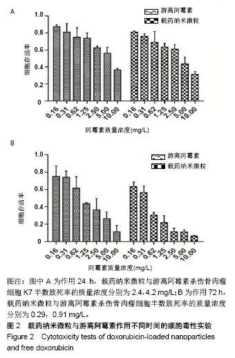
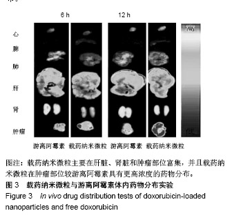
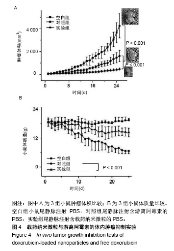
结果与结论:①药物作用24 h时,载药纳米微粒与游离阿霉素杀伤骨肉瘤细胞的半数致死率质量浓度分别为2.4,4.2 mg/L;72 h时,载药纳米微粒与游离阿霉素杀伤骨肉瘤细胞的半数致死率质量浓度分别为0.29,0.91 mg/L;②载药纳米微粒主要在肝脏、肾脏和肿瘤部位富集;③治疗周期内,实验组平均肿瘤体积明显小于对照组、空白组(P < 0.001),实验组平均体质量大于对照组(P < 0.001);④结果表明,该载药体系可很好地实现阿霉素体内外控制释放,明显提高化疗药物对骨肉瘤的抑制能力。

关键词: 骨肉瘤, 阿霉素, 壳聚糖, 纳米微粒, 多糖, 药物控制释放, 纳米系统, 肿瘤抑制

Abstract:

BACKGROUND: A large number of recent studies have shown that the nanocarrier system can achieve drug release or activation in tumor tissues, increase local drug concentration, reduce drug accumulation in normal tissues, and reduce adverse reactions.
OBJECTIVE: To analyze the cytotoxicity of doxorubicin-loaded chitosan nanoparticles and their inhibitory effect on osteosarcoma.
METHODS: Cytotoxicity tests: Murine osteosarcoma cell line K7 was interfered with PBS containing doxorubicin-loaded chitosan nanoparticles (referred to as drug-loaded nanoparticles) and PBS containing free doxorubicin (doxorubicin concentrations 0.16, 0.31, 0.62, 1.25, 2.5, 5,10 mg/L) for 24 and 72 hours. Cell viability was measured by MTT assay. In vivo drug distribution test: Balb/c mouse (Changchun Institute of Biological Product Co., Ltd., China) models of tumor were established by subcutaneous injection of murine osteosarcoma cell line K7. When tumor volume reached 200 mm3, mice in the experimental and control groups were respectively injected with PBS containing doxorubicin-loaded nanoparticles and PBS containing free doxorubicin via the tail vein. The mice were sacrificed at 6 and 12 hours after drug administration and the fluorescence of doxorubicin in various organs was observed. In vivo tumor growth inhibition test: Balb/c mouse models of tumor were established by subcutaneous injection of murine osteosarcoma cell line K7. When tumor volume reached 50 mm3, mice in the experimental and control groups were respectively injected with PBS containing doxorubicin-loaded nanoparticles and PBS containing free doxorubicin via the tail vein. PBS was injected in the blank control group, once every 4 days, for a total of 6 times. Mouse body mass and tumor volume were daily measured. Animal experiments were approved by Animal Ethics Committee of Jilin University, China.
RESULTS AND CONCLUSION: The median lethal concentration of doxorubicin-loaded nanoparticles and free doxorubicin was 2.4 and 4.2 mg/L respectively at 24 hours after drug application, and it was 0.29 and 0.91 mg/L respectively at 72 hours after drug application. Doxorubicin-loaded nanoparticles mainly accumulated in the liver, kidney, and tumor. During the treatment period, average tumor volume in the experimental group was significantly smaller than that in the control and blank control groups (P < 0.001). The average mouse body mass in the experimental group was significantly greater than that in the control group (P < 0.001). The results show that the drug-loaded system can well control in vivo release of doxorubicin and greatly enhance the inhibitory capacity of chemotherapy drugs against osteosarcoma.

Key words: osteosarcoma, doxorubicin, chitosan, nanoparticles, polysaccharide, controlled drug release, nanosystem, tumor suppression

中图分类号: