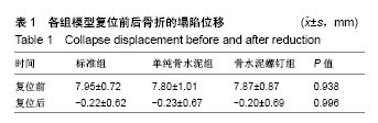
| [1] Lee MH,Hsu CJ,Lin KC,et al.Comparison of outcome of unilateral locking plate and dual plating in the treatment of bicondylar tibial plateau fractures.J Orthop Surg Res. 2014; 9:62.[2] Yang G,Zhu Y,Luo C,et al.Morphological characteristics of Schatzker type Ⅳtibial plateau fractures: a computer tomography based study.Int Orthop.2012;36(11):2355-2360.[3] Haider SJ,Pean CA,Davidovitch RI,et al.Functional Outcomes of Isolated Medial Tibial Plateau Fractures.J Knee Surg. 2016; 29(5):414-422.[4] Sciadini MF,Sims SH.Proximal tibial intra-articular osteotomy for treatment of complex Schatzker type Ⅳ tibial plateau fractures with lateral joint line impaction: description of surgical technique and report of nine cases.J Orthop Trauma. 2013;27(1):e18-23.[5] Wahlquist M,Iaguilli N,Ebraheim N,et al.Medial tibial plateau fractures: a new classification system. J Trauma. 2007;63(6): 1418-1421.[6] Chang SM,Wang X,Zhou JQ,et al.Posterior coronal plating of bicondylar tibial plateau fractures through posteromedial and anterolateral approaches in a healthy floating supine position. Orthopedics. 2012;35(7):583-588.[7] 陈文龙,张英琪,张世民.Schatzker Ⅳ型胫骨平台骨折及手术入路研究[J].国际骨科学杂志,2014,35(2): 97-99.[8] 王军,赵春鹏,李庭,等.骨折脱位型胫骨平台骨折发生率及内侧和后内侧骨块影像学特点[J].中华创伤杂志, 2015,31(5):427-430.[9] 胡孙君,张世民,张英琪,等.胫骨平台后外侧象限骨折手术入路的深层解剖及后外侧与后内侧比较[J].中国临床解剖学杂志, 2015, 33(5):497-501.[10] Berkes MB,Little MT,Schottel PC,et al.Outcomes of Schatzker II tibial plateau fracture open reduction internal fixation using structural bone allograft.J Orthop Trauma.2014;28(2):97-102.[11] 胡勇,尹宗生,张辉,等.累及后柱的胫骨平台骨折的手术治疗[J].中华骨科杂志,2012,32(12):1138-1144.[12] Hoekstra H,Rosseels W,Luo CF,et al.A combined posterior reversed L-shaped and anterolateral approach for two column tibial plateau fractures in Caucasians: A technical note. Injury. 2015;46(12): 2516-2519.[13] Brunner A,Horisberger M,Ulmar B,et al.Classi?cation systems for tibial plateau fractures; Does computed tomography scanning improve their reliability? Injury.2010;41:173-178.[14] Luo CF,Sun H,Zhang B,et al.Three-column fixation for complex tibial plateau fractures.J Orthop Trauma.2010;24(11): 683-692.[15] Chang SM,Zhang YQ,Yao MW,et al.Schatzker type Ⅳ medial tibial plateau fractures: a computed tomography- based morphological subclassification. Orthopedics.2014; 37(8):e699-706.[16] Ruffolo MR,Gettys FK,Montijo HE,et al.Complications of highenergy bicondylar tibial plateau fractures treated with dual plating through 2 incisions.J Orthop Trauma. 2015; 29(2):85-90.[17] Bagherifard A,Jabalameli M,Hadi H,et al.Surgical Management of Tibial Plateau Fractures With 3.5 mm Simple Plates.Trauma Mon.2016;21(2):e26733.[18] Qiu WJ,Zhan Y,Sun H,et al.A posterior reversed L-shaped approach for the tibial plateau fractures-A prospective study of complications (95 cases).Injury.2015;46(8):1613-1618. |
.jpg)
.jpg)