中国组织工程研究 ›› 2017, Vol. 21 ›› Issue (34): 5499-5505.doi: 10.3969/j.issn.2095-4344.2017.34.015

• 生物材料模型构建 biomaterial modeling • 上一篇    下一篇

角膜修复材料有效性评价动物模型的建立和验证

韩倩倩1,李  青2,王宝全2,杨昭鹏1,王春仁1
  

  1. 1中国食品药品检定研究院,北京市  102629;2青岛中皓生物工程有限公司,山东省青岛市  266000
  • 收稿日期:2017-10-23 出版日期:2017-12-08 发布日期:2018-01-04
  • 通讯作者: 王宝全,青岛中皓生物工程有限公司,山东省青岛市 266000
  • 作者简介:韩倩倩,女,1982年生,山东省济南市人,汉族,2011年中国科学院遗传与发育生物学研究所毕业,博士,副研究员,主要从事生物材料、组织工程和医疗器械产品的安全有效性评价技术和标准化工作。
  • 基金资助:

    国家重点研发计划:组织诱导型生物材料及产品安全有效性评价关键技术研究(2016YFC1103202)2016年- 2020年负责人:韩倩倩

Establishment and validation of an animal model for evaluating the effectiveness of corneal repair materials

Han Qian-qian1, Li Qing2, Wang Bao-quan2, Yang Zhao-peng1, Wang Chun-ren1
  

  1. 1National Institutes for Food and Drug Control, Beijing 102629, China; 2Zhonghao Biological Engineering Co., Ltd., Qingdao 266000, Shandong Province, China
  • Received:2017-10-23 Online:2017-12-08 Published:2018-01-04
  • Contact: Wang Bao-quan, Zhonghao Biological Engineering Co., Ltd., Qingdao 266000, Shandong Province, China
  • About author:Han Qian-qian, M.D., Associate investigator, National Institutes for Food and Drug Control, Beijing 102629, China
  • Supported by:
     the National Key Research and Development Program of China, No. 2016YFC1103202

摘要:

文章快速阅读:


文题释义:
板层角膜移植:是以角膜部分组织为操作对象进行的手术,只切除有病变的角膜浅层组织,深层比较完好的受主角膜仍然保留作为移植床,然后取同样大小和厚度的角膜材料浅层角膜片,缝于患者角膜的创面上。
Ⅲ类植入性医疗器械:是最高级别的医疗器械,也是必须严格控制的医疗器械,是指植入人体,用于支持、维持生命,对人体具有潜在危险,对其安全性、有效性必须严格控制的医疗器械。
 
背景:角膜修复材料可修复角膜损伤,这种产品在进入临床试验之前需要评估其有效性,但现阶段尚缺乏标准化的有效性评估方法。
目的:建立并验证兔角膜真菌感染模型和角膜移植实验方法。
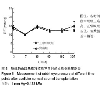
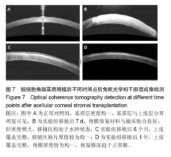
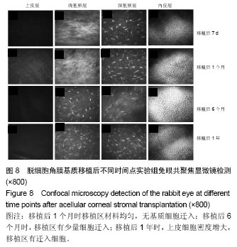
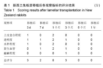
方法:取新西兰大白兔12只,建立左眼角膜真菌感染模型(实验组),右眼作为正常对照。模型建立2周后,左眼进行脱细胞猪角膜基质移植,移植后定期进行裂隙灯显微镜、角膜厚度、眼压、共聚焦显微镜及光学相干断层成像等检测,评估角膜修复材料透明度、上皮愈合程度、水肿程度、角膜新生血管程度、角膜修复材料融解程度;移植后3,6,12个月,进行角膜病理组织观察。
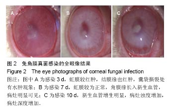
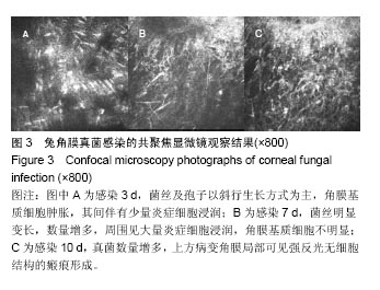
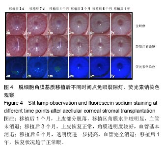
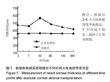
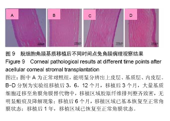
结果与结论:①实验组移植后1个月时角膜厚度明显增高,移植3-6个月时角膜厚度开始稳定,1年时趋于稳定,趋向于正常眼;②实验组眼压始终接近于正常眼;③光学相干断层成像显示,移植后7 d,角膜修复材料与植床贴合良好;移植后6个月上皮覆盖完整,移植区植片厚度较为均一;移植后1年恢复情况趋于正常眼;④共聚焦显微镜显示,移植后1个月时,移植区材料均匀;移植后6个月时,移植区有少量细胞迁入;移植后1年时,上皮细胞密度增大,移植区有迁入细胞,但少于正常眼;⑤移植后1年,角膜修复材料几近完全透明,可认为此角膜修复材料治疗感染性角膜溃疡有效;未发生排斥反应,表明该角膜修复材料的治愈性良好;⑥移植后3个月,大量基质细胞迁移至角膜角膜替代物中,移植区域胶原纤维排列整齐、致密,无明显瘢痕及降解现象;移植后6个月时,移植区域已基本恢复至正常角膜状态;移植后1年,移植区域已恢复至正常角膜状态,生物相容性很好;⑦结果表明,实验建立的兔角膜真菌感染模型和角膜移植实验方法可作为评价此类角膜材料有效性的动物模型和方法。

关键词: 生物材料, 材料相容性, 角膜修复材料, 角膜基质替代物, 临床前有效性评价动物模型, 角膜移植

Abstract:

BACKGROUND: Corneal repair materials can be used as an alternative of human donor corneas to repair corneal injuries, but their evaluation of effectiveness is necessary before entering clinical trials. Unfortunately, there is no standardized method for effectiveness evaluation until now.
OBJECTIVE: To establish and validate a corneal fungal infection model in rabbits and the corneal transplantation method.
METHODS: Twelve New Zealand White rabbits were selected to establish a corneal fungal infection model in the left eye (experimental) and a normal control in the right eye. Two weeks after modeling, acellular porcine corneal stroma was transplanted into the left eye. After transplantation, slit lamp microscope test, corneal thickness detection, intraocular pressure measurement, confocal microscopy test and optical coherence tomography were performed. Then the degree of transparency, degree of epithelium healing, degree of edema, degree of corneal neovascularization and degree of material thawing were evaluated. The corneal pathological sections with hematoxylin-eosin staining were observed at 3, 6 and 12 months after surgery.
RESULTS AND CONCLUSION: (1) The corneal thickness increased significantly at 1 month after transplantation, varied slightly within 3-6 months, and became close to the normal value at 1 year. (2) The intraocular pressure of the left eye was close to normal eyes. (3) Findings from the optical coherence tomography showed that the repair materials fit well with the implantation bed at 7 days after transplantation; the transplanted area was fully covered with epithelial cells at 6 months after transplantation, and the uniform thickness of the repair material in the transplanted area was detected; the grafted cornea was restored to normal cornea at 1 year after transplantation. (4) Under the confocal microscope, the repair materials in the transplanted area were evenly spread at 1 month after transplantation; few cells migrated into the transplanted area at 6 months after transplantation; the density of epithelial cells was increased, and there were migrated cells in the transplanted cells, but the cell number was less than that of normal eyes at 1 year after transplantation. (5) The corneal repair material was almost completely transparent at 1 year after transplantation, indicating its effectiveness in the treatment of infectious corneal ulcers. No rejection occurred, indicating that the corneal healing material is well-curative. (6) At 3 months after transplantation, a large number of stromal cells migrated to the corneal substitute, and the collagen fibers in the transplanted area were arranged neatly and densely without obvious scarring and degradation. At 6 months after transplantation, the transplanted area basically recovered. At 1 year after transplantation, the transplanted area was fully restored to the normal cornea state with good biocompatibility. Our experimental findings indicate that the rabbit model of corneal fungal infection and the corneal transplantation method can be used to evaluate the effectiveness of such corneal materials.

Key words: Corneal Transplantation, Corneal Stroma, Models, Animal, Tissue Engineering

中图分类号: