| [1]Marston WA,Hanft J,Norwood P,et al.The efficacy and safety of Dermagraft in improving the healing of chronic diabetic foot ulcers: results of a prospective randomized trial. Diabetes Care.2003;26(6):1701-1705.[2]Anders S,Volz M,Frick H,et al. A randomized,controlled trial com- paring autologous matrix-induced chondrogenesis (AMIC)to micro-fracture: Analysis of 1- and 2-year follow-up data of 2 centers.Open Orthop J.2013;7: 133-143.[3]Schüettler KF,Struewer J,Rominer MB,et al.Repair of a chondral de- fect using a cell free sca old in a young patient—a case report of suc-cessful sca old transformation and colonization.BMC Surgery.2013;13:11-17.[4]张皑峰,欧喜超,杨朝阳,等.应用结合bFGF的壳聚糖导管修复大鼠坐骨神经损伤的实验研究[J].中国康复理论与实践, 2008, 14(12):1133-1135.[5]Marques PA,Gon alves G,Singh MK,et al. Graphene oxide and hy-droxyapatite as fillers of polylactic acid nanocomposites: preparation and characterization.J Nanosci Nanotechnol.2012;12(8):6686-6692.[6]Gu Y,Chen L,Yang HL,et al. Evaluation of an injectable silk fibroin enhanced calcium phosphate cement loaded with human recombinant bone morphogenetic protein-2 in ovine lumbar interbody fusion.J Biomed Mater Res A.2011;97(2): 177-185.[7]Mirahmadi F,Tafazzoli-Shadpour M,Shokrgozar MA,et al.Enhanced mechanical properties of thermosensitive chitosan hydrogel by silk fi-bers for cartilage tissue engineering.Mater Sci Eng C Mater Biol Appl.2013;33(8): 4786-4794.[8]刘中宁,姜婷,王衣祥.富血小板血浆促进人牙髓细胞的体内矿化潜能[J].北京大学学报:医学版,2011,43(2):276-279.[9]方厂云,曹莹,夏宇,等.大鼠牙乳头细胞与纳米羟基磷灰石的体外复合培养[J].中南大学学报:医学版,2007,32(1):114:118.[10]Kim NR,Lee DH,Chung PH,et al.Distinct differentiation properties of human dental pulp cells on collagen,gelatin,and chitosan scaffolds. Oral Surg Oral Med Oral Pathol Oral Radiol Endod.2009;108(5):e94-100.[11]张文涛,刘建华,王慧明,等.重组人BMP-2修饰的β磷酸三钙/胶原材料制备及其诱导成牙性能的初步研究[J].中国修复重建外科杂志,2011,25(2):149-154.[12]王瑛慧,王万永,贾智.APA微囊包裹的牙髓干细胞成牙本质作用的研究[J].天津医科大学学报,2006,12(4):548-550.[13]包柳郁,金岩,史俊南,等.人牙源性间充质细胞体内立体培养模型的建立--构建组织工程化牙本质和牙髓组织[J].牙体牙髓牙周病学杂志,2004,14(11):603-606.[14]李珊,樊明文,梁素霞.成人牙髓干细胞与壳聚糖-磷酸三钙复合材料相容性试验研究[J].口腔医学研究,2006,22(3):264-266.[15]陈建洪,唐倩,王萍,等.应用SGBG/PHBV复合材料构建牙本质牙髓复合体样结构的初步研究[J].临床医学工程, 2011,18(9): 1339-1340.[16]Zeng W,Huang JH,Luo ZJ,et al.Ionically cross linked chitosan microspheres for controlled release of bioactive nerve factor. Int J Pharm.2011;421(2):283-290.[17]焦旭文,何仲义,李军平,等.NGF 和 GM1 联 合应用对去细胞异种神经支架移植后神经再生和修复的影响[J].神经解剖学杂志, 2007,23(5):495-500.[18]Lian XF,Wang W,Zhang L,et al.Co application of muscle derived stem cells and FK-506 influences nerve regeneration and recovery following acellular nerve allograft implantation.Zhongguo Zu Zhi Gong Cheng Yan Jiu Yu Lin Chuang Kang Fu. 2010;14(3):389-392. [19]谭太贵,马景鉴,张文治,等.神经干细胞在PLGA、壳聚糖和明胶膜表面的生长规律[J].中国生物医学工程学报, 2006,25(3): 377-381.[20]许文静,赵喆,赵斌,等.不同数量BMSCs对大鼠背根神经节生长影响的实验研究[J].中国修复重建外科杂志, 2011,25(10): 1245-1249.[21]袁普卫,贺西京,王国毓,等.嗅鞘细胞移植对脊髓损伤后损伤区髓鞘相关轴突生长抑制因子受体表达的影响[J].中国骨伤, 2011, 24(1):51-54.[22]van der Zande M,Vandebriel RJ,Van Doren E,et al. Distribution,elimination,and toxicity of silver nanoparticles and silver ions in rats after 28-day oral exposure.ACS Nano. 2012; 6(8):7427-7442.[23]Lin JJ,Lin WC,Li SD,et al.Evaluation of the antibacterial activity and biocompatibility for silver nanoparticles immobilized on nano silicate platelets.CS Appl Mater Interfaces. 2013;5(2):433-443.[24]胡莹,刘笑涵,王靖宇,等.低强度脉冲超声波辐照与泡沫TiC / Ti 对犬节段性骨缺修复的促进作用[J].中国比较医学杂志, 2013, 23(4):44-47.[25][31]Hu N,Feng C,Jiang Y,et al.Regulative Effect of Mir-205 on Osteogenic Differentiation of Bone Mesenchymal Stem Cells ( BMSCs) : Possible Role of SATB2/Runx2 and ERK/MAPK Pathway.Int J Mol Sci.2015;16(5):10491-10506.[26]Yang H,Aprecio RM,Zhou X,et al.Therapeutic Effect of TSG-6 Engineered iPSC-Derived MSCs on Experimental Periodokntitis in Rats: A Pilot Study.PloS One.2014;9(6): e100285.[27]Liu Y,Goldberg AJ,Dennis JE,et al.One-step derivation of mesenchymal stem cell ( MSC) -like cells from human pluripotent stem cells on a fibrillar collagen coating.PloS One. 2012;7:e33225.[28]Ganji F,Abroun S,Baharvand H,et al.Differentiation potential of o bombay human-induced pluripotent stem cells and human embryonic stem cells into fetal erythroid-like cells.Cell J.2015;16(4):426-439. |
.jpg)
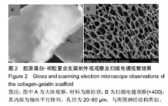
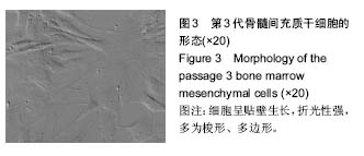
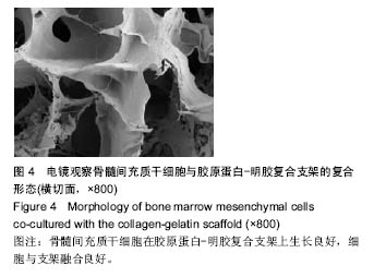
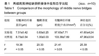
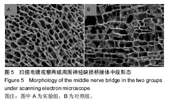
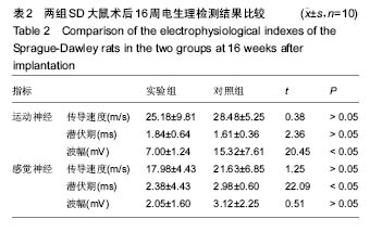
.jpg) 1.5 主要观察指标 观察胶原蛋白-明胶复合支架材料构建及扫描电镜下外观形态;观察骨髓间充质干细胞的分离、培养及形态;观察骨髓间充质干细胞与胶原蛋白-明胶复合支架的复合形态;观察两组SD大鼠术后状态;观察两组神经植入后16周桥接体中段形态(在扫描电镜下完成相应的测量);观察两组神经植入后16周电生理检测结果[19]。
1.5 主要观察指标 观察胶原蛋白-明胶复合支架材料构建及扫描电镜下外观形态;观察骨髓间充质干细胞的分离、培养及形态;观察骨髓间充质干细胞与胶原蛋白-明胶复合支架的复合形态;观察两组SD大鼠术后状态;观察两组神经植入后16周桥接体中段形态(在扫描电镜下完成相应的测量);观察两组神经植入后16周电生理检测结果[19]。.jpg)