[1] YAO Q, WU X, TAO C, et al. Osteoarthritis: pathogenic signaling pathways and therapeutic targets. Signal Transduct Target Ther. 2023;8(1):56.
[2] GLYN-JONES S, PALMER AJ, AGRICOLA R, et al. Osteoarthritis. Lancet. 2015; 386(9991):376-387.
[3] BIJLSMA JW, BERENBAUM F, LAFEBER F P. Osteoarthritis: an update with relevance for clinical practice. Lancet. 2011;377(9783):2115-2126.
[4] ALEXANDER PG, GOTTARDI R, LIN H, et al. Three-dimensional osteogenic and chondrogenic systems to model osteochondral physiology and degenerative joint diseases. Exp Biol Med (Maywood). 2014;239(9):1080-1095.
[5] SACITHARAN PK. Ageing and Osteoarthritis. Subcell Biochem.2019; 91:123-159.
[6] 谢俊雄, 邓志钦, 张宁峰, 等. 关节腔渗透压调控骨关节炎的研究进展 [J]. 生物骨科材料与临床研究,2021,18(1):55-58.
[7] LIN Z, DENG Z, LIU J, et al. Chloride Channel and Inflammation-Mediated Pathogenesis of Osteoarthritis. J Inflamm Res. 2022;15:953-964.
[8] OROZCO GA, TANSKA P, FLOREA C, et al. A novel mechanobiological model can predict how physiologically relevant dynamic loading causes proteoglycan loss in mechanically injured articular cartilage. Sci Rep. 2018;8(1):15599.
[9] ZIMMERMAN BK, NIMS RJ, CHEN A, et al. Direct Osmotic Pressure Measurements in Articular Cartilage Demonstrate Nonideal and Concentration-Dependent Phenomena. J Biomech Eng. 2021;143(4):041007.
[10] STEINECKER-FROHNWIESER B, LOHBERGER B, TOEGEL S, et al. Activation of the Mechanosensitive Ion Channels Piezo1 and TRPV4 in Primary Human Healthy and Osteoarthritic Chondrocytes Exhibits Ion Channel Crosstalk and Modulates Gene Expression. Int J Mol Sci. 2023;24(9):7868.
[11] WANG S, LI W, ZHANG P, et al. Mechanical overloading induces GPX4-regulated chondrocyte ferroptosis in osteoarthritis via Piezo1 channel facilitated calcium influx. J Adv Res. 2022;41:63-75.
[12] QUATMAN CE, HARRIS JD, HEWETT T E. Biomechanical outcomes of cartilage repair of the knee. J Knee Surg. 2012;25(3):197-206.
[13] HUANG X, DAS R, PATEL A, et al. Physical Stimulations for Bone and Cartilage Regeneration. Regen Eng Transl Med. 2018;4(4):216-237.
[14] DENG Z, CHEN X, LIN Z, et al. The Homeostasis of Cartilage Matrix Remodeling and the Regulation of Volume-Sensitive Ion Channel. Aging Dis. 2022;13(3):787-800.
[15] CHEN S, HE T, ZHONG Y, et al. Roles of focal adhesion proteins in skeleton and diseases. Acta Pharm Sin B. 2023;13(3):998-1013.
[16] SUMSUZZMAN DM, KHAN ZA, CHOI J, et al. Assessment of functional roles and therapeutic potential of integrin receptors in osteoarthritis: A systematic review and meta-analysis of preclinical studies. Ageing Res Rev. 2022;81:101729.
[17] PROFF A, NAZET U, SCHRÖDER A, et al. Mechanical Stress Induces Sodium Entry and Osmoprotective Responses in Murine Synovial Fibroblasts. Cells. 2024; 13(6):496.
[18] FU S, MENG H, INAMDAR S, et al. Activation of TRPV4 by mechanical, osmotic or pharmaceutical stimulation is anti-inflammatory blocking IL-1β mediated articular cartilage matrix destruction. Osteoarthritis Cartilage. 2021;29(1):89-99.
[19] MOORE A C, BURRIS D L. Tribological rehydration of cartilage and its potential role in preserving joint health. Osteoarthritis Cartilage. 2017;25(1):99-107.
[20] SZAFRANSKI JD, GRODZINSKY AJ, BURGER E, et al. Chondrocyte mechanotransduction: effects of compression on deformation of intracellular organelles and relevance to cellular biosynthesis. Osteoarthritis Cartilage. 2004; 12(12):937-946.
[21] WESTLUND KN, KOCHUKOV MY, LU Y, et al. Impact of central and peripheral TRPV1 and ROS levels on proinflammatory mediators and nociceptive behavior. Mol Pain. 2010;6:46.
[22] COPP G, ROBB KP, VISWANATHAN S. Culture-expanded mesenchymal stromal cell therapy: does it work in knee osteoarthritis? A pathway to clinical success. Cell Mol Immunol. 2023;20(6):626-650.
[23] LI Q, YU H, ZHAO F, et al. 3D Printing of Microenvironment-Specific Bioinspired and Exosome-Reinforced Hydrogel Scaffolds for Efficient Cartilage and Subchondral Bone Regeneration. Adv Sci (Weinh). 2023;10(26):e2303650.
[24] LEE MS, LIN EC, SIVAPATHAM A, et al. Autologous iPSC- and MSC-derived chondrocyte implants for cartilage repair in a miniature pig model. Stem Cell Res Ther. 2025;16(1):86.
[25] YOU B, ZHOU C, YANG Y. MSC-EVs alleviate osteoarthritis by regulating microenvironmental cells in the articular cavity and maintaining cartilage matrix homeostasis. Ageing Res Rev. 2023;85:101864.
[26] HWANG JJ, RIM YA, NAM Y, et al. Recent Developments in Clinical Applications of Mesenchymal Stem Cells in the Treatment of Rheumatoid Arthritis and Osteoarthritis. Front Immunol. 2021;12:631291.
[27] WANG W, CAO W. Treatment of osteoarthritis with mesenchymal stem cells. Sci China Life Sci. 2014;57(6):586-595.
[28] VANGSNESS CT JR, FARR J 2ND, BOYD J, et al. Adult human mesenchymal stem cells delivered via intra-articular injection to the knee following partial medial meniscectomy: a randomized, double-blind, controlled study. J Bone Joint Surg Am. 2014;96(2):90-98.
[29] ENGLER AJ, SEN S, SWEENEY HL, et al. Matrix elasticity directs stem cell lineage specification. Cell. 2006;126(4):677-689.
[30] 张晓梅, 彭旭, 魏诗航, 等. 力学刺激调控骨髓间充质细胞向软骨的分化 [J]. 中国组织工程研究,2016,20(45):6834-6840.
[31] NEUHOFER W, KüPER C, LICHTNEKERT J, et al. Focal adhesion kinase regulates the activity of the osmosensitive transcription factor TonEBP/NFAT5 under hypertonic conditions. Front Physiol. 2014;5:123.
[32] LóPEZ-RODRíGUEZ C, ARAMBURU J, JIN L, et al. Bridging the NFAT and NF-kappaB families: NFAT5 dimerization regulates cytokine gene transcription in response to osmotic stress. Immunity. 2001;15(1):47-58.
[33] VAICIULEVICIUTE R, BIRONAITE D, UZIELIENE I, et al. Cardiovascular Drugs and Osteoarthritis: Effects of Targeting Ion Channels. Cells. 2021;10(10):2572.
[34] SCHUIRINGA G H, PASTRAMA M, ITO K, et al. Towards a load bearing hydrogel: A proof of principle in the use of osmotic pressure for biomimetic cartilage constructs. J Mech Behav Biomed Mater. 2023;137:105552.
[35] SAVADIPOUR A, NIMS RJ, KATZ DB, et al. Regulation of chondrocyte biosynthetic activity by dynamic hydrostatic pressure: the role of TRP channels. Connect Tissue Res. 2022;63(1):69-81.
[36] 张熊劲夫, 陈奕达, 程歆怡, 等. 年轻大鼠骨髓间充质干细胞来源外泌体逆转老龄大鼠骨髓间充质干细胞衰老[J]. 中国组织工程研究,2025,29(36):7709-7718.
[37] MIAO K, ZHOU Y, HE X, et al. Microenvironment-responsive bilayer hydrogel microspheres with gelatin-shell for osteoarthritis treatment. Int J Biol Macromol. 2024;261(Pt 2):129862.
[38] SUN K, ZHANG X, HOU L, et al. TRPM2-mediated feed-forward loop promotes chondrocyte damage in osteoarthritis via calcium-cGAS-STING-NF-κB pathway.J Adv Res. 2024:S2090-1232(24)00499-5. doi: 10.1016/j.jare.2024.11.007.
[39] ZHANG S, ZHANG B, LIAO Z, et al. Hnrnpk protects against osteoarthritis through targeting WWC1 mRNA and inhibiting Hippo signaling pathway. Mol Ther. 2024; 32(5):1461-1478.
[40] LI G, LIU S, CHEN Y, et al. An injectable liposome-anchored teriparatide incorporated gallic acid-grafted gelatin hydrogel for osteoarthritis treatment. Nat Commun. 2023;14(1):3159.
[41] LI X, MEI W, HUANG Z, et al. Casticin suppresses monoiodoacetic acid-induced knee osteoarthritis through inhibiting HIF-1α/NLRP3 inflammasome signaling. Int Immunopharmacol. 2020;86:106745.
[42] HUANGFU WC, MATSUMOTO K, NINOMIYA-TSUJI J. Osmotic stress blocks NF-kappaB-dependent inflammatory responses by inhibiting ubiquitination of IkappaB. FEBS Lett. 2007;581(29):5549-5554.
[43] ZHANG Y, CHEN H, WU J, et al. Deficiency of Cbfβ in articular cartilage leads to osteoarthritis-like phenotype through Hippo/Yap, TGFβ, and Wnt/β-catenin signaling pathways. Int J Biol Sci. 2024;20(6):1965-1977.
[44] LEE J, LEE J, LEE S, et al. Genetic deficiency of nuclear factor of activated T cells 5 attenuates the development of osteoarthritis in mice. Joint Bone Spine. 2022;89(1):105273.
[45] JEVOTOVSKY DS, ALFONSO AR, EINHORN TA, et al. Osteoarthritis and stem cell therapy in humans: a systematic review. Osteoarthritis Cartilage. 2018;26(6): 711-729.
[46] HUANG K, LI Q, LIN H, et al. Cartilage-Penetrating Framework Nucleic Acid Nanoparticles Ameliorate Osteoarthritis by Promoting Drug Delivery and Chondrocyte Uptake. Adv Sci (Weinh). 2025;12(26):e2502661.
[47] JIA Y, LE H, WANG X, et al. Double-edged role of mechanical stimuli and underlying mechanisms in cartilage tissue engineering. Front Bioeng Biotechnol. 2023;11:1271762.
[48] KATZ JN, ARANT KR, LOESER RF. Diagnosis and Treatment of Hip and Knee Osteoarthritis: A Review. Jama. 2021;325(6):568-578.
[49] GAZENDAM A, EKHTIARI S, BOZZO A, et al. Intra-articular saline injection is as effective as corticosteroids, platelet-rich plasma and hyaluronic acid for hip osteoarthritis pain: a systematic review and network meta-analysis of randomised controlled trials. Br J Sports Med. 2021;55(5):256-261.
[50] SOHN HS, CHOI JW, JHUN J, et al. Tolerogenic nanoparticles induce type II collagen-specific regulatory T cells and ameliorate osteoarthritis. Sci Adv. 2022; 8(47):eabo5284.
[51] MIAO MZ, LEE JS, YAMADA KM, et al. Integrin signalling in joint development, homeostasis and osteoarthritis. Nat Rev Rheumatol. 2024;20(8):492-509.
[52] DAI B, ZHU Y, LI X, et al. Blockage of Osteopontin-Integrin β3 Signaling in Infrapatellar Fat Pad Attenuates Osteoarthritis in Mice. Adv Sci (Weinh). 2023; 10(22):e2300897.
[53] BAI H, ZHANG Q. Activation of NLRP3 Inflammasome and Onset of Alzheimer’s Disease. Front Immunol. 2021;12:701282.
[54] ZHOU R, FU W, VASYLYEV D, et al. Ion channels in osteoarthritis: emerging roles and potential targets. Nat Rev Rheumatol. 2024;20(9):545-564.
[55] LI W, ZHONG Y, LIN Z, et al. Forsythoside A mitigates osteoarthritis and inhibits chondrocyte senescence by promoting mitophagy and suppressing NLRP3 inflammasome via the Nrf2 pathway. Phytomedicine. 2024;135:156052.
[56] ZUO G, ZHUANG P, YANG X, et al. Regulating Chondro-Bone Metabolism for Treatment of Osteoarthritis via High-Permeability Micro/Nano Hydrogel Microspheres. Adv Sci (Weinh). 2024;11(5):e2305023.
[57] LEI Y, WANG X, LIAO J, et al. Shear-responsive boundary-lubricated hydrogels attenuate osteoarthritis. Bioact Mater. 2022;16:472-484.
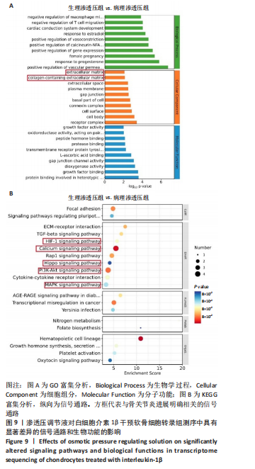
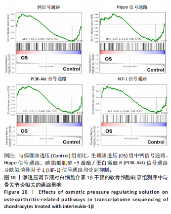
|