中国组织工程研究 ›› 2026, Vol. 30 ›› Issue (25): 6554-6565.doi: 10.12307/2026.416

• 组织构建实验造模 experimental modeling in tissue construction • 上一篇    下一篇

黄芪-桃仁缓解慢性肾脏病大鼠肾脏纤维化的作用机制

刘佳勇1,姚静静2,刘诗雨1,唐  艺1,董佳凝1,张  鑫1,侯岚炜1,康建英3,赵怡蕊3   

  1. 1山西中医药大学第三临床学院,山西省晋中市   030619;2山西中医药大学太行本草研究院,山西省晋中市   030619;3山西省中西医结合医院,山西省太原市   030013
  • 收稿日期:2025-10-09 修回日期:2025-12-31 出版日期:2026-09-08 发布日期:2026-04-22
  • 通讯作者: 赵怡蕊,主任医师,山西省中西医结合医院肾病科,山西省太原市 030013
  • 作者简介:第一作者:刘佳勇,男,1998年生,黑龙江省鸡西市人,汉族,山西中医药大学在读硕士,主要从事中西医结合防治肾病方向研究。
  • 基金资助:
    山西省中医药科技创新工程项目(14000023218T200000289),项目负责人:赵怡蕊;山西省医学重点科研项目(2022XM09),项目负责人:赵怡蕊;2024年科技创新能力培育计划“国家自然科学基金培育专项”项目 (2024PY-NS-008),项目负责人:赵怡蕊;山西省中医药管理局科研课题(2024ZYYC028),项目负责人:康建英

Mechanism by which astragalus-peach kernel alleviates renal fibrosis in chronic kidney disease rats

Liu Jiayong1, Yao Jingjing2, Liu Shiyu1, Tang Yi1, Dong Jianing1, Zhang Xin1, Hou Lanwei1, Kang Jianying3, Zhao Yirui3   

  1. 1The Third Clinical College, Shanxi University of Traditional Chinese Medicine, Jinzhong 030619, Shanxi Province, China; 2Taihang Materia Medica Research Institute, Shanxi University of Traditional Chinese Medicine, Jinzhong 030619, Shanxi Province, China; 3Shanxi Provincial Hospital of Integrated Traditional Chinese and Western Medicine, Taiyuan 030013, Shanxi Province, China
  • Received:2025-10-09 Revised:2025-12-31 Online:2026-09-08 Published:2026-04-22
  • Contact: Zhao Yirui, Chief physician, Shanxi Provincial Hospital of Integrated Traditional Chinese and Western Medicine, Taiyuan 030013, Shanxi Province, China
  • About author:Liu Jiayong, MS candidate, The Third Clinical College, Shanxi University of Traditional Chinese Medicine, Jinzhong 030619, Shanxi Province, China
  • Supported by:
    Shanxi Province Traditional Chinese Medicine Science and Technology Innovation Project, No. 14000023218T200000289 (to ZYR); Shanxi Province Key Medical Research Project, No. 2022XM09 (to ZYR); 2024 Science and Technology Innovation Capacity Building Program—“National Natural Science Foundation Cultivation Project,” No. 2024PY-NS-008 (to ZYR); Shanxi Provincial Administration of Traditional Chinese Medicine Research Project, No. 2024ZYYC028 (to KJY)

摘要:


文题释义:

c-Myc:为重要的原癌基因,与人类癌症的发生高度相关,可参与调控细胞生长、增殖、代谢、凋亡等诸多过程。近年来有研究证实,c-Myc异常表达会引发肾小管损伤、促进炎症因子释放、加速慢性肾脏病的进展。
转化生长因子β/Smad3信号通路:是肾纤维化进展的核心信号传导通路,转化生长因子β通过促进其下游因子Smad3的磷酸化调控肾脏损伤的分子表达。转化生长因子β/Smad3信号通路可促进上皮-间充质转化、加速肌成纤维细胞形成,促进α-平滑肌肌动蛋白表达与Ⅰ型胶原合成,最终导致细胞外基质大量沉积在肾小球、肾小管间质等区域,造成肾脏结构损伤和功能丧失。

背景:课题组前期研究发现黄芪-桃仁可以缓解慢性肾脏病的进展,但作用机制有待进一步验证。
目的:探究黄芪-桃仁防治慢性肾脏病大鼠肾纤维化的作用及机制。
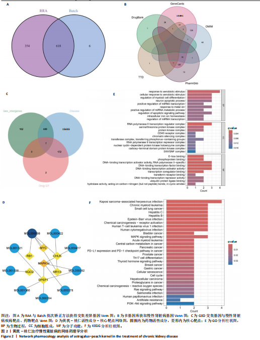
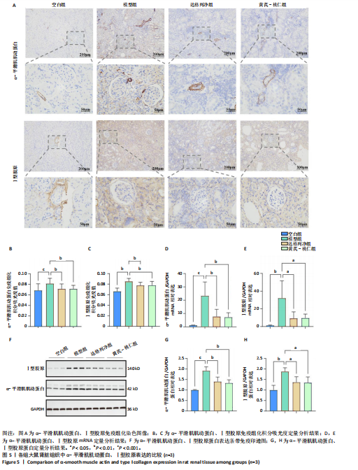
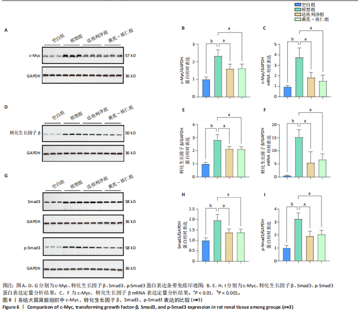
方法:①在GEO数据库(是由美国国立生物技术信息中心创建并维护的公共基因表达数据库,主要用于存储和共享高通量基因组数据,提供免费下载和分析工具,为开放数据库,研究已获得相关机构审查委员会的批准)中检索并筛选慢性肾病相关表达谱芯片数据集,结合网络药理学筛选慢性肾脏病核心靶点,将获得的关键基因进行分子对接验证。②将40只SD大鼠随机分为4组,空白组(n=10)不造模,模型组(n=10)、达格列净组(n=10)、黄芪-桃仁组(n=10)通过灌胃2%腺嘌呤溶液建立慢性肾脏病模型。造模成功后次日,空白组、模型组给予生理盐水灌胃,达格列净组给予达格列净灌胃,黄芪-桃仁组给予黄芪-桃仁(质量比为1∶1)灌胃,1次/d,连续给药8周。末次给药结束后,检测大鼠动脉血血清中肌酐、尿素氮水平,观察肾脏组织苏木精-伊红染色、Masson染色与α-平滑肌肌动蛋白、Ⅰ型胶原免疫组化染色,RT-qRCR检测肾脏组织中α-平滑肌肌动蛋白、Ⅰ型胶原、转化生长因子β、c-Myc mRNA表达,Western blot检测肾脏组织中α-平滑肌肌动蛋白、Ⅰ型胶原、转化生长因子β、c-Myc、Smad3、p-Smad3蛋白表达。 
结果与结论:①GEO数据库结合网络药理学分析共获得黄芪-桃仁9个药物活性成分和7个核心疾病靶点(c-Myc、RB1、CHUK、MAPK14、DPEP1、NR1I3、NQO2);KEGG富集分析显示Ras-MAPK-c-Myc信号通路与慢性肾脏病相关;分子对接验证提示c-Myc与核心药物成分均有较强结合能力。②与空白组相比,模型组大鼠肾小球结构数量减少、结构异常,肾小管有大量炎性细胞浸润,大量成纤维细胞增殖,肾间质中有胶原纤维异常募集,血清中肌酐、尿素氮水平以及α-平滑肌肌动蛋白、Ⅰ型胶原的mRNA和蛋白表达上调(P < 0.05);与模型组相比,黄芪-桃仁组肾组织形态更趋完整,炎性细胞浸润明显降低,肾组织成纤维细胞增殖减少,血清中肌酐、尿素氮水平以及α-平滑肌肌动蛋白、Ⅰ型胶原的mRNA和蛋白表达降低(P < 0.05)。与空白组相比,模型组c-Myc、转化生长因子β mRNA和蛋白表达以及Smad3、p-Smad3蛋白表达升高(P < 0.05);与模型组相比,c-Myc、转化生长因子β mRNA和蛋白表达以及Smad3、p-Smad3蛋白表达降低(P < 0.05)。③结果表明黄芪-桃仁可延缓SD大鼠慢性肾脏病的进展,治疗机制可能与c-Myc/转化生长因子β/Smad3信号通路有关。 

https://orcid.org/0009-0008-3173-7255 (刘佳勇) 


中国组织工程研究杂志出版内容重点:干细胞;骨髓干细胞;造血干细胞;脂肪干细胞;肿瘤干细胞;胚胎干细胞;脐带脐血干细胞;干细胞诱导;干细胞分化;组织工程

关键词: 黄芪, 桃仁, 慢性肾脏病, 肾纤维化, c-Myc, 转化生长因子β, α-平滑肌肌动蛋白, Ⅰ型胶原

Abstract: BACKGROUND: Our previous studies have indicated that astragalus–peach kernel alleviates the progression of chronic kidney disease yet its precise mechanism remains to be elucidated.
OBJECTIVE: To investigate the therapeutic effects and underlying mechanisms of astragalus-peach kernel in preventing renal fibrosis in chronic kidney disease rats.
METHODS: (1) Gene expression profile chip datasets related to chronic kidney disease were retrieved and filtered via the Gene Expression Omnibus (GEO) database (this public gene expression database, established and maintained by the National Center for Biotechnology Information, which is primarily used for storing and sharing high-throughput genomic data. The database offers free download and analysis tools, serves as an open-access resource, and has obtained approval from relevant institutional review boards for its research). Disease-related core targets were screened via the GEO database combined with network pharmacology analyses. Key genes obtained were validated through molecular docking. (2) Forty Sprague-Dawley rats were randomly assigned into blank, model, dapagliflozin, and astragalus–peach kernel groups (n=10 each). A chronic kidney disease model was established by intragastric administration of a 2% adenine solution in the latter three groups. Starting the day after the induction of chronic kidney disease, the blank and model groups received saline via gastric lavage, while the dapagliflozin group received dapagliflozin via gastric lavage, and the astragalus–peach kernel group received astragalus–peach kernel (with a mass ratio of 1:1) via gastric lavage, once daily for 8 consecutive weeks. Following the final administration, serum creatinine and blood urea nitrogen levels in rat arterial blood were measured.  Renal tissue was examined via hematoxylin-eosin staining, Masson staining, and immunohistochemical staining for α-smooth muscle actin and type I collagen. RT-qPCR was used to detect mRNA expression of α-smooth muscle actin, type I collagen, transforming growth factor-β, and c-Myc in renal tissue. Western blot analysis was performed to detect protein expression levels of α-smooth muscle actin, type I collagen, transforming growth factor-β, c-Myc, Smad3, and p-Smad3 in renal tissue. 
RESULTS AND CONCLUSION: (1) Through GEO-based network pharmacology analysis, nine active components of astragalus–peach kernel and seven core disease targets (c-Myc, RB1, CHUK, MAPK14, DPEP1, NR1I3, NQO2) were identified. Kyoto Encyclopedia of Genes and Genomes enrichment analysis indicated that the Ras–MAPK–c-Myc pathway is associated with chronic kidney disease. Molecular docking suggested strong binding affinity between c-Myc and the core active compounds. (2) Compared with the blank group, the model group exhibited reduced glomerular number, structural abnormalities, pronounced inflammatory cell infiltration in renal tubules, extensive fibroblast proliferation in the renal tissue, and abnormal accumulation of collagen fibers in the renal interstitium. Serum creatinine and blood urea nitrogen levels, as well as the mRNA and protein levels of α-smooth muscle actin and type I collagen, were significantly elevated (P < 0.05). Compared with the model group, the astragalus–peach kernel group showed more intact renal histology, reduced inflammatory infiltration, and decreased fibroblast proliferation in the renal tissue. The astragalus–peach kernel treatment suppressed the elevations in serum creatinine and blood urea nitrogen levels and both mRNA and protein expression of α-smooth muscle actin and type I collagen (P < 0.05). Compared with the blank group, the model group showed significantly elevated mRNA and protein levels of c-Myc and transforming growth factor-β, as well as increased protein levels of Smad3 and p-Smad3 (P < 0.05). Compared with the model group, the astragalus–peach kernel treatment significantly downregulated mRNA and protein levels of c-Myc and transforming growth factor-β, as well as reduced protein levels of Smad3 and p-Smad3 (P < 0.05). These findings suggest that astragalus–peach kernel may slow chronic kidney disease progression in Sprague-Dawley rats, potentially through the modulation of the c-Myc/transforming growth factor-β/Smad3 signaling pathway. 

Key words: astragalus, peach kernel, chronic kidney disease, renal fibrosis, c-Myc, transforming growth factor-β, α-smooth muscle actin, type I collagen 

中图分类号: