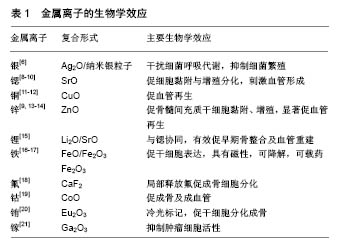
| [1] Kaur G, Pandey OP, Singh K, et al. A review of bioactive glasses: Their structure, properties, fabrication and apatite formation. J Biomed Mater Res A. 2014;102(1):254-274. [2] Wang X, Li X, Ito A, et al. Synthesis and characterization of hierarchically macroporous and mesoporous CaO-MO-SiO(2)-P(2)O(5) (M=Mg, Zn, Sr) bioactive glass scaffolds. Acta Biomater. 2011;7(10):3638-3644. [3] Qin Z, Zhang J, Hu Y, et al. Organic compounds inhibiting S. epidermidis adhesion and biofilm formation. Ultramicroscopy. 2009;109(8):881-888.[4] Percival SL, Bowler PG, Russell D. Bacterial resistance to silver in wound care. J Hosp Infect. 2005;60(1):1-7.[5] 王会,黄文旵,罗仕华,等.具有抗菌及骨修复的硼酸盐玻璃支架的性能研究[J].稀有金属材料与工程,2014,43(S1):54-58.[6] Seuss S, Heinloth M, Boccaccini AR. Development of bioactive composite coatings based on combination of PEEK, bioactive glass and Ag nanoparticles with antibacterial properties. Surf Coat Technol. 2016;301:100-105.[7] Zhu H, Hu C, Zhang F, et al. Preparation and antibacterial property of silver-containing mesoporous 58S bioactive glass. Mater Sci Eng C Mater Biol Appl. 2014;42:22-30. [8] Arepalli SK, Tripathi H, Hira SK, et al. Enhanced bioactivity, biocompatibility and mechanical behavior of strontium substituted bioactive glasses. Mater Sci Eng C Mater Biol Appl. 2016;69:108-116. [9] Zhao S, Zhang J, Zhu M, et al. Three-dimensional printed strontium-containing mesoporous bioactive glass scaffolds for repairing rat critical-sized calvarial defects. Acta Biomater. 2015;12:270-280. [10] Jia W, Lau GY, Huang W, et al. Bioactive Glass for Large Bone Repair. Adv Healthc Mater. 2015;4(18):2842-2848. [11] Wu C, Zhou Y, Xu M, et al. Copper-containing mesoporous bioactive glass scaffolds with multifunctional properties of angiogenesis capacity, osteostimulation and antibacterial activity. Biomaterials. 2013;34(2):422-433. [12] Wang H, Zhao S, Xiao W, et al. Influence of Cu doping in borosilicate bioactive glass and the properties of its derived scaffolds. Mater Sci Eng C Mater Biol Appl. 2016;58:194-203.[13] Samira J, Saoudi M, Abdelmajid K, et al. Accelerated bone ingrowth by local delivery of Zinc from bioactive glass: oxidative stress status, mechanical property, and microarchitectural characterization in an ovariectomized rat model. Libyan J Med. 2015;10(1):28572. [14] Badr-Mohammadi MR, Hesaraki S, Zamanian A. Mechanical properties and in vitro cellular behavior of zinc-containing nano-bioactive glass doped biphasic calcium phosphate bone substitutes. J Mater Sci Mater Med. 2014;25(1):185-197. [15] Khan PK, Mahato A, Kundu B, et al. Influence of single and binary doping of strontium and lithium on in vivo biological properties of bioactive glass scaffolds. Sci Rep. 2016;6: 32964.[16] Goh YF, Akram M, Alshemary AZ, et al. Synthesis, characterization and in vitro study of magnetic biphasic calcium sulfate-bioactive glass. Mater Sci Eng C Mater Biol Appl. 2015;53:29-35. [17] Wu C, Fan W, Zhu Y, et al. Multifunctional magnetic mesoporous bioactive glass scaffolds with a hierarchical pore structure. Acta Biomater. 2011;7(10):3563-3572.[18] Gentleman E, Stevens MM, Hill RG, et al. Surface properties and ion release from fluoride-containing bioactive glasses promote osteoblast differentiation and mineralization in vitro. Acta Biomater. 2013;9(3):5771-5779. [19] Hoppe A, Brandl A, Bleiziffer O, et al. In vitro cell response to Co-containing 1,393 bioactive glass. Mater Sci Eng C Mater Biol Appl. 2015;57:157-163.[20] Wu C, Xia L, Han P, et al. Europium-Containing Mesoporous Bioactive Glass Scaffolds for Stimulating in Vitro and in Vivo Osteogenesis. ACS Appl Mater Interfaces. 2016;8(18): 11342-11354.[21] Keenan TJ, Placek LM, Coughlan A, et al. Structural characterization and anti-cancerous potential of gallium bioactive glass/hydrogel composites. Carbohydr Polym. 2016; 153:482-491. [22] Yang F, Yang D, Tu J, et al. Strontium enhances osteogenic differentiation of mesenchymal stem cells and in vivo bone formation by activating Wnt/catenin signaling. Stem Cells. 2011;29(6):981-991. [23] Marie PJ, Felsenberg D, Brandi ML. How strontium ranelate, via opposite effects on bone resorption and formation, prevents osteoporosis. Osteoporos Int. 2011;22(6): 1659-1667. [24] Basini G, Grasselli F, Bussolati S, et al. Hypoxia stimulates the production of the angiogenesis inhibitor 2-methoxyestradiol by swine granulosa cells. Steroids. 2011;76(13):1433-1436. [25] Ahluwalia A, Tarnawski AS. Critical role of hypoxia sensor--HIF-1α in VEGF gene activation. Implications for angiogenesis and tissue injury healing. Curr Med Chem. 2012;19(1):90-97.[26] Shepherd DV, Kauppinen K, Brooks RA, et al. An in vitro study into the effect of zinc substituted hydroxyapatite on osteoclast number and activity. J Biomed Mater Res A. 2014;102(11):4136-4141. [27] Ferensztajn-Rochowiak E, Rybakowski JK. The effect of lithium on hematopoietic, mesenchymal and neural stem cells. Pharmacol Rep. 2016;68(2):224-230. [28] Andronescu E, Ficai M, Voicu G, et al. Synthesis and characterization of collagen/hydroxyapatite: magnetite composite material for bone cancer treatment. J Mater Sci Mater Med. 2010;21(7):2237-2242. [29] Yang J K, Yu JH, Kim J, et al. Preparation of superparamagnetic nanocomposite particles for hyperthermia therapy application. Mater Sci Eng A. 2007;449:477-479.[30] Pei J, Li B, Gao Y, et al. Fluoride decreased osteoclastic bone resorption through the inhibition of NFATc1 gene expression. Environ Toxicol. 2014;29(5):588-595. [31] Burke FM, Ray NJ, McConnell RJ. Fluoride-containing restorative materials. Int Dent J. 2006;56(1):33-43.[32] Yang X, Ricciardi BF, Hernandez-Soria A, et al. Callus mineralization and maturation are delayed during fracture healing in interleukin-6 knockout mice. Bone. 2007;41(6): 928-936. [33] Li NH, Ouchi Y, Okamoto Y, et al. Effect of parathyroid hormone on release of interleukin 1 and interleukin 6 from cultured mouse osteoblastic cells. Biochem Biophys Res Commun. 1991;179(1):236-242.[34] Miao G, Chen X, Mao C, et al. Synthesis and characterization of europium-containing luminescent bioactive glasses and evaluation of in vitro bioactivity and cytotoxicity. J Solgel Sci Technol. 2014;69(2):250-259.[35] Mawani Y, Orvig C. Improved separation of the curcuminoids, syntheses of their rare earth complexes, and studies of potential antiosteoporotic activity. J Inorg Biochem. 2014;132: 52-58. [36] Shi M, Chen Z, Farnaghi S, et al. Copper-doped mesoporous silica nanospheres, a promising immunomodulatory agent for inducing osteogenesis. Acta Biomater. 2016;30:334-344. [37] Shi M, Xia L, Chen Z, et al. Europium-doped mesoporous silica nanosphere as an immune-modulating osteogenesis/ angiogenesis agent. Biomaterials. 2017;144:176-187. [38] Minandri F, Bonchi C, Frangipani E, et al. Promises and failures of gallium as an antibacterial agent. Future Microbiol. 2014;9(3):379-397. [39] Keenan TJ, Placek LM, Mcginnity TL, et al. Relating ion release and pH to in vitro cell viability for gallium-inclusivebioactive glasses. J Mater Sci. 2016;51(2): 1107-1120.[40] Hardes J, Ahrens H, Gebert C, et al. Lack of toxicological side-effects in silver-coated megaprostheses in humans. Biomaterials. 2007;28(18):2869-2875.[41] Hung YH, Bush AI, Cherny RA. Copper in the brain and Alzheimer's disease. J Biol Inorg Chem. 2010;15(1):61-76. [42] Zamani A, Omrani GR, Nasab MM. Lithium's effect on bone mineral density. Bone. 2009;44(2):331-334. [43] Balena R, Kleerekoper M, Foldes JA, et al. Effects of different regimens of sodium fluoride treatment for osteoporosis on the structure, remodeling and mineralization of bone. Osteoporos Int. 1998;8(5):428-435. |
.jpg)

.jpg)
.jpg)