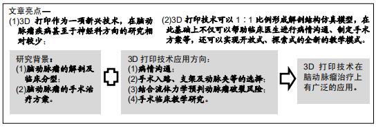
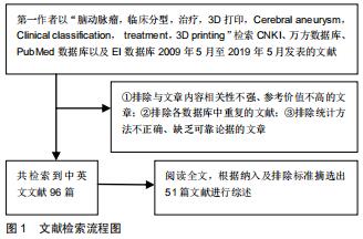
|
[1] 呼铁民,杨立军,孟杰,等.显微手术夹闭及血管内介入栓塞术治疗高分级大脑中动脉瘤破裂的疗效及安全性研究[J].中国全科医学,2015,18(30): 3671-3674.
[2] 何春柳,张乃崇,赖廷海,等.血管栓塞介入治疗脑动脉瘤的方法分析与疗效探究[J].中国医药科学,2019,9(3):206-209.
[3] 陈加源,曹杰,许卫国,等.双能量 CT 血管造影和二维、三维数字减影血管造影在诊断脑动脉瘤中的比较[J].广东医学,2014, 35(11):1699-1702.
[4] 金国良,王建莉,袁紫刚,等.3D打印颅内动脉瘤模型及其临床应用[J].中华神经医学杂志,2017,16(1):75-77.
[5] CIBIS M, POTTERS WV, GIJSEN FJH, et al. Wall shear stress calculations based on 3D cine phase contrast MRI and computational fluid dynamics: a comparison study in healthy carotid arteries. NMR Biomed. 2014;27(7):826-834.
[6] KAKISIS JD, ANTONOPOULOS CN, MANTAS G, et al. Cranial nerve injury after carotid endarterectomy: incidence, risk factors, and time trends. Eur J Vasc Endovasc Surg. 2017;53(3):320-335.
[7] 阎春森. CT、MR诊断颈动脉狭窄和粥样硬化斑块的应用价值对比[J].影像技术,2019,31(1):41-43.
[8] YANG Y, ZHOU Z, LIU R, et al. Application of 3D visualization and 3D printing technology on ERCP for patients with hilar cholangiocarcinoma. Exp Ther Med. 2018;15(4):3259-3264.
[9] Groth C, Kravitz ND, Jones PE, et al. Three-dimensional printing technology. J Clin Oahod. 2014;48(8):475,485.
[10] 王宁.血管内介入栓塞术与开颅术治疗脑动脉瘤对比观察[J].陕西医学杂志,2018,47(3):56-58.
[11] LIU JKC, KSHETTRY VR, RECINOS PF, et al. Establishing a surgicalskills laboratorv and dissection curriculum for neurosurgical residency training. J Neurosurg.2015;123(5):1331-1338.
[12] 黄星,刘祯,王旋,等.3D打印技术在神经外科手术中的应用[J].中华神经医学杂志,2018,17(10):1014-1018.
[13] MCCARRON MO, NICOLL JA. Cerebral amyloid angiopathy and thrombolysis-related intracerebral haemorrhage. Lancet Neurol. 2004; 3(8):484-492.
[14] 张宪哲,李永豪,等.血管内介入栓塞术与开颅夹闭术治疗脑动脉瘤临床对比研究[J].河北医药,2016,38(24):3755-3757.
[15] DELLA PUPPA A, VOLPIN F, GIOFFRE GA, et al. Microsurgical clipping of intracranial aneurysms assisted by green indocyanine videoangiography (ICGV) and ultrasonic perivascular microflow probe measurement. Clin Neurol Neurosurg.2014;116(1):35-40.
[16] MOHR JP, MICHAEL KP, CHRISTIAN SM, et al. Management with or without interventional therapy for unruptured brain arteriovenous malformations (Aruba): a multicentre, non-blinded, randomised trial. Lancet. 2014;383(9917):614-621.
[17] LI J, LAN ZG, LIU Y, et al. Large and giant ventral paraclinoid carotid aneurysms: Surgical techniques, complications and outcomes. Clin Neurol Neurosurg. 2012;114(7):907-913.
[18] EL BELTAGY M, MUROI C, ROTH P, et al. Recurrent intracranial aneurysms after successful neck clipping. World Neurosurg.2010; 74(4/5):472-477.
[19] 张茂,陈建龙.早期介入栓塞治疗脑动脉瘤破裂的疗效及安全性评价[J].临床和实验医学杂志,2016,15(21):2135-2137.
[20] 敬谢攀,王森岗,徐彬,等.早期显微手术夹闭瘤颈治疗脑动脉瘤破裂出血的临床效果分析[J].转化医学电子杂志,2018,5(12):38-39.
[21] 巴特尔,徐亮,李杨.血管内介入栓塞术治疗脑动脉瘤的远期随访观察[J]. 中国实用神经疾病杂志,2016,19(13):86-87.
[22] 张睿,陈灿中.开颅夹闭与血管介入栓塞术治疗老年脑动脉瘤患者的临床效果观察[J].检验医学与临床,2017,14(Z1):299-300.
[23] 曹彦鹏.血管栓塞术及显微手术夹闭术治疗脑动脉瘤的疗效和安全性[J].中国医药科学,2016,6(22):160-162.
[24] 尤雪梅,孟军鹏,侯有荣,等.不同血管介入治疗方案对颅内动脉瘤的临床效果比较[J].医学临床研究,2017,34(12):2318-2320.
[25] 王萌萌,赵英红,孙存杰,等.3D打印技术在颅内动脉瘤介入治疗中的辅助应用[J].实用放射学杂志,2018,34(5):798-800.
[26] WURM G, LEHNER M, TOMANCOK B, et al. Cerebrovascular Biomodeling for Aneurysm Surgery: Simulation-Based Training by Means of Rapid Prototyping Technologies.Surg Innov. 2011;18(3): 294-306.
[27] 周路球,娄明武,陈国昌,等.640层3D-CTA联合3D打印脑动脉瘤成像的临床价值[J].南方医科大学学报,2017,37(9):1222-1227.
[28] 兰青,爱林,张檀,等.通过3D打印技术制备颅脑实体模型[J].中华医学杂志,2016,96(30):2434-2437.
[29] NAMBA K, HIGAKI A, KANEKO N, et al. Mierocatheter shaping for intracranial aneurysm coiling using the 3-dimensional printing rapid prototyping technology: preliminary result in the first 10 consecutive cases. World Neurosurg. 2015;84(1):178-186.
[30] ZHENG Y, LIU Y, LENG B, et al. Periprocedural complications associated with endovascular treatment of intracranial aneurysms in 1764 cases. Neurointerv Surg. 2016;8:152-157.
[31] BURKHARDT JK, NEIDERT MC, MOHME M, et al. Initial clinical status and spot sign are associated with intraoperative aneurysm rupture in patients undergoing surgical clipping for aneurysmal subarachnoid hemorrhage. Neurol Surg A Cent Eur Neurosurg. 2016;77:130-138.
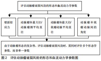
[32] 戴璇,乔爱科.计算流体力学在脑动脉瘤诊治中的应用[J].医用生物力学,2016,31(5):461-466.
[33] CHIVUKULA VK, LEVITT MR, CLARK A, et al. Reconstructing patient-specific cerebral aneurysm vasculature for in vitro investigations and treatment efficacy assessments. J Clin Neurosci. 2018;10(103):1-7.
[34] 谭衍,边远,陆弘盈,等.3D打印技术在颅内动脉瘤介入栓塞治疗中的应用[J].中国医学装备,2017,14(12):64-67.
[35] VALENCIA A, BURDILE PS, IGNAT M, et al. Fluid struc tural analysis of human cerebral aneurysm using their own wall mechanical properties. Comput Math Methods Med. 2013; 2013:293128.
[36] BAHAROGLU MI, SCHIRMER CM, HOIT DA, et al. An eurysm in flow-angle as a discriminant for rupture in side wall cerebral aneurysms. Morphometric and computational fluid dynamic analysis. Stroke.2010;41(7):1423-1430.
[37] XIANG J, NATARAJAN SK, TREMME M, et al. Hemody namic-morphologic discriminants for intracranial aneurysm rupture. Stroke.2011;42(1):144-152.
[38] JING L, FAN J, WANG Y, et al. Morphologic and hemo dynamic analysis in the patients with multiple intracranial aneurysms: ruptured versus unruptured. PLoS One. 2015; 10(7): e0132494.
[39] WIEBERS DO, WHISNANT JP, HUSTON J, et al. Unruptured intracranial aneurysms: natural history, clinical outcome, and risks of surgical and endovascular treatment. Lancet. 2003; 362(9378):103-110.
[40] CHUNG B, CEBRAL JR. CFD for evaluation and treatment planning of aneurysms: review of proposed clinical uses and their challenges. Ann Biomed Eng. 2015;43(1):122-138.
[41] 李欣,徐智,修典荣,等.多元化教学方法在八年制医学生外科总论教学中的应用研究[J].中国高等医学教育,2015,29(4):87-88.
[42] 苏星,黄庆锋,孙树清,等.3D打印技术在脑血管病临床教学中的应用[J].交通医学,2016,30(2):549-550.
[43] RYAN JR, ALMEFTY KK, NAKAJI P, et al. Cerebral aneurysm clipping surgery simulation using patient-specific 3d printing and silicone casting. World Neurosurg. 2016;88:175-181.
[44] ABLA AA, LAWTON MT. Three-dimensional hollow intracranial aneurysm models and their potential role for teaching, simulation, and training. World Neurosurgery. 2015; 83(1):35-36.
[45] WANG JL, YUAN ZG, QIAN GL, et al. 3D printing of intracranial aneurysm based on intracranial digital subtraction angiography and its clinical application. Medicine. 2018; 97(24):e11103.
[46] 李涤尘,刘佳煜,王延杰,等.4D打印—智能材料的增材制造技术[J].机电工程技术,2014,43(5):1-9.
[47] 王亚男,王芳辉.4D打印的研究进展及应用展望[J].航空材料学报,2018, 38(2):70-76.
[48] 宋波,卓林蓉,温银堂,等.4D打印技术的现状与未来[J].电加工与模具, 2018,53(6):1-7,30.
[49] 王群.4D打印及其军事应用前景[J].国防科技,2016,37(4):36-39.
[50] WEI H, ZHANG Q, YAO Y, et al. Direct-write fabrication of 4D active shape-changing structures based on a shape memory polymer and its nanocomposite. ACS Appl Mater Interfaces. 2017;9(1):876-883.
[51] 刘宇清,吕翱,黄绳跃,等.基于颅脑CTA 3D打印技术在颅内动脉瘤手术治疗中的应用[J].中国微侵袭神经外科杂志,2016,21(3):245-248.
|