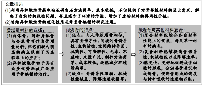
[1] DE TULLIO I, CAPUTI S, PERFETTI G, et al. A Human Clinical and Histomorphometrical Study on Different Resorbable and Non-Resorbable Bone Substitutes Used in Post-Extractive Sites. Preliminary Results.Materials(Basel).2019;12(15).pii: E2408. doi: 10.3390/ma12152408.
[2] TRAINI T, PIATTELLI A, CAPUTI S, et al. Regeneration of human bone using different bone substitute biomaterials.Clin Implant Dent Relat Res.2015;17(1):150-162.
[3] XI Y, MIAO X, LI Y, et al. BMP2-mimicking peptide modified with E7 coupling to calcined bovine bone enhanced bone regeneration associating with activation of the Runx2/SP7 signaling axis. 2020; 108(1):80-93.doi:10.1002/jbm.b.34368.
[4] SANTOS KOTAKE BG, GONZAGA MG, COUTINHO-NETTO J, et al. Bone repair of critical-sized defects in Wistar rats treated with autogenic, allogenic or xenogenic bone grafts alone or in combination with natural latex fraction F1.Biomed Mater.2018;13(2):025022.
[5] STACCHI C, LOMBARDI T, OREGLIA F, et al. Histologic and Histomorphometric Comparison between Sintered Nanohydroxyapatite and Anorganic Bovine Xenograft in Maxillary Sinus Grafting: A Split-Mouth Randomized Controlled Clinical Trial.Biomed Res Int.2017;2017:9489825.
[6] RAMIREZ FERNANDEZ MP, GEHRKE SA, PEREZ ALBACETE MARTINEZ C, et al.SEM-EDX Study of the Degradation Process of Two Xenograft Materials Used in Sinus Lift Procedures.Materials (Basel).2017;10(5).pii: E542. doi: 10.3390/ma10050542.
[7] WANG W, YEUNG KWK. Bone grafts and biomaterials substitutes for bone defect repair: A review.Bioact Mater.2017;2(4):224-247.
[8] BORG TD, MEALEY BL. Histologic healing following tooth extraction with ridge preservation using mineralized versus combined mineralized-demineralized freeze-dried bone allograft: a randomized controlled clinical trial. J Periodontol.2015;86(3):348-355.
[9] MCALLISTER BS, HAGHIGHAT K. Bone augmentation techniques.J Periodontol.2007;78(3):377-396.
[10] YAMAMURA H, DA SILVA VHP, RUIZ PLM, et al. Physico-chemical characterization and biocompatibility of hydroxyapatite derived from fish waste.J Mech Behav Biomed Mater.2018;80:137-142.
[11] PRAKASAM M, LOCS J, SALMA-ANCANE K, et al. Fabrication, Properties and Applications of Dense Hydroxyapatite: A Review.J Funct Biomater.2015;6(4):1099-1140.
[12] SZCZEŚ A, YAN Y, CHIBOWSKI E, et al. Properties of natural and synthetic hydroxyapatite and their surface free energy determined by the thin-layer wicking method.ApplSurf Sci.2018;434:1232-1238.
[13] LOWE B, VENKATESAN J, ANIL S, et al. Preparation and characterization of chitosan-natural nano hydroxyapatite-fucoidan nanocomposites for bone tissue engineering.Int J Biol Macromol. 2016;93(Pt B):1479-1487.
[14] ESMAEILKHANIAN A, SHARIFIANJAZI F, ABOUCHENARI A, et al. Synthesis and Characterization of Natural Nano-hydroxyapatite Derived from Turkey Femur-Bone Waste.Appl Biochem Biotechnol. 2019;189(3):919-932.
[15] RAMÍREZ FERNÁNDEZ MP, MAZÓN P, GEHRKE SA, et al. Comparison of Two Xenograft Materials Used in Sinus Lift Procedures: Material Characterization and In Vivo Behavior.Materials (Basel). 2017; 10(6).pii: E623. doi:10.3390/ma10060623.
[16] AARTHY S, THENMUHIL D, DHARUNYA G, et al.Exploring the effect of sintering temperature on naturally derived hydroxyapatite for bio-medical applications. J Mater Sci Mater Med.2019;30(2):21.
[17] HEIDARI F, BAHROLOLOOM ME, VASHAEE D, et al. In situ preparation of iron oxide nanoparticles in natural hydroxyapatite/chitosan matrix for bone tissue engineering application.Ceram Int.2015;41(2):3094-3100.
[18] APALANGYA V, RANGARI V, JEELANI S, et al. Rapid microwave synthesis of needle-liked hydroxyapatite nanoparticles via template directing ball-milled spindle-shaped eggshell particles.Ceram Int. 2018;44(6):7165-7171.
[19] SUNIL BR,JAGANNATHAM M.Producing hydroxyapatite from fish bones by heat treatment.Mater Lett.2016;185:411-414.
[20] PAUL S, PAL A, CHOUDHURY AR, et al. Effect of trace elements on the sintering effect of fish scale derived hydroxyapatite and its bioactivity. Ceram Int.2017;43(17):15678-15684.
[21] FERNANDES HR, GADDAM A, REBELO A, et al. Bioactive Glasses and Glass-Ceramics for Healthcare Applications in Bone Regeneration and Tissue Engineering.Materials (Basel).2018;11(12). pii: E2530.doi: 10.3390/ma11122530.
[22] BIGHETTI ACC, CESTARI TM, SANTOS PS, et al. In vitro and in vivo assessment of CaP materials for bone regenerative therapy. The role of multinucleated giant cells/osteoclasts in bone regeneration.J Biomed Mater Res B Appl Biomater.2020;108(1):282-297.
[23] TSAI WC, LIAO CJ, WU CT, et al. Clinical result of sintered bovine hydroxyapatite bone substitute: analysis of the interface reaction between tissue and bone substitute.J Orthop Sci.2010;15(2):223-232.
[24] FIENITZ T, MOSES O, KLEMM C, et al. Histological and radiological evaluation of sintered and non-sintered deproteinized bovine bone substitute materials in sinus augmentation procedures. A prospective, randomized-controlled, clinical multicenter study.Clin Oral Investig. 2017;21(3):787-794.
[25] LONDONO-RESTREPO SM, JERONIMO-CRUZ R, RUBIO-ROSAS E, et al. The effect of cyclic heat treatment on the physicochemical properties of bio hydroxyapatite from bovine bone.J Mater Sci Mater Med. 2018;29(5):52.
[26] JOSCHEK S, NIES B, KROTZ R, et al. Chemical and Physicochemical Characterization of Porous Hydroxapatite Ceramics Made of Natural Bone.Biomaterials.2000;21:1645-1658.
[27] ZHANG J, BARBIERI D, TEN HOOPEN H, et al.Microporous calcium phosphate ceramics driving osteogenesis through surface architecture.J Biomed Mater Res A.2015;103(3):1188-1199.
[28] JIN QM, TAKITA H, KOHGO T, et al. Effects of Geometry of Hydroxyapatite as Cell Substratum in BMP-Induced Ectopic Bone Formation.J Biomed Mater Res.2016;51(2000):491-499.
[29] DEWIDAR MM, LIM JK. Properties of solid core and porous surface Ti–6Al–4V implants manufactured by powder metallurgy.J Alloys Compd.2008;454(1-2):442-446.
[30] RINCÓN-LÓPEZ JA, HERMANN-MUÑOZ JA, GIRALDO-BETANCUR AL, et al. Synthesis Characterization and In Vitro Study of Synthetic and Bovine Derived Hydroxyapatite Ceramics A Comparison.Materials (Basel).2018;11(3).pii: E333.
[31] ZHANG YJ, LU JJ. A Mild and Efficient Biomimetic Synthesis of Rodlike Hydroxyapatite Particles with a High Aspect Ratio Using Polyvinylpyrrolidone As Capping Agent.Cryst Growth Des.2008;8(7): 2101-2107.
[32] XU AT, QI WT, LIN MN, et al. The optimization of sintering treatment on bovine-derived bone grafts for bone regeneration: in vitro and in vivo evaluation.J Biomed Mater Res B Appl Biomater. 2020;108(1):272-281.
[33] MATÉ SÁNCHEZ DE VAL J, MAZÓN P, PIATTELLI A, et al. Comparison among the physical properties of calcium phosphate-based bone substitutes of natural or synthetic origin.Int J Appl Ceram Technol. 2018;15(4):930-937.
[34] RAMIREZ FERNANDEZ MP, GEHRKE SA, MAZON P, et al. Implant Stability of Biological Hydroxyapatites Used in Dentistry. Materials(Basel).2017;10(6).pii: E644. doi: 10.3390/ma10060644..
[35] MATE SANCHEZ DE VAL JE, CALVO-GUIRADO JL, GOMEZ-MORENO G, et al. Influence of hydroxyapatite granule size, porosity, and crystallinity on tissue reaction in vivo. Part B: a comparative study with biphasic synthetic biomaterials.Clin Oral Implants Res.2018;29(11):1077-1084.
[36] LONDOÑO-RESTREPO SM, RAMIREZ-GUTIERREZ CF, DEL REAL A, et al. Study of bovine hydroxyapatite obtained by calcination at low heating rates and cooled in furnace air.J Mater Sci. 2016;51(9): 4431-4441.
[37] RESMIM CM, DALPASQUALE M, VIELMO NIC, et al. Study of physico-chemical properties and in vitro antimicrobial activity of hydroxyapatites obtained from bone calcination.Prog Biomater. 2019;8(1):1-9.
[38] FEITOSA GT, SANTOS MVB, BARRETO HM, et al. Hydroxyapatites Obtained from Different Routes and their Antimicrobial Properties. Mater Sci Forum.2016;869:890-895.
[39] LI B, WEBSTER TJ. Bacteria antibiotic resistance: New challenges and opportunities for implant-associated orthopedic infections.J Orthop Res.2018;36(1):22-32.
[40] INZANA JA, SCHWARZ EM, KATES SL, et al. Biomaterials approaches to treating implant-associated osteomyelitis.Biomaterials. 2016;81:58-71.
[41] ROSETI L, PARISI V, PETRETTA M, et al. Scaffolds for Bone Tissue Engineering: State of the art and new perspectives.Mater Sci Eng C Mater Biol Appl.2017;78:1246-1262.
[42] LEE JW, CHU SG, KIM HT, et al. Osteogenesis of Adipose-Derived and Bone Marrow Stem Cells with Polycaprolactone/Tricalcium Phosphate and Three-Dimensional Printing Technology in a Dog Model of Maxillary Bone Defects.Polymers(Basel).2017;9(9).pii:E450.
[43] YIN HM, QIAN J, ZHANG J, et al. Engineering Porous Poly(lactic acid) Scaffolds with High Mechanical Performance via a Solid State Extrusion/Porogen Leaching Approach.Polymers (Basel).2016;8(6).pii: E213. doi:10.3390/8060213.
[44] YU X, TANG X, GOHIL SV, et al. Biomaterials for Bone Regenerative Engineering.Adv Healthc Mater.2015;4(9):1268-1285.
[45] KOHRI M, MIKI K, WAITE DE, et al. In vitro stability of biphasic calcium phosphate ceramics. Biomaterials.1990;14(4):299-304.
[46] FRAYSSINET P, TROUILLET JL, ROUQUET N, et al. Osseointegration of macroporous calcium phosphate ceramics having a different chemical composition.Biomaterials.1993;14(6):423-429.
[47] PICCIRILLO C, SILVA MF, PULLAR RC, et al. Extraction and characterisation of apatite- and tricalcium phosphate-based materials from cod fish bones.Mater Sci Eng C Mater Biol Appl. 2013;33(1): 103-110.
[48] ZHU Q, ABLIKIM Z, CHEN T, et al. The preparation and characterization of HA/β-TCP biphasic ceramics from fish bones. Ceramic Int. 2017;43(15):12213-12220.
[49] PAL A, PAUL S, CHOUDHURY AR, et al.Synthesis of hydroxyapatite from Lates calcarifer fish bone for biomedical applications.Mater Lett. 2017;203:89-92.
[50] BAKAN F, SEZEN M, GECGIN M, et al. Structural and Chemical Analysis of Hydroxyapatite (HA)-Boron Nitride (BN) Nanocomposites Sintered Under Different Atmospheric Conditions.Microsc Microanal. 2017;23(5):891-899.
[51] UNAL S, EKREN N, SENGIL AZ, et al. Synthesis, characterization, and biological properties of composites of hydroxyapatite and hexagonal boron nitride.J Biomed Mater Res B Appl Biomater. 2018; 106(6):2384-2392.
[52] COZZA N, MONTE F, BONANI W, et al. Bioactivity and mineralization of natural hydroxyapatite from cuttlefish bone and Bioglass((R)) co-sintered bioceramics.J Tissue Eng Regen Med.2018;12(2): e1131-e1142.
[53] MEKA SRK, JAIN S, CHATTERJEE K. Strontium eluting nanofibers augment stem cell osteogenesis for bone tissue regeneration.Colloids Surf B Biointerfaces.2016;146:649-656.
[54] KIM SS, SUN PARK M, JEON O, et al. Poly(lactide-co-glycolide)/ hydroxyapatite composite scaffolds for bone tissue engineering. Biomaterials.2006;27(8):1399-1409.
[55] CHENG D, LIANG Q, LI Y, et al. Strontium incorporation improves the bone-forming ability of scaffolds derived from porcine bone.Colloids Surf B Biointerfaces.2018;162:279-287.
[56] PETTINICCHIO M, TRAINI T, MURMURA G, et al. Histologic and histomorphometric results of three bone graft substitutes after sinus augmentation in humans.Clin Oral Investig.2012;16(1):45-53.
[57] RUIXIN L, CHENG X, YINGJIE L, et al. Degradation behavior and compatibility of micro, nanoHA/chitosan scaffolds with interconnected spherical macropores.Int J Biol Macromol.2017;103: 385-394.
[58] YUAN H, NING CHEN N, LÜ XY, et al. Experimental study of natural hydroxyapatite_chitosancomposite on reconstructin.J Nanjing Med Univ.2008;22(6):372-375.
[59] SHEIKH Z, JAVAID MA, HAMDAN N, et al. Bone Regeneration Using Bone Morphogenetic Proteins and Various Biomaterial Carriers. Materials (Basel).2015;8(4):1778-1816.
[60] ZHANG H, WANG F, DING L, et al. A meta analysis of lumbar spinal fusion surgery using bone morphogenetic proteins and autologous iliac crest bone graft.PLoS One.2014;9(6):e97049.
[61] WILLIAMS BJ, SMITH JS, FU KM, et al. Does bone morphogenetic protein increase the incidence of perioperative complications in spinal fusion? A comparison of 55,862 cases of spinal fusion with and without bone morphogenetic protein.Spine(Phila Pa 1976). 2011;36(20): 1685-1691.
[62] XIAO W, FU H, RAHAMAN MN, et al. Hollow hydroxyapatite microspheres: a novel bioactive and osteoconductive carrier for controlled release of bone morphogenetic protein-2 in bone regeneration. Acta Biomater.2013;9(9):8374-8383.
[63] KIEKER CA, GERHART TN, SHELLING SH, et al. Long-Term Healing of Bone Using Recombinant Human Bone Morphogenetic Protein 2.Clin Orthop Relat Res.1995;(318):222-230.
[64] FALCIGNO L, D'AURIA G, CALVANESE L, et al. Osteogenic properties of a short BMP-2 chimera peptide.J Pept Sci.2015;21(9):700-709.
[65] WU M, CHEN G, LI YP. TGF-β and BMP signaling in osteoblast, skeletal development, and bone formation, homeostasis and disease. one Res.2016;4:16009.
[66] DIAS FJ, ISSA JP, COUTINHO-NETTO J, et al. Morphometric and high resolution scanning electron microscopy analysis of low-level laser therapy and latex protein (Hevea brasiliensis) administration following a crush injury of the sciatic nerve in rats.J Neurol Sci. 2015;349(1-2):129-137.
[67] MACHADO EG, ISSA JP, FIGUEIREDO FA, et al. A new heterologous fibrin sealant as scaffold to recombinant human bone morphogenetic protein-2 (rhBMP-2) and natural latex proteins for the repair of tibial bone defects.Acta Histochem.2015;117(3):288-296.
[68] PATEL PP, BUCKLEY C, TAYLOR BL, et al. Mechanical and biological evaluation of a hydroxyapatite-reinforced scaffold for bone regeneration. J Biomed Mater Res A.2019;107(4):732-741.
[69] SIMS A, MARTIN J. Coupling the activities of bone formation and resorption: a multitude of signals within the basic multicellular unit. Bonekey Rep.2014;3:1-10.
|