中国组织工程研究 ›› 2017, Vol. 21 ›› Issue (26): 4113-4118.doi: 10.3969/j.issn.2095-4344.2017.26.003

• 组织工程骨及软骨材料 tissue-engineered bone and cartilage materials • 上一篇    下一篇

人脱细胞羊膜与骨髓间充质干细胞复合体修复关节软骨缺损

姜良斌1,2,韦标方2,冯  志2,岳永彬1,2 
  

  1. 1广州中医药大学,广东省广州市  510006;2临沂市人民医院,山东省临沂市  276000
  • 收稿日期:2017-04-09 出版日期:2017-09-18 发布日期:2017-09-28
  • 通讯作者: 韦标方,主任医师,教授,临沂市人民医院,山东省临沂市 276000
  • 作者简介:姜良斌,男,1984年生,山东省临沂市人,汉族,广州中医药大学在读博士,主要从事中西医结合治疗股骨头坏死研究。
  • 基金资助:

    山东省科技发展计划(2014GSF119022)

Repair of articular cartilage defects with human acellular amniotic membrane/bone marrow mesenchymal stem cell composite

Jiang Liang-bin1, 2, Wei Biao-fang2, Feng Zhi2, Yue Yong-bin1, 2 
  

  1. 1Guangzhou University of Chinese Medicine, Guangzhou 510006, Guangdong Province, China; 2Linyi People’s Hospital, Linyi 276000, Shandong Province, China
  • Received:2017-04-09 Online:2017-09-18 Published:2017-09-28
  • Contact: Wei Biao-fang, Chief physician, Professor, Linyi People’s Hospital, Linyi 276000, Shandong Province, China
  • About author:Jiang Liang-bin, Studying for doctorate, Guangzhou University of Chinese Medicine, Guangzhou 510006, Guangdong Province, China
  • Supported by:

    the Scientific Development Plan of Shandong Province, No. 2014GSF119022

摘要:

文章快速阅读:


文题释义:
人羊膜:是一种半透明的生物膜,源于天然,主要特点如下:①含有促进细胞增殖及分化的成分,促贴壁物质(胶原蛋白和层粘连蛋白)等较多;②抗炎、抗微生物、抗纤维化、抗瘢痕形成,机械性能较合理,表达生长因子如转化生长因子,这为细胞增殖、分化提供了丰富的营养物质;③促进骨髓间充质干细胞向软骨转化。
软骨单位:是关节软骨最基本的解剖功能结构,它是软骨细胞与细胞外基质联系的重要“桥梁”,它由一个或几个软骨细胞与其细胞周围基质构成,是关节软骨的结构、功能和代谢的基础。
 
背景:骨髓间充质干细胞在软骨组织工程研究中作为一种常见的种子细胞被广泛应用于软骨缺损修复。
目的:以人脱细胞羊膜作为细胞支架负载兔骨髓间充质干细胞用以修复兔股骨髁间窝软骨缺损。  
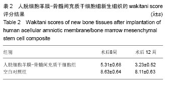
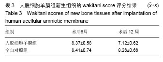
方法:将兔骨髓间充质干细胞接种于人脱细胞羊膜上,体外共培养2周。建立兔股骨髁间窝区关节软骨缺损模型,右膝软骨缺损处设为空白对照组,不植入任何材料,左膝软骨缺损处为实验侧,分别植入人脱细胞羊膜-骨髓间充质干细胞和单纯人脱细胞羊膜。术后第8,12周,取膝关节软骨缺损部位新生组织,行大体观察、组织学检测,评估新生软骨质量。
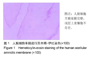
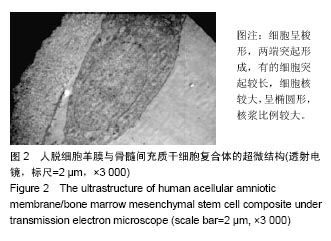
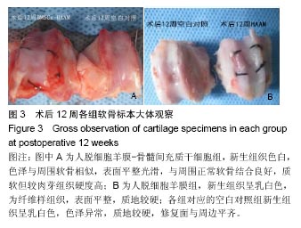
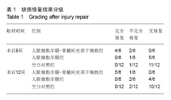
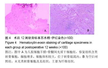
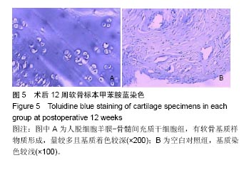
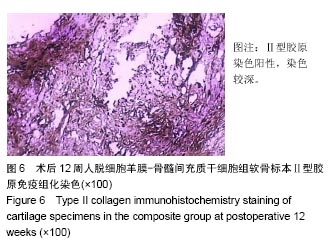
结果与结论:①大体观察结果:人脱细胞羊膜-骨髓间充质干细胞组有类软骨形成,质地较软,与周围正常软骨结合良好;人脱细胞羊膜组未形成软骨组织。空白对照组缺损区只由纤维样组织填充;②组织学观察结果:人脱细胞羊膜-骨髓间充质干细胞组新生出软骨细胞及软骨陷窝,并形成软骨基质,Ⅱ型胶原免疫组织化学染色呈阳性。软骨基质甲苯胺蓝染色较深。人脱细胞羊膜组仅有极少量的软骨细胞,甲苯胺蓝染色较浅,Ⅱ型胶原免疫组织化学染色呈阴性,无软骨基质形成。空白对照组为纤维组织修复,甲苯胺蓝染色较浅;③结果表明,人脱细胞羊膜有利于骨髓间充质干细胞的软骨化,软骨缺损处由类软骨组织填充,能够有效修复关节软骨缺损。

关键词: 生物材料, 软骨生物材料, 羊膜, 骨髓间充质干细胞, 软骨缺损,

Abstract:

BACKGROUND: Bone marrow mesenchymal stem cells (BMSCs) as common seed cells have been widely used in tissue-engineered cartilage repair.  OBJECTIVE: To use human amniotic membrane as a cell scaffold to carry rabbit BMSCs in order to repair articular cartilage defects in the femoral intercondylar fossa of rabbits.

METHODS: Rabbit BMSCs were inoculated onto the human acellular amniotic membrane (HAAM) and co-cultured for 2 weeks. Articular defect models were made in the femoral intercondylar fossa of rabbits. The defects of the right knees served as blank control. BMSCs/HAAM composite was transplanted into the defect of the left knee joint as composite group, and HAAM was implanted into the defect of the left knee joint as HAAM group. These rabbits were killed at 8 and 12 weeks after implantation and the newly cartilage samples were evaluated grossly and histologically and then graded.

RESULTS AND CONCLUSION: Gross observation showed the defects were filled with cartilaginous tissues in the composite group, and there were no cartilage tissues in the HAAM group, while only fibrous tissues were seen in the blank control group. Histologically, the defect region was full of chondrocytes in the composite group, immunohistochemistry staining indicated that collagen II was rich in the tissue, and furthermore, the cartilage matrix was stained deeply by toluidine blue. In the the HAAM group, there were few chondrocytes, toluidine blue staining was weakly positive, and immunohistochemistry staining was negative, indicating there was no cartilage matrix. In the blank control group, the defects were filled of fibroblasts and toluidine blue staining was weakly positive. To conclude, the BMSCs/HAAM is a good scaffold for BMSCs chondrogenic differentiation to effectively repair articular cartilage defects.

Key words: Amnion, Bone Marrow, Mesenchymal Stem Cells, Cartilage, Articular, Tissue Engineering

中图分类号: