| [1] Falanga V. Wound healing and its impairment in the diabetic foot. Lancet. 2005;366(9498):1736-1743.[2] King A, Balaji S, Keswani SG, et al. The role of stem cells in wound angiogenesis. Adv Wound Care. 2014;3(10):614-625.[3] Sen CK, Gordillo GM, Roy S, et al. Human skin wounds: a major and snowballing threat to public health and the economy. Wound Repair Regen. 2009;17(6):763-771.[4] Demidova-Rice TN, Durham JT, Herman IM. Wound healing angiogenesis: innovations and challenges in acute and chronic wound healing. Adv Wound Care. 2012;1(1):17-22.[5] Chong MS, Ng WK, Chan JK. Concise review: endothelial progenitor cells in regenerative medicine: applications and challenges. Stem Cells Transl Med. 2016;5(4):530-538.[6] Asai J, Takenaka H, Ii M, et al. Topical application of ex vivo expanded endothelial progenitor cells promotes vascularisation and wound healing in diabetic mice. Int Wound J. 2013;10(5):527-533.[7] Bai YY, Wang L, Chang D, et al. Synergistic Effects of Transplanted Endothelial Progenitor Cells and RWJ 67657 in Diabetic Ischemic Stroke Models. Stroke. 2015;46(7): 1938-1946.[8] Yin Y, Liu H, Wang F, et al. Transplantation of cryopreserved human umbilical cord blood-derived endothelial progenitor cells induces recovery of carotid artery injury in nude rats. Stem Cell Res Ther. 2015;6:37.[9] Urbich C, Heeschen C, Aicher A, et al. Relevance of monocytic features for neovascularization capacity of circulating endothelial progenitor cells. Circulation. 2003;108(20):2511-2516.[10] Rigato M, Bittante C, Albiero M, et al. Circulating Progenitor Cell Count Predicts Microvascular Outcomes in Type 2 Diabetic Patients. J Clin Endocrinol Metab. 2015;100(7): 2666-2672.[11] Heublein H, Bader A, Giri S. Preclinical and clinical evidence for stem cell therapies as treatment for diabetic wounds. Drug Discov Today. 2015;20(6):703-717.[12] Pawitan JA. Prospect of stem cell conditioned medium in regenerative medicine. Biomed Res Int. 2014;2014:1-14.[13] Lin RZ, Moreno-Luna R, Li D, et al. Human endothelial colony-forming cells serve as trophic mediators for mesenchymal stem cell engraftment via paracrine signaling. Proc Natl Acad Sci U S A. 2014;111(28):10137-10142.[14] Flex A, Biscetti F, Iachininoto MG, et al. Human cord blood endothelial progenitors promote post-ischemic angiogenesis in immunocompetent mouse model. Thromb Res. 2016;141: 106-111.[15] Asahara T. Isolation of putative progenitor endothelial cells for angiogenesis. Science. 1997;275(5302):964-966.[16] Hur J, Yoon C H, Kim H S, et al. Characterization of Two Types of Endothelial Progenitor Cells and Their Different Contributions to Neovasculogenesis. Arterioscler Thromb Vasc Biol. 2004;24(2):288-293.[17] Rehman J. Peripheral Blood "Endothelial Progenitor Cells" Are Derived From Monocyte/Macrophages and Secrete Angiogenic Growth Factors. Circulation. 2003;107(8): 1164-1169.[18] Urbich C, Aicher A, Heeschen C, et al. Soluble factors released by endothelial progenitor cells promote migration of endothelial cells and cardiac resident progenitor cells. J Mol Cell Cardiol. 2005;39(5):733-742.[19] Yang Z, von Ballmoos MW, Faessler D, et al. Paracrine factors secreted by endothelial progenitor cells prevent oxidative stress-induced apoptosis of mature endothelial cells. Atherosclerosis. 2010;211(1):103-109.[20] Cheng CC, Chang SJ, Chueh YN, et al. Distinct angiogenesis roles and surface markers of early and late endothelial progenitor cells revealed by functional group analyses. BMC Genomics. 2013;14:182.[21] Guillevic O, Ferratge S, Pascaud J, et al. A novel molecular and functional stemness signature assessing human cord blood-derived endothelial progenitor cell immaturity. PLoS One. 2016;11(4):e0152993.[22] 乔威,冉峰,刘长建.人外周血内皮祖细胞的分离、培养及鉴定[J].中国组织工程研究,2013,17(36):6508-6514.[23] Yoder MC, Mead LE, Prater D, et al. Redefining endothelial progenitor cells via clonal analysis and hematopoietic stem/progenitor cell principals. Blood. 2007;109(5): 1801-1809.[24] Pelosi E, Castelli G, Testa U. Endothelial progenitors. Blood Cells, Molecules, and Diseases.2014;52(4):186-194.[25] 彭艳,程培,徐勇.脐血内皮祖细胞尾静脉与局部注射治疗糖尿病下肢缺血[J].中国组织工程研究与临床康复, 2011,15(19): 3499-3502.[26] Park S, Moon S, Lee H, et al. A comparison of human cord blood- and embryonic stem cell-derived endothelial progenitor cells in the treatment of chronic wounds. Biomaterials. 2013;34(4):995-1003.[27] Schmidt-Lucke C, Rossig L, Fichtlscherer S, et al. Reduced number of circulating endothelial progenitor cells predicts future cardiovascular events: proof of concept for the clinical importance of endogenous vascular repair. Circulation. 2005;111(22):2981-2987.[28] Cantinieaux D, Quertainmont R, Blacher S, et al. Conditioned medium from bone marrow-derived mesenchymal stem cells improves recovery after spinal cord injury in rats: an original strategy to avoid cell transplantation. PLoS One. 2013;8(8): e69515.[29] Li L F, Liu YY, Yang CT, et al. Improvement of ventilator-induced lung injury by IPS cell-derived conditioned medium via inhibition of PI3K/Akt pathway and IP-10-dependent paracrine regulation. Biomaterials. 2013; 34(1):78-91.[30] Chang CP, Chio CC, Cheong CU, et al. Hypoxic preconditioning enhances the therapeutic potential of the secretome from cultured human mesenchymal stem cells in experimental traumatic brain injury. Clin Sci (Lond). 2013; 124(3):165-176.[31] Hynes B, Kumar AH, O'Sullivan J, et al. Potent endothelial progenitor cell-conditioned media-related anti-apoptotic, cardiotrophic, and pro-angiogenic effects post-myocardial infarction are mediated by insulin-like growth factor-1. Eur Heart J. 2013;34(10):782-789.[32] Bhang S H, Lee S, Shin J Y, et al. Efficacious and clinically relevant conditioned medium of human adipose-derived stem cells for therapeutic angiogenesis. Mol Ther. 2014;22(4): 862-872.[33] Kim HO, Choi S, Kim H. Mesenchymal stem cell-derived secretome and microvesicles as a cell-free therapeutics for neurodegenerative disorders. Tissue Eng Regen Med. 2013;10(3):93-101.[34] Bae YU, Choi JH, Nagy A, et al. Antisenescence effect of mouse embryonic stem cell conditioned medium through a PDGF/FGF pathway. FASEB J. 2016;30(3):1276-1286.[35] He T. Transplantation of Circulating Endothelial Progenitor Cells Restores Endothelial Function of Denuded Rabbit Carotid Arteries. Stroke. 2004;35(10):2378-2384.[36] Dumortier J, Streblow DN, Moses AV, et al. Human Cytomegalovirus Secretome Contains Factors That Induce Angiogenesis and Wound Healing. J Virol. 2008;82(13): 6524-6535.[37] Lacoste E, Martineau I, Gagnon G. Platelet concentrates: effects of calcium and thrombin on endothelial cell proliferation and growth factor release. J Periodontol. 2003; 74(10):1498-507.[38] 折涛,胡大海,张彦刚,等.胰岛素干预后脂肪干细胞旁分泌对人血管内皮细胞的作用[J].中华烧伤杂志,2011,27(1):32-36.[39] Bertrand-Duchesne MP, Grenier D, Gagnon G. Epidermal growth factor released from platelet-rich plasma promotes endothelial cell proliferation in vitro. J Periodontal Res. 2010; 45(1):87-93.[40] 王卿,王大虎,王爱学,等.银屑病患者损伤处皮肤间充质干细胞对T细胞增殖的影响[J].中国组织工程研究, 2016,20(23): 3451-3456.[41] Kwon HM, Hur SM, Park KY, et al. Multiple paracrine factors secreted by mesenchymal stem cells contribute to angiogenesis. Vascul Pharmacol. 2014;63(1):19-28.[42] Miya M, Maeshima A, Mishima K, et al. Enhancement of in vitro human tubulogenesis by endothelial cell-derived factors: implications for in vivo tubular regeneration after injury. Am J Physiol Renal Physiol. 2011;301(2):F387-F395.[43] Wang J, Wang Y, Wang S, et al. Bone marrow-derived mesenchymal stem cell-secreted IL-8 promotes the angiogenesis and growth of colorectal cancer. Oncotarget. 2015;6(40):42825-42837 |
.jpg) 文题释义:
内皮克隆形成细胞:内皮祖细胞的亚群,来源于骨髓,是内皮细胞的前体细胞,参与出生后血管生成。在体外培养时呈典型的“铺路石”样生长,具有克隆性,增殖能力较强。研究发现,内皮克隆形成细胞可参与生理性或病理性的新血管形成,在治疗缺血性疾病的研究中具有可观的前景。
条件培养基:在细胞培养过程中,细胞可能会向培养基中分泌某些活性物质,如生长因子及趋化因子等,这种培养过某种细胞的培养基,可用于培养其他细胞或作为其他细胞培养基的添加成分,即为条件培养基。研究发现,除了自身分化为成熟细胞,干细胞还可通过分泌某些细胞活性因子改善局部微环境,对损伤组织产生修复作用。因此,干细胞条件培养基在再生医学领域有着广阔的应用前景。
文题释义:
内皮克隆形成细胞:内皮祖细胞的亚群,来源于骨髓,是内皮细胞的前体细胞,参与出生后血管生成。在体外培养时呈典型的“铺路石”样生长,具有克隆性,增殖能力较强。研究发现,内皮克隆形成细胞可参与生理性或病理性的新血管形成,在治疗缺血性疾病的研究中具有可观的前景。
条件培养基:在细胞培养过程中,细胞可能会向培养基中分泌某些活性物质,如生长因子及趋化因子等,这种培养过某种细胞的培养基,可用于培养其他细胞或作为其他细胞培养基的添加成分,即为条件培养基。研究发现,除了自身分化为成熟细胞,干细胞还可通过分泌某些细胞活性因子改善局部微环境,对损伤组织产生修复作用。因此,干细胞条件培养基在再生医学领域有着广阔的应用前景。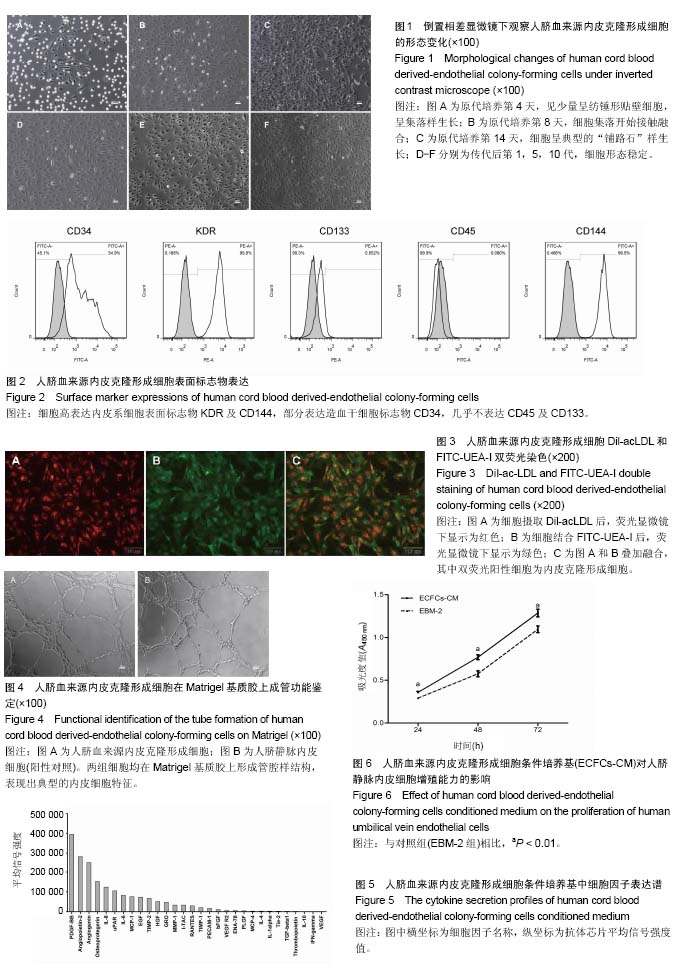
.jpg) 文题释义:
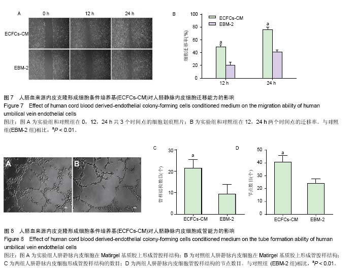
内皮克隆形成细胞:内皮祖细胞的亚群,来源于骨髓,是内皮细胞的前体细胞,参与出生后血管生成。在体外培养时呈典型的“铺路石”样生长,具有克隆性,增殖能力较强。研究发现,内皮克隆形成细胞可参与生理性或病理性的新血管形成,在治疗缺血性疾病的研究中具有可观的前景。
条件培养基:在细胞培养过程中,细胞可能会向培养基中分泌某些活性物质,如生长因子及趋化因子等,这种培养过某种细胞的培养基,可用于培养其他细胞或作为其他细胞培养基的添加成分,即为条件培养基。研究发现,除了自身分化为成熟细胞,干细胞还可通过分泌某些细胞活性因子改善局部微环境,对损伤组织产生修复作用。因此,干细胞条件培养基在再生医学领域有着广阔的应用前景。
文题释义:
内皮克隆形成细胞:内皮祖细胞的亚群,来源于骨髓,是内皮细胞的前体细胞,参与出生后血管生成。在体外培养时呈典型的“铺路石”样生长,具有克隆性,增殖能力较强。研究发现,内皮克隆形成细胞可参与生理性或病理性的新血管形成,在治疗缺血性疾病的研究中具有可观的前景。
条件培养基:在细胞培养过程中,细胞可能会向培养基中分泌某些活性物质,如生长因子及趋化因子等,这种培养过某种细胞的培养基,可用于培养其他细胞或作为其他细胞培养基的添加成分,即为条件培养基。研究发现,除了自身分化为成熟细胞,干细胞还可通过分泌某些细胞活性因子改善局部微环境,对损伤组织产生修复作用。因此,干细胞条件培养基在再生医学领域有着广阔的应用前景。