中国组织工程研究 ›› 2016, Vol. 20 ›› Issue (51): 7654-7659.doi: 10.3969/j.issn.2095-4344.2016.51.008
• 肌肉肌腱韧带组织构建 tissue construction of the muscle, tendon and ligament • 上一篇 下一篇
新型韧带横截面积测量仪器的设计与应用
祝建飞1,成永忠2,侯汪洋1,程 灏1,程 玲1,温建民3,陈 诚1,蔡静怡1
- 1中国中医科学院望京医院,北京市 100102;中国中医科学院望京医院,2创伤一科,3关节二科,北京市 100102
A new ligament cross-sectional area measuring instrument: design and application
Zhu Jian-fei1, Cheng Yong-zhong2, Hou Wang-yang1, Cheng Hao1, Cheng Ling1, Wen Jian-min3, Chen Cheng1, Cai Jing-yi1
- 1Wangjing Hospital of CACMS, Beijing 100102, China; 2First Department of Traumatic Orthopaedics, 3Second Department of Joint, Wangjing Hospital of CACMS, Beijing 100102, China
摘要:
文章快速阅读:
.jpg) 文题释义:
踝关节特点:关节囊前后较薄,两侧较厚,并有韧带加强。胫侧副韧带为一强韧的三角形韧带,又名三角韧带,位于关节的内侧。起自内踝,呈扇形向下止于距、跟、舟三骨。由于附着部不同,由后向前可分为四部:距胫后韧带、跟胫韧带、胫舟韧带和位于其内侧的距胫前韧带。三角韧带主要限制足的背屈,前部纤维则限制足的跖屈。腓侧副韧带位于关节的外侧,由从前往后排列有距腓前、跟腓、距腓后3条独立的韧带组成,连结于外踝与距、跟骨之间。距腓后韧带可防止小腿骨向前脱位。当足过度跖屈内翻时,易损伤距腓前韧带及跟腓韧带。
游标卡尺:是一种测量长度、内外径、深度的量具。游标卡尺由主尺和附在主尺上能滑动的游标两部分构成。主尺一般以毫米为单位,而游标上则有10、20或50个分格,根据分格的不同,游标卡尺可分为十分度游标卡尺、二十分度游标卡尺、五十分度格游标卡尺等,游标为10分度的有9 mm,20分度的有19 mm,50分度的有49 mm。游标卡尺的主尺和游标上有两副活动量爪,分别是内测量爪和外测量爪,内测量爪通常用来测量内径,外测量爪通常用来测量长度和外径。
文题释义:
踝关节特点:关节囊前后较薄,两侧较厚,并有韧带加强。胫侧副韧带为一强韧的三角形韧带,又名三角韧带,位于关节的内侧。起自内踝,呈扇形向下止于距、跟、舟三骨。由于附着部不同,由后向前可分为四部:距胫后韧带、跟胫韧带、胫舟韧带和位于其内侧的距胫前韧带。三角韧带主要限制足的背屈,前部纤维则限制足的跖屈。腓侧副韧带位于关节的外侧,由从前往后排列有距腓前、跟腓、距腓后3条独立的韧带组成,连结于外踝与距、跟骨之间。距腓后韧带可防止小腿骨向前脱位。当足过度跖屈内翻时,易损伤距腓前韧带及跟腓韧带。
游标卡尺:是一种测量长度、内外径、深度的量具。游标卡尺由主尺和附在主尺上能滑动的游标两部分构成。主尺一般以毫米为单位,而游标上则有10、20或50个分格,根据分格的不同,游标卡尺可分为十分度游标卡尺、二十分度游标卡尺、五十分度格游标卡尺等,游标为10分度的有9 mm,20分度的有19 mm,50分度的有49 mm。游标卡尺的主尺和游标上有两副活动量爪,分别是内测量爪和外测量爪,内测量爪通常用来测量内径,外测量爪通常用来测量长度和外径。摘要
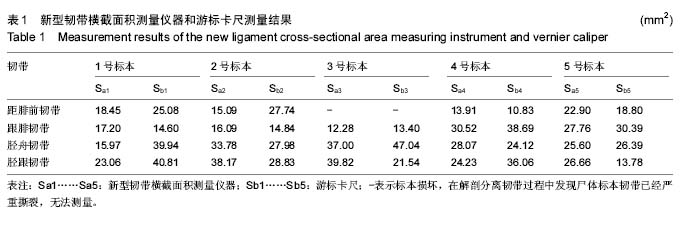
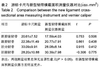
背景:目前足部有限元模型的材料特性和参数大多参考国外文献,尚未见国内开展材料参数的测试研究工作的报道。国内常用的方法用游标卡尺测量韧带、肌腱的宽度和厚度计算截面积。
目的:设计一种测量韧带横截面积的新仪器,提高测量精度。
方法:通过设计一种新型韧带横截面积测量仪器,分别应用该新型仪器与传统游标卡尺对5具新鲜尸体踝关节韧带横截面积进行测量,然后对两种测量方法进行对比分析。
结果与结论:①游标卡尺测量组距腓前韧带、跟腓韧带、胫舟韧带、胫跟韧带的横截面积平均值分别为(20.61±7.52)、(22.38±11.49)、(33.09±9.91)、(28.20±10.88) mm2,新型韧带横截面积测量仪器为(17.59±4.03)、(20.77±7.91)、(28.08±8.14)、(30.39±7.98) mm2,两种方法测量结果差异均无显著性意义,结果相当。②结果说明,该新型韧带横截面积测量仪操作简便,测量结果较准确可靠,填补了韧带横截面积测量实验无专用仪器的空白,其应用价值值得进一步研究。
中国组织工程研究杂志出版内容重点:组织构建;骨细胞;软骨细胞;细胞培养;成纤维细胞;血管内皮细胞;骨质疏松;组织工程
ORCID: 0000-0001-7333-2012(祝建飞)
中图分类号:

.jpg)
.jpg) 文题释义:
踝关节特点:关节囊前后较薄,两侧较厚,并有韧带加强。胫侧副韧带为一强韧的三角形韧带,又名三角韧带,位于关节的内侧。起自内踝,呈扇形向下止于距、跟、舟三骨。由于附着部不同,由后向前可分为四部:距胫后韧带、跟胫韧带、胫舟韧带和位于其内侧的距胫前韧带。三角韧带主要限制足的背屈,前部纤维则限制足的跖屈。腓侧副韧带位于关节的外侧,由从前往后排列有距腓前、跟腓、距腓后3条独立的韧带组成,连结于外踝与距、跟骨之间。距腓后韧带可防止小腿骨向前脱位。当足过度跖屈内翻时,易损伤距腓前韧带及跟腓韧带。
游标卡尺:是一种测量长度、内外径、深度的量具。游标卡尺由主尺和附在主尺上能滑动的游标两部分构成。主尺一般以毫米为单位,而游标上则有10、20或50个分格,根据分格的不同,游标卡尺可分为十分度游标卡尺、二十分度游标卡尺、五十分度格游标卡尺等,游标为10分度的有9 mm,20分度的有19 mm,50分度的有49 mm。游标卡尺的主尺和游标上有两副活动量爪,分别是内测量爪和外测量爪,内测量爪通常用来测量内径,外测量爪通常用来测量长度和外径。
文题释义:
踝关节特点:关节囊前后较薄,两侧较厚,并有韧带加强。胫侧副韧带为一强韧的三角形韧带,又名三角韧带,位于关节的内侧。起自内踝,呈扇形向下止于距、跟、舟三骨。由于附着部不同,由后向前可分为四部:距胫后韧带、跟胫韧带、胫舟韧带和位于其内侧的距胫前韧带。三角韧带主要限制足的背屈,前部纤维则限制足的跖屈。腓侧副韧带位于关节的外侧,由从前往后排列有距腓前、跟腓、距腓后3条独立的韧带组成,连结于外踝与距、跟骨之间。距腓后韧带可防止小腿骨向前脱位。当足过度跖屈内翻时,易损伤距腓前韧带及跟腓韧带。
游标卡尺:是一种测量长度、内外径、深度的量具。游标卡尺由主尺和附在主尺上能滑动的游标两部分构成。主尺一般以毫米为单位,而游标上则有10、20或50个分格,根据分格的不同,游标卡尺可分为十分度游标卡尺、二十分度游标卡尺、五十分度格游标卡尺等,游标为10分度的有9 mm,20分度的有19 mm,50分度的有49 mm。游标卡尺的主尺和游标上有两副活动量爪,分别是内测量爪和外测量爪,内测量爪通常用来测量内径,外测量爪通常用来测量长度和外径。