中国组织工程研究 ›› 2016, Vol. 20 ›› Issue (33): 4940-4948.doi: 10.3969/j.issn.2095-4344.2016.33.011
• 角膜组织构建 corneal tissue construction • 上一篇 下一篇
阻断血管内皮生长因子C促进角膜移植后新生淋巴管和血管的“分离生长”
叶 辉1,严 浩2,钟 蕾1,王 涛1,邓 娟1,凌士奇1
- 1中山大学附属第三医院眼科,广东省广州市 510630;2广东医学院附属南山医院眼科,广东省深圳市 510080
-
收稿日期:2016-05-15出版日期:2016-08-12发布日期:2016-08-12 -
通讯作者:凌士奇,博士,中山大学附属第三医院眼科,广东省广州市 510630 -
作者简介:叶辉,男,1968年生,广东省人,1998年中山大学毕业,硕士,主治医师,主要从事角膜方面的研究。 并列第一作者:严浩,男,1968年生,湖北省人,2004年华中科技大学毕业,博士,主任医师,主要从事角膜方面的研究。 -
基金资助:国家自然科学基金项目(81070711),广东省自然科学基金项目(S2013010016324);广东省科技计划项目(2014A020212393);深圳市科技计划项目(JCYJ20150402152130186)
Lymphatic vessels growing apart from blood vessels in transplanted corneas after the blockade of vascular endothelial growth factor C
Ye Hui1, Yan Hao2, Zhong Lei1, Wang Tao1, Deng Juan1, Ling Shi-qi1
- 1 Department of Ophthalmology, The Third Affiliated Hospital, Sun Yat-sen University, Guangzhou 510630, Guangdong Province, China
2 Department of Ophthalmology, Nanshan Hospital of Guangdong Medical College, Shenzhen 510080, Guangdong Province, China
-
Received:2016-05-15Online:2016-08-12Published:2016-08-12 -
Contact:Ling Shi-qi, M.D., Department of Ophthalmology, The Third Affiliated Hospital, Sun Yat-sen University, Guangzhou 510630, Guangdong Province, China -
About author:Ye Hui, Master, Attending physician, Department of Ophthalmology, The Third Affiliated Hospital, Sun Yat-sen University, Guangzhou 510630, Guangdong Province, China Yan Hao, M.D., Chief physician, Department of Ophthalmology, Nanshan Hospital of Guangdong Medical College, Shenzhen 510080, Guangdong Province, China Ye Hui and Yan Hao contributed equally to this work. -
Supported by:the National Natural Science Foundation of China, No. 81070711; the Natural Science Foundation of Guangdong, China, No. S2013010016324; the Science and Technology Project of Guangdong, China, No. 2014A020212393; and the Science and Technology Project of Shenzhen, China, No. JCYJ20150402152130186
摘要:
.jpg) 文题释义:
角膜新生淋巴管:正常角膜无淋巴管,但在炎症、烧伤、移植后均可出现角膜新生淋巴管。角膜新生淋巴管属于毛细微淋巴管,无色透明,临床上难以辨认。淋巴管可以回收组织间液,避免组织水肿。除此之外,角膜淋巴管可以加速角膜抗原呈递的速度,在角膜免疫中起着重要的作用。
血管内皮生长因子C:是一种与血管内皮生长因子同源的内皮细胞生长因子,其受体为血管内皮生长因子受体3和血管内皮生长因子受体2。血管内皮生长因子C介导了淋巴管的发生、生长和增生,是目前公认的淋巴管诱生因子。
文题释义:
角膜新生淋巴管:正常角膜无淋巴管,但在炎症、烧伤、移植后均可出现角膜新生淋巴管。角膜新生淋巴管属于毛细微淋巴管,无色透明,临床上难以辨认。淋巴管可以回收组织间液,避免组织水肿。除此之外,角膜淋巴管可以加速角膜抗原呈递的速度,在角膜免疫中起着重要的作用。
血管内皮生长因子C:是一种与血管内皮生长因子同源的内皮细胞生长因子,其受体为血管内皮生长因子受体3和血管内皮生长因子受体2。血管内皮生长因子C介导了淋巴管的发生、生长和增生,是目前公认的淋巴管诱生因子。中图分类号:
引用本文
叶 辉,严 浩2,钟 蕾,王 涛,邓 娟,凌士奇. 阻断血管内皮生长因子C促进角膜移植后新生淋巴管和血管的“分离生长”[J]. 中国组织工程研究, 2016, 20(33): 4940-4948.
Ye Hui, Yan Hao, Zhong Lei, Wang Tao, Deng Juan, Ling Shi-qi. Lymphatic vessels growing apart from blood vessels in transplanted corneas after the blockade of vascular endothelial growth factor C[J]. Chinese Journal of Tissue Engineering Research, 2016, 20(33): 4940-4948.
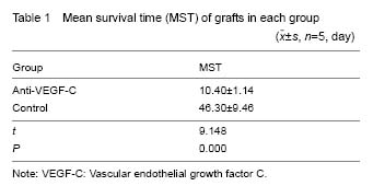
Compared with the graft survival time for the control group, the graft survival time for the anti-VEGF-C group was significantly increased (P < 0.05, Table 1). In addtion, the scores of RI were much higher in the control group in comparison with that in the anti-VEGF-C group (Table 2), suggesting that intraperitoneal injection of a monoclonal anti-VEGF-C antibody was effective in preventing corneal allograft rejection.
| [1] Maruyama K, Ii M, Cursiefen C, et al. Inflammation- induced lymphangiogenesis in the cornea arises from CD11b-positive macrophages. J Clin Invest. 2005; 115(9):2363-2372. [2] Cursiefen C, Chen L, Dana MR, et al. Corneal lymphangiogenesis: evidence, mechanisms, and implications for corneal transplant immunology. Cornea. 2003;22(3):273-281. [3] Yamagami S, Dana MR. The critical role of lymph nodes in corneal alloimmunization and graft rejection. Invest Ophthalmol Vis Sci. 2001;42(6):1293-1298. [4] Yamagami S, Dana MR, Tsuru T. Draining lymph nodes play an essential role in alloimmunity generated in response to high-risk corneal transplantation. Cornea. 2002;21(4):405-409. [5] Ling S, Qi C, Li W, et al. Crucial role of corneal lymphangiogenesis for allograft rejection in alkali-burned cornea bed. Clin Experiment Ophthalmol. 2009;37(9):874-883. [6] Dietrich T, Bock F, Yuen D, et al. Cutting edge: lymphatic vessels, not blood vessels, primarily mediate immune rejections after transplantation. J Immunol. 2010;184(2):535-539. [7] Ling SQ, Liu C, Li WH, et al. Corneal lymphangiogenesis correlates closely with hemangiogenesis after keratoplasty. Int J Ophthalmol. 2010;3(1):76-79. [8] Cursiefen C, Schlötzer-Schrehardt U, Küchle M, et al. Lymphatic vessels in vascularized human corneas: immunohistochemical investigation using LYVE-1 and podoplanin. Invest Ophthalmol Vis Sci. 2002;43(7): 2127-2135. [9] Liu YC, Peng Y, Lwin NC, et al. A biodegradable, sustained-released, prednisolone acetate microfilm drug delivery system effectively prolongs corneal allograft survival in the rat keratoplasty model. PLoS One. 2013;8(8):e70419. [10] Chang LK, Garcia-Cardeña G, Farnebo F, et al. Dose-dependent response of FGF-2 for lymphangiogenesis. Proc Natl Acad Sci U S A. 2004; 101(32):11658-11663. [11] Chang L, Kaipainen A, Folkman J. Lymphangiogenesis new mechanisms. Ann N Y Acad Sci. 2002;979:111-119. [12] Holland EJ, Olsen TW, Sterrer J, et al. Suppression of graft rejection using 15-deoxyspergualin in the allogeneic rat penetrating keratoplasty model. Cornea. 1994;13(1):28-32. [13] Zhang W, Pan Z, Zhai C. Efficacy of topical cyclosporine A on keratoplasty rejection in rats. Zhonghua Yan Ke Za Zhi. 2001;37(2):140-143. [14] Yuen D, Grimaldo S, Sessa R, et al. Role of angiopoietin-2 in corneal lymphangiogenesis. Invest Ophthalmol Vis Sci. 2014;55(5):3320-3327. [15] Tang X, Sun J, Du L, et al. Neuropilin-2 contributes to LPS-induced corneal inflammatory lymphangiogenesis. Exp Eye Res. 2016;143:110-119. [16] Ling S, Lin H, Liang L, et al. Development of new lymphatic vessels in alkali-burned corneas. Acta Ophthalmol. 2009;87(3):315-322. [17] Mouta C, Heroult M. Inflammatory triggers of lymphangiogenesis. Lymphat Res Biol. 2003;1(3):201-218. [18] Yan H, Qi C, Ling S, et al. Lymphatic vessels correlate closely with inflammation index in alkali burned cornea. Curr Eye Res. 2010;35(8):685-697. [19] Yan H, Yuan J, Peng R, et al. The Blockade of Vascular Endothelial Growth Factor C Effectively Inhibits Corneal Lymphangiogenesis and Promotes Allograft Survival. J Ocul Pharmacol Ther. 2015;31(9):546-554. [20] Oh SJ, Jeltsch MM, Birkenhäger R, et al. VEGF and VEGF-C: specific induction of angiogenesis and lymphangiogenesis in the differentiated avian chorioallantoic membrane. Dev Biol. 1997;188(1):96-109. [21] Skobe M, Hawighorst T, Jackson DG, et al. Induction of tumor lymphangiogenesis by VEGF-C promotes breast cancer metastasis. Nat Med. 2001;7(2):192-198. [22] Jeltsch M, Kaipainen A, Joukov V, et al. Hyperplasia of lymphatic vessels in VEGF-C transgenic mice. Science. 1997;276(5317):1423-1425. [23] Mäkinen T, Jussila L, Veikkola T, et al. Inhibition of lymphangiogenesis with resulting lymphedema in transgenic mice expressing soluble VEGF receptor-3. Nat Med. 2001;7(2):199-205. [24] Ling S, Qi C, Li W, et al. The expression of vascular endothelial growth factor C in transplanted corneas. Curr Eye Res. 2009;34(7):553-561. [25] Oh SJ, Jeltsch MM, Birkenhäger R, et al. VEGF and VEGF-C: specific induction of angiogenesis and lymphangiogenesis in the differentiated avian chorioallantoic membrane. Dev Biol. 1997;188(1):96-109. [26] Kriehuber E, Breiteneder-Geleff S, Groeger M, et al. Isolation and characterization of dermal lymphatic and blood endothelial cells reveal stable and functionally specialized cell lineages. J Exp Med. 2001;194(6):797-808. [27] Lymboussaki A, Achen MG, Stacker SA, et al. Growth factors regulating lymphatic vessels. Curr Top Microbiol Immunol. 2000;251:75-82. [28] Cao Y, Linden P, Farnebo J, et al. Vascular endothelial growth factor C induces angiogenesis in vivo. Proc Natl Acad Sci U S A. 1998;95(24):14389-14394. |
| [1] | 张同同, 王中华, 文 杰, 宋玉鑫, 刘 林. 3D打印模型在颈椎肿瘤手术切除与重建中的应用[J]. 中国组织工程研究, 2021, 25(9): 1335-1339. |
| [2] | 李珊珊, 郭笑霄, 尤 冉, 杨秀芬, 赵 露, 陈 曦, 王艳玲. 感光细胞替代治疗视网膜变性疾病[J]. 中国组织工程研究, 2021, 25(7): 1116-1121. |
| [3] | 曾燕华, 郝延磊. 许旺细胞体外培养及纯化的系统性综述[J]. 中国组织工程研究, 2021, 25(7): 1135-1141. |
| [4] | 刘立华, 孙 伟, 王云亭, 高福强, 程立明, 李子荣, 王江宁. 头颈部开窗减压治疗L1型激素性股骨头坏死:单中心前瞻性临床研究[J]. 中国组织工程研究, 2021, 25(6): 906-911. |
| [5] | 蒋 欣, 乔良伟, 孙 东, 李 明, 房 军, 曲青山. 肾移植患者血清中长链非编码RNA PGM5-AS1表达及调控人肾小球内皮细胞的作用[J]. 中国组织工程研究, 2021, 25(5): 741-745. |
| [6] | 杨 鑫, 金 喆, 冯 旭, 卢 兵. 沈阳市居民对器官、眼组织及遗体捐献的认知及意愿调查[J]. 中国组织工程研究, 2021, 25(5): 779-784. |
| [7] | 徐东紫, 张 婷, 欧阳昭连. 心脏组织工程领域全球专利竞争态势分析[J]. 中国组织工程研究, 2021, 25(5): 807-812. |
| [8] | 吴子健, 胡昭端, 谢有琼, 王 峰, 李 佳, 李柏村, 蔡国伟, 彭 锐. 3D打印技术与骨组织工程研究文献计量及研究热点可视化分析[J]. 中国组织工程研究, 2021, 25(4): 564-569. |
| [9] | 常文辽, 赵 杰, 孙晓亮, 王 锟, 吴国锋, 周 剑, 李树祥, 孙 晗. 人工骨膜的材料选择、理论设计及生物仿生功能[J]. 中国组织工程研究, 2021, 25(4): 600-606. |
| [10] | 刘 旒, 周箐竹, 龚 桌, 刘博言, 杨 斌, 赵 娴. 胶原/无机材料构建组织工程骨的特点及制造技术[J]. 中国组织工程研究, 2021, 25(4): 607-613. |
| [11] | 刘 飞, 崔宇韬, 刘 贺. 局部抗生素递送系统治疗骨髓炎的优势与问题[J]. 中国组织工程研究, 2021, 25(4): 614-620. |
| [12] | 叶海民, 丁凌华, 孔维豪, 黄祖泰, 熊 龙. 多级微管结构骨支架载体促进成骨成血管作用及机制[J]. 中国组织工程研究, 2021, 25(4): 621-625. |
| [13] | 李晓壮, 段 浩, 王伟舟, 唐志宏, 王旸昊, 何 飞. 骨组织工程材料治疗骨缺损疾病在体内实验中的应用[J]. 中国组织工程研究, 2021, 25(4): 626-631. |
| [14] | 张振坤, 李 喆, 李 亚, 王莹莹, 王亚苹, 周馨魁, 马珊珊, 关方霞. 海藻酸盐基水凝胶/敷料在创面愈合中的应用:持续、动态与顺序释放[J]. 中国组织工程研究, 2021, 25(4): 638-643. |
| [15] | 陈佳娜, 邱燕玲, 聂敏海, 刘旭倩. 组织工程支架材料修复口腔颌面部软组织缺损[J]. 中国组织工程研究, 2021, 25(4): 644-650. |
中国组织工程研究杂志出版内容重点:组织构建;骨细胞;软骨细胞;细胞培养;成纤维细胞;血管内皮细胞;骨质疏松;组织工程
Total RNA was isolated from the samples using TRIzol® Reagent (Gibco-BRL Life Technologies, Gaithersburg, MD, USA). RNA was prepared following the protocol from the manufacturer. The RNA pellets were washed with 75% ethanol, centrifuged, and dried. The residual DNA was removed by treatment with DNase I. Pellets were resuspended in 30 μL of DEPC-treated water followed by the addition of 50 mmol/L Tris, pH 7.5, 10 mmol/L MgCl2, 20 U of RNase-free DNase I, and 20 U of RNas in a total volume of 60 μL. Samples were incubated at 37 ℃ for 25 minutes. Then the RNA was cleaned using RNeasy Mini Kits (Qiagen, Valencia, CA, USA) following the manufacturer’s protocol. The RNA concentration and purity were determined by measuring optical density at 260 nm and 280 nm using a spectrophotometer (Metash Instru-ments Co., Ltd., Shanghai, China). The cDNA was generated from total RNA samples by using the Taqman Reverse Transcription Reagents kit (Applied Biosystems, Foster City, CA, USA). To prepare the cDNA, the total RNA from each sample was first incubated at 25 ℃ for 10 minutes, and then reverse transcribed at 48 ℃ for 30 minutes. Real-time RT-PCR was performed using a SYBR® green dye (Applied Biosystems) with the ABI PRISM 7900HT (Applied Biosystems). The primers for VEGF-C were 5’-GTA AAA CTT GCT GCT GCA CAT T-3’ (sense) and 5’-GAA CGT CTA ATA ATT GAA TGA ACT TGT CT-3’(antisense) (GenBank accession no. AY032729). The DNA polymerase was first activated at 95 ℃ for 10 minutes, denatured at 95 ℃ for 15 seconds, and annealed/extended at 60 ℃ for 1 minute, for 40 cycles according to the manufacturer’s protocol. The products were sequenced to confirm that the correct gene sequence was being amplified. All PCR reactions were performed in triplicate. Relative quantitation of gene expression was performed using the standard curve method (User Bulletin number 2, ABI PRISM 7700 Sequence Detection System; Applied Biosystems). For a comparison of the transcript levels between samples, standard curves were prepared for both the target gene and the endogenous reference (18S ribosomal RNA). For each experimental sample, the amounts of target and endogenous reference were determined from the appropriate standard curves. Then, the target amount was divided by the endogenous reference amount to obtain a normalized target value. Each of the experimental normalized sample values was divided by the normalized control sample value to generate the relative expression levels.
中国组织工程研究杂志出版内容重点:组织构建;骨细胞;软骨细胞;细胞培养;成纤维细胞;血管内皮细胞;骨质疏松;组织工程
中国组织工程研究杂志出版内容重点:组织构建;骨细胞;软骨细胞;细胞培养;成纤维细胞;血管内皮细胞;骨质疏松;组织工程
.jpg) 文题释义:
角膜新生淋巴管:正常角膜无淋巴管,但在炎症、烧伤、移植后均可出现角膜新生淋巴管。角膜新生淋巴管属于毛细微淋巴管,无色透明,临床上难以辨认。淋巴管可以回收组织间液,避免组织水肿。除此之外,角膜淋巴管可以加速角膜抗原呈递的速度,在角膜免疫中起着重要的作用。
血管内皮生长因子C:是一种与血管内皮生长因子同源的内皮细胞生长因子,其受体为血管内皮生长因子受体3和血管内皮生长因子受体2。血管内皮生长因子C介导了淋巴管的发生、生长和增生,是目前公认的淋巴管诱生因子。
中国组织工程研究杂志出版内容重点:组织构建;骨细胞;软骨细胞;细胞培养;成纤维细胞;血管内皮细胞;骨质疏松;组织工程
文题释义:
角膜新生淋巴管:正常角膜无淋巴管,但在炎症、烧伤、移植后均可出现角膜新生淋巴管。角膜新生淋巴管属于毛细微淋巴管,无色透明,临床上难以辨认。淋巴管可以回收组织间液,避免组织水肿。除此之外,角膜淋巴管可以加速角膜抗原呈递的速度,在角膜免疫中起着重要的作用。
血管内皮生长因子C:是一种与血管内皮生长因子同源的内皮细胞生长因子,其受体为血管内皮生长因子受体3和血管内皮生长因子受体2。血管内皮生长因子C介导了淋巴管的发生、生长和增生,是目前公认的淋巴管诱生因子。
中国组织工程研究杂志出版内容重点:组织构建;骨细胞;软骨细胞;细胞培养;成纤维细胞;血管内皮细胞;骨质疏松;组织工程角膜淋巴管有利于角膜抗原的运输,从而加速了抗原提呈的进程,在角膜免疫中起着重要的作用。然而,由于角膜移植后角膜新生血管和淋巴管呈“平行”生长,因而很难对淋巴管在移植免疫中所起的具体作用大小作出准确的评估。 中国组织工程研究杂志出版内容重点:组织构建;骨细胞;软骨细胞;细胞培养;成纤维细胞;血管内皮细胞;骨质疏松;组织工程
| 阅读次数 | ||||||
|
全文 |
|
|||||
|
摘要 |
|
|||||