中国组织工程研究 ›› 2016, Vol. 20 ›› Issue (29): 4341-4348.doi: 10.3969/j.issn.2095-4344.2016.29.012
• 口腔组织构建 oral tissue construction • 上一篇 下一篇
下颌运动轨迹描记仪在不同牙合型人群中的应用
刘 玉1,张 琪2,李天舒2,付贵源2,徐 琳2,易 龙2,王佳莹2,古再丽阿依2,何 媛2,高 璐2
- 1大连市第五人民医院口腔科,辽宁省大连市 116021;2大连医科大学口腔医学院,辽宁省大连市 116044
Application of mandibular kinesiography in dental occlusion
Liu Yu1, Zhang Qi2, Li Tian-shu2, Fu Gui-yuan2, Xu Lin2, Yi Long2, Wang Jia-ying2, Güzelay2, He Yuan2, Gao Lu2
- 1Department of Stomatology, the Fifth People's Hospital of Dalian, Dalian 116021, Liaoning Province, China; 2College of Stomatology, Dalian Medical University, Dalian 116044, Liaoning Province, China
摘要:
文章快速阅读:
.jpg) 文题释义:
下颌运动轨迹描记仪:是一种磁电量转换方式的描记仪,能从矢状面、额状面和水平面精确地观测下颌中切牙切点运动的轨迹,是一种快速、准确操作、简便无损伤性,不限制下颌运动的三维描记仪。
错牙合:上下颌牙齿正常接触关系的偏差。常被分为3种主要形式(Angle分类):第Ⅰ类:上下颌磨牙为正常关系,但前牙拥挤或错位;第Ⅱ类:下颌和下颌磨牙后移,面部侧面观呈凸出形;第Ⅲ类:下颌和下颌磨牙与上颌磨牙的关系为前突,反时一个或更多的下颌牙的颊尖位于相应的上颌牙牙尖的外侧。
文题释义:
下颌运动轨迹描记仪:是一种磁电量转换方式的描记仪,能从矢状面、额状面和水平面精确地观测下颌中切牙切点运动的轨迹,是一种快速、准确操作、简便无损伤性,不限制下颌运动的三维描记仪。
错牙合:上下颌牙齿正常接触关系的偏差。常被分为3种主要形式(Angle分类):第Ⅰ类:上下颌磨牙为正常关系,但前牙拥挤或错位;第Ⅱ类:下颌和下颌磨牙后移,面部侧面观呈凸出形;第Ⅲ类:下颌和下颌磨牙与上颌磨牙的关系为前突,反时一个或更多的下颌牙的颊尖位于相应的上颌牙牙尖的外侧。
文题释义:
下颌运动轨迹描记仪:是一种磁电量转换方式的描记仪,能从矢状面、额状面和水平面精确地观测下颌中切牙切点运动的轨迹,是一种快速、准确操作、简便无损伤性,不限制下颌运动的三维描记仪。
错牙合:上下颌牙齿正常接触关系的偏差。常被分为3种主要形式(Angle分类):第Ⅰ类:上下颌磨牙为正常关系,但前牙拥挤或错位;第Ⅱ类:下颌和下颌磨牙后移,面部侧面观呈凸出形;第Ⅲ类:下颌和下颌磨牙与上颌磨牙的关系为前突,反时一个或更多的下颌牙的颊尖位于相应的上颌牙牙尖的外侧。摘要
背景:错牙合畸形者牙列牙弓的不协调直接影响着下颌运动的发挥,甚至会造成颞下颌关节的超负荷,最终发生病变。
目的:测量各类牙合型人群的下颌运动特点。
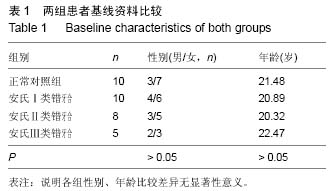
方法:①选取符合实验标准的自愿者33人,平均年龄21.71岁,男性1人,女性32人。分为4组,正常对照组10人,安氏Ⅰ类错牙合组10人,安氏Ⅱ类错牙合组8人,安氏Ⅲ类错牙合组5人;②用描记仪测量各组下颌各运动的轨迹,以及下颌姿势位到牙尖交错位的运动轨迹;③用SPSS17.0软件进行统计学分析,得出各组人群下颌运动轨迹特点和差异。
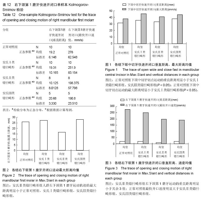
结果与结论:①开闭口、左侧、右侧、前伸运动时,安氏Ⅱ类错牙合畸形组人群右下颌第1磨牙运动轨迹的最大距离与其他3组比较,差异有显著性意义(P < 0.05);②做快速开闭口运动时,正常对照组下颌中切牙切点运动的垂直距离与安氏Ⅰ类错牙合畸形组、安氏Ⅲ类错牙合畸形组比较差异有显著性意义(< 0.05);正常对照组最大距离与安氏Ⅰ类错牙合畸形组比较差异有显著性意义(P < 0.05);安氏Ⅱ类错牙合畸形组右下颌第1磨牙运动的垂直距离与其余3组比较差异有显著性意义(P < 0.05);正常对照组最快开口速度与安氏Ⅱ类错牙合畸形组、安氏Ⅲ类错牙合畸形组比较差异有显著性意义(P < 0.05);③各组人群息止牙合间隙不存在显著性差异。结果提示,不同牙合型人群下颌中切牙运动轨迹、下颌第1磨牙运动轨迹和运动速度不同,因而推断错牙合畸形可以影响下颌运动的方向、范围和速度。
中国组织工程研究杂志出版内容重点:组织构建;骨细胞;软骨细胞;细胞培养;成纤维细胞;血管内皮细胞;骨质疏松;组织工程
ORCID: 0000-0003-2729-2616(刘玉)
中图分类号:


.jpg) 文题释义:
下颌运动轨迹描记仪:是一种磁电量转换方式的描记仪,能从矢状面、额状面和水平面精确地观测下颌中切牙切点运动的轨迹,是一种快速、准确操作、简便无损伤性,不限制下颌运动的三维描记仪。
错牙合:上下颌牙齿正常接触关系的偏差。常被分为3种主要形式(Angle分类):第Ⅰ类:上下颌磨牙为正常关系,但前牙拥挤或错位;第Ⅱ类:下颌和下颌磨牙后移,面部侧面观呈凸出形;第Ⅲ类:下颌和下颌磨牙与上颌磨牙的关系为前突,反时一个或更多的下颌牙的颊尖位于相应的上颌牙牙尖的外侧。
文题释义:
下颌运动轨迹描记仪:是一种磁电量转换方式的描记仪,能从矢状面、额状面和水平面精确地观测下颌中切牙切点运动的轨迹,是一种快速、准确操作、简便无损伤性,不限制下颌运动的三维描记仪。
错牙合:上下颌牙齿正常接触关系的偏差。常被分为3种主要形式(Angle分类):第Ⅰ类:上下颌磨牙为正常关系,但前牙拥挤或错位;第Ⅱ类:下颌和下颌磨牙后移,面部侧面观呈凸出形;第Ⅲ类:下颌和下颌磨牙与上颌磨牙的关系为前突,反时一个或更多的下颌牙的颊尖位于相应的上颌牙牙尖的外侧。