| [1] Kerr JF,Rosen MD.Apoptosis:A basic biological phenomennon with widespread mplication in tissue kinetics. Br J Cancer.1972;26:239-257.
[2] 袁箭锋,常芸.运动训练与心血管系统细胞凋亡的现象[J].中国运动医学杂志,2001,20(3):301-303.
[3] 常芸,袁箭峰,祁永梅,等.运动心脏重塑与微损伤发生中的细胞凋亡现象[J].中国运动医学杂志,2003,22(4):344-349.
[4] 郭勇力,刘霞,张文峰,等.不同强度运动对大鼠心肌细胞凋亡的影响[J].中国临床康复,2004,8(9):1732-1733.
[5] 袁海平,刘学.大鼠不同强度运动对心肌细胞凋亡及其抗氧化能力的影响[J].体育科学,2002,22(1):107-109.
[6] Tanaka M, Ito H, Adachi S,et al.Hypoxia induces apoptosis with enhanced expression of Fas antigen messenger RNA in cultured neonatal rat cardiomyoeytes.Cire Res 2004;75: 426-433.
[7] 洪欣,董宏彬,杜宏伟,等.低氧及低氧预处理时心肌细胞HIF-1α与凋亡相关蛋白表达的变化[J].中国应用生理学杂志,2005,21(4): 423-426.
[8] 王雪冰,路瑛丽,冯连世,等.不同低氧训练对大鼠腓肠肌糖酵解酶活性的影响[J]中国运动医学杂志, 2014,12(5):76-79.
[9] 邢维新.运动训练与心肌细胞凋亡研究的新进展[J].绵阳师范学院学报,2011,30(2):127-130.
[10] 张萍,邱健,李志樑.等,压力超负荷时左室心肌凋亡和fas蛋白的表达[J].中国临床康复,2003,7(15):2150-2151.
[11] 黄黎月,庄国洪,丘劲华,等.DcR3重组蛋白对糖尿病大鼠心肌组织凋亡相关分子的表达及细胞凋亡的作用[J].中国免疫学杂志, 2015,4(8):43-47.
[12] 李强,郭壮波,伍光颖,等.阿托伐他汀对急性心肌梗死大鼠内皮微颗粒及心肌细胞凋亡的影响[J].中国病理生理杂志,2015,2(6): 56-59.
[13] 孙宇泽,李艳丽,赵勇,等.人生长素对雌性大鼠心力衰竭心脏重塑及心肌细胞凋亡的影响[J].医学研究杂志,2015,6(8):73-77.
[14] Leri A.Insulin-like growth factor-1induces Mdnd and down-regulates P53,atenuating the myocyte renin- angiotensin system and stretch-mediated apoptosis.Am J Pathol.2009;152(2):567-580.
[15] 杨海平.低氧、运动对大鼠骨骼肌细胞凋亡及bcl-2、bax表达的影响[J].中国运动医学杂志,2006,11(6):706-709.
[16] 林喜秀,瞿树林,周桔,等.低氧训练对大鼠心、肝、肾、海马组织细胞凋亡的影响及其机制研究[J].中国运动医学杂志,2012,2(2): 146-156.
[17] Wang GL, Jiang BH, Rue EA, et al. hypoxia inducible factor-1 is abasic helixloop -helix-PAS heterodimer regulated by cellular O2 tension. Proc Natn Acadsci USA.2005;92:5510-5514.
[18] 王宁琦,胡扬,赵华,等.低氧训练中血氧饱和度作为运动强度评价指标的可行性研究[J].中国运动医学杂志, 2014,10(6):44-48.
[19] 李靖,张漓,冯连世,等.高原或低氧训练对肥胖青少年减体重效果及血糖代谢相关指标的影响[J].中国运动医学杂,2014,05(8): 32-36.
[20] 徐建方,冯连世,张漓,等.常压低氧耐力训练对肥胖大鼠Visfatin水平的影响[J].体育科学,2014,2(6):45-48.
[21] 张奕奕, 薛一涛,李焱,等.心力衰竭与细胞凋亡及中医药研究进展[J].中国中医急症,2015,7(8):73-77.
[22] 田振军,张可,陈婷,等.运动锻炼上调M3R对MI大鼠心脏产生保护效应及其机制探讨[J].体育科学,2015,5(6):43-47.
[23] Cheng W, Li B, Kajstura J, et al. Stretch-induced programmed myocyte cell death.Clin Invest.2005; 96:2247-2259.
[24] Carmeliet P, Dor Y, Herbert JM, et at. Role of HIF-1αin hypoxia-mediated aopptosis,cellproliferation and tumor angiogenesis.nature.1998,394(6692):485-490. |


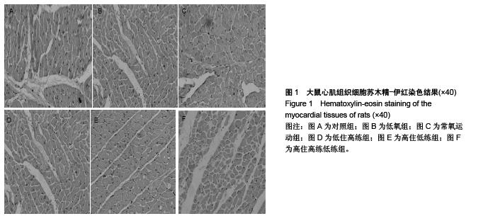
.jpg)

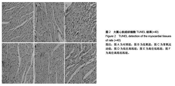
.jpg)

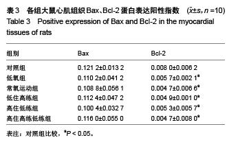
.jpg)

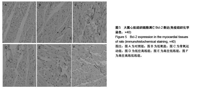
.jpg)