中国组织工程研究 ›› 2015, Vol. 19 ›› Issue (18): 2906-2910.doi: 10.3969/j.issn.2095-4344.2015.18.021

• 器官移植动物模型 organ transplantation and animal model • 上一篇    下一篇

成人头面部模型标本:内眦动脉的定位观测及临床意义

马春晓1,刘媛媛2,任珊珊2,王  芳1,卢小生2   

  1. 1潍坊医学院,山东省潍坊市  261000;2潍坊医学院附属医院,山东省潍坊市  261000
  • 收稿日期:2015-02-15 出版日期:2015-04-30 发布日期:2015-04-30
  • 通讯作者: 卢小生,博士,副主任医师,副教授,硕士生导师,潍坊医学院附属医院整形外科,山东省潍坊市 261000
  • 作者简介:马春晓,女,1988年生,山东省滨州市人,汉族,潍坊医学院在读硕士。

Adult head and face models: localization observation of the angular artery and its clinical significance

Ma Chun-xiao1, Liu Yuan-yuan2, Ren Shan-shan2, Wang Fang1, Lu Xiao-sheng2   

  1. 1Weifang Medical University, Weifang 261000, Shandong Province, China; 2Affiliated Hospital of Weifang Medical University, Weifang 261000, Shandong Province, China
  • Received:2015-02-15 Online:2015-04-30 Published:2015-04-30
  • Contact: Lu Xiao-sheng, M.D., Associate chief physician, Associate professor, Master’s supervisor, Affiliated Hospital of Weifang Medical University, Weifang 261000, Shandong Province, China
  • About author:Ma Chun-xiao, Studying for master’s degree, Weifang Medical University, Weifang 261000, Shandong Province, China

摘要:

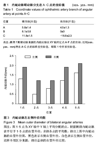
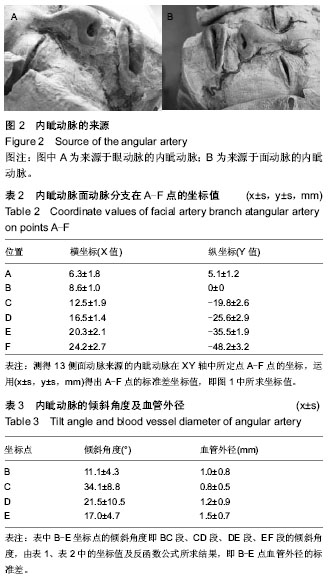
背景:鼻唇沟皮瓣在临床手术中应用较广,面动脉的解剖学研究已广泛应用于临床,内眦动脉解剖对鼻唇沟区手术日益重要,但目前缺乏对内眦动脉的解剖分析。 
目的:对内眦动脉进行解剖,为鼻唇沟皮瓣的手术提供解剖学依据。
方法:解剖20侧成人头面部尸体标本,以内眦连线为X轴,面中线为Y轴,建立坐标轴,定点A-F点测量内眦动脉的位置。
结果与结论:①内眦动脉在BC、CD、DE、EF段的倾斜角度分别为(11.1±4.3)°,(34.1±8.8)°,(21.5±10.5)°,(17.0±4.7)°。②内眦动脉来源于面动脉多于眼动脉,并且右侧血管直径要大于左侧。③来源于眼动脉的内眦动脉起始于由内眦连线与面中线交点正上方10 mm处向两侧延伸8.1 mm位置,起始点管径为(0.7±0.2) mm,全程共20.1 mm。④来源于面动脉的内眦动脉起始于内眦连线与面中线交点正下方40 mm处向两侧延伸25.8 mm位置,起始点管径为(0.9±0.3) mm,走行至鼻翼最外侧点的距离为(5.0±1.2) mm,全程共68.7 mm。由解剖结果得出内眦动脉的体表投影,可为鼻唇沟皮瓣的相关手术提供解剖学基础。


中国组织工程研究
杂志出版内容重点:肾移植肝移植移植;心脏移植;组织移植;皮肤移植;皮瓣移植;血管移植;器官移植组织工程


全文链接:

关键词: 实验动物, 组织构建实验模型, 内眦动脉, 尸体模型, 鼻唇沟皮瓣, 血管灌注, 定位, 体表投影

Abstract:

BACKGROUND: Nasolabial fold flap has been widely used in clinical surgery. The facial artery anatomy has been widely used in clinical research. Angular artery dissection is becoming more and more important to nasolabial groove area surgery, but at present, there is a lack of anatomical analysis of internal angular artery.
OBJECTIVE: To study the anatomy of the angular artery, and to provide anatomical data for protecting the nasolabial flap during surgery.
METHODS: Twenty sides of adult cadaver specimens on head and face were dissected. A reference coordinate system was made based on the line between the connection of two medial angles of eyes (axis X) and the facial midline line (axis Y). The location of the angular artery was measured taking A-F as reference points.
RESULTS AND CONCLUSION: (1) The slant angles of the angular artery on BC section, CD section, DE section and EF section were (11.1±4.3)°, (34.1±8.8)°, (21.5±10.5)°, and (17.0±4.7)°, respectively. (2) The angular artery sourced from facial artery was more than it sourced from ophthalmic artery. The diameter of right blood vessel was larger than that of left side. (3) The angular artery sourced from ophthalmic artery comes from the location which extended 8.1 mm to both sides from the point which was 10 mm up from the intersection of facial medial angle of eyes connection and midline. The blood vessel diameter of the starting point was (0.7±0.2) mm. The whole range was 20.1 mm. (4) The angular artery sourced from facial artery comes from the location which extended 25.8 mm to both sides from the point which was 40 mm down to the intersection of facial medial angle of eyes connection and midline. The blood vessel diameter of the starting 
point was (0.9±0.3) mm. Point to the wing of nose the lateral distance was (5.0±1.2) mm. The whole range was 68.7 mm. The surface projecting of angular artery coming from research results provided anatomic basis for surgery of nasolabial flap.


中国组织工程研究
杂志出版内容重点:肾移植肝移植移植;心脏移植;组织移植;皮肤移植;皮瓣移植;血管移植;器官移植组织工程


全文链接:

Key words: Autopsy, Manikins, Surgical Flaps

中图分类号: