中国组织工程研究 ›› 2018, Vol. 22 ›› Issue (8): 1241-1246.doi: 10.3969/j.issn.2095-4344.0143
• 组织构建细胞学实验 cytology experiments in tissue construction • 上一篇 下一篇
解剖学观察血管灌注后头颅模型眉下区上睑支的来源及走行区域
李旭风1,刘媛媛2,任珊珊2,王 爱1,卢小生2
- 1潍坊医学院,山东省潍坊市 261000;2潍坊医学院附属医院,山东省潍坊市 261000
Origin and trend of the upper eyelid artery in the eyelid region of a skull model after blood reperfusion: an anatomical observation
Li Xu-feng1, Liu Yuan-yuan2, Ren Shan-shan2, Wang Ai1, Lu Xiao-sheng2
- 1Weifang Medical University, Weifang 261000, Shandong Province, China; 2Affiliated Hospital of Weifang Medical University, Weifang 261000, Shandong Province, China
摘要:
文章快速阅读:
.jpg) 文题释义:
皮瓣移植:皮瓣是由具有血液供应的皮肤及其附着的皮下脂肪组织所形成。在皮瓣形成与转移过程中,必须有一部分与本体(供皮瓣区)相连,此相连的部分称为蒂部,以保持血液供应,其他部分在表面及深面均与本体分离,转移到另一创面后(受皮瓣区),暂时仍由蒂部血运供应营养,等受皮瓣区创面血管长入皮瓣,建立新的血运后,再将蒂部切断,始完成皮瓣转移的全过程,故又名带蒂皮瓣,但局部皮瓣或岛状皮瓣转移后则不需要断蒂。
眶上动脉:自眶上孔或眶上切迹出眶以后分为深、浅两支,其中眶上动脉的浅支分布于上眼睑区域。
文题释义:
皮瓣移植:皮瓣是由具有血液供应的皮肤及其附着的皮下脂肪组织所形成。在皮瓣形成与转移过程中,必须有一部分与本体(供皮瓣区)相连,此相连的部分称为蒂部,以保持血液供应,其他部分在表面及深面均与本体分离,转移到另一创面后(受皮瓣区),暂时仍由蒂部血运供应营养,等受皮瓣区创面血管长入皮瓣,建立新的血运后,再将蒂部切断,始完成皮瓣转移的全过程,故又名带蒂皮瓣,但局部皮瓣或岛状皮瓣转移后则不需要断蒂。
眶上动脉:自眶上孔或眶上切迹出眶以后分为深、浅两支,其中眶上动脉的浅支分布于上眼睑区域。
文题释义:
皮瓣移植:皮瓣是由具有血液供应的皮肤及其附着的皮下脂肪组织所形成。在皮瓣形成与转移过程中,必须有一部分与本体(供皮瓣区)相连,此相连的部分称为蒂部,以保持血液供应,其他部分在表面及深面均与本体分离,转移到另一创面后(受皮瓣区),暂时仍由蒂部血运供应营养,等受皮瓣区创面血管长入皮瓣,建立新的血运后,再将蒂部切断,始完成皮瓣转移的全过程,故又名带蒂皮瓣,但局部皮瓣或岛状皮瓣转移后则不需要断蒂。
眶上动脉:自眶上孔或眶上切迹出眶以后分为深、浅两支,其中眶上动脉的浅支分布于上眼睑区域。摘要
背景:上睑皮瓣转移相关血管如颞浅动脉、滑车上动脉、眶上动脉主干均有详细报道,上睑支血管解剖对眉下区手术日益重要,但目前缺乏对上睑支动脉的解剖分析。
目的:解剖测量位于眉下区的上睑支动脉血管位置,为邻近皮瓣转移提供解剖学依据。
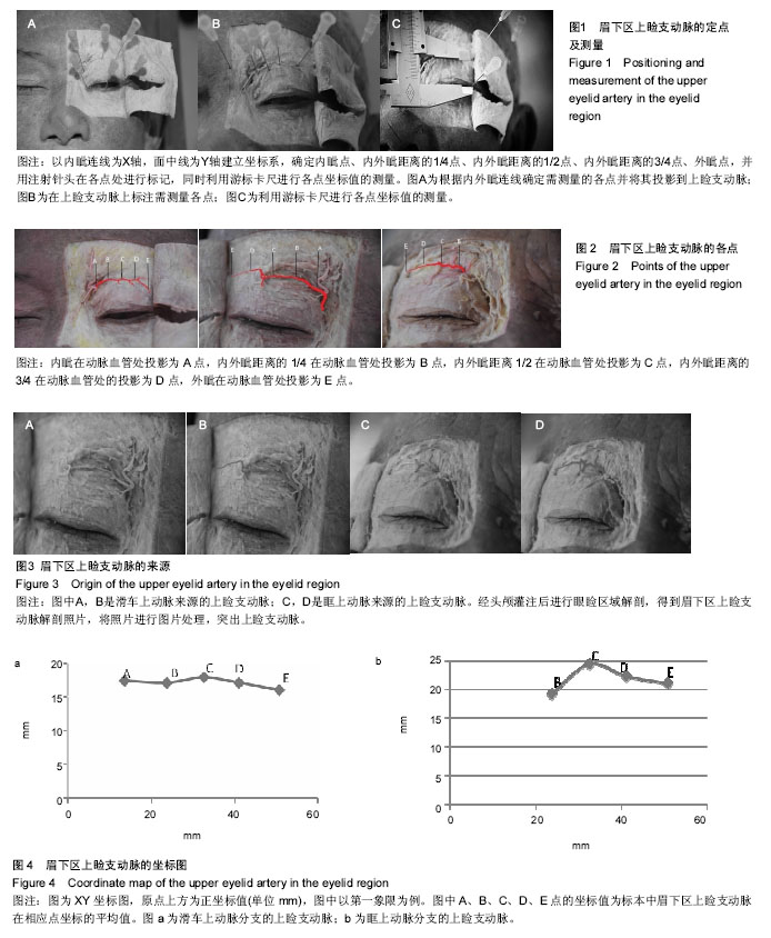
方法:解剖20侧成人头颅模型标本,以内眦连线为X轴,面中线为Y轴建立坐标系,用注射器将红色乳胶溶液经颈总动脉灌注头颅模型,定点(A-E)测量眉下区上睑支动脉的位置。
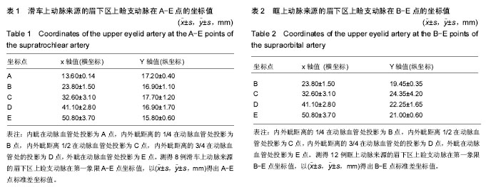
结果与结论:①眉下区上睑支动脉主要是滑车上动脉或眶上动脉的分支,与X轴大致平行;②来源于滑车上动脉的上睑支起始处位于内眦投影处,血管全长约为24.50 mm,发出点管径为0.51 mm,向外眦延伸并且血管管径逐渐缩小;③来源于眶上动脉的上睑支起始于瞳孔和内眦交点1/2投影处,血管全长为23- 24.6 mm,发出点管径0.55±0.05 mm;④由解剖经血管灌注后的头颅模型得出眉下区上睑支的来源及走行区域,为上睑皮肤的皮瓣转移提供详细的解剖学依据。
中国组织工程研究杂志出版内容重点:组织构建;骨细胞;软骨细胞;细胞培养;成纤维细胞;血管内皮细胞;骨质疏松;组织工程
ORCID: 0000-0003-2218-0033(李旭风)
中图分类号:
.jpg) 文题释义:
皮瓣移植:皮瓣是由具有血液供应的皮肤及其附着的皮下脂肪组织所形成。在皮瓣形成与转移过程中,必须有一部分与本体(供皮瓣区)相连,此相连的部分称为蒂部,以保持血液供应,其他部分在表面及深面均与本体分离,转移到另一创面后(受皮瓣区),暂时仍由蒂部血运供应营养,等受皮瓣区创面血管长入皮瓣,建立新的血运后,再将蒂部切断,始完成皮瓣转移的全过程,故又名带蒂皮瓣,但局部皮瓣或岛状皮瓣转移后则不需要断蒂。
眶上动脉:自眶上孔或眶上切迹出眶以后分为深、浅两支,其中眶上动脉的浅支分布于上眼睑区域。
文题释义:
皮瓣移植:皮瓣是由具有血液供应的皮肤及其附着的皮下脂肪组织所形成。在皮瓣形成与转移过程中,必须有一部分与本体(供皮瓣区)相连,此相连的部分称为蒂部,以保持血液供应,其他部分在表面及深面均与本体分离,转移到另一创面后(受皮瓣区),暂时仍由蒂部血运供应营养,等受皮瓣区创面血管长入皮瓣,建立新的血运后,再将蒂部切断,始完成皮瓣转移的全过程,故又名带蒂皮瓣,但局部皮瓣或岛状皮瓣转移后则不需要断蒂。
眶上动脉:自眶上孔或眶上切迹出眶以后分为深、浅两支,其中眶上动脉的浅支分布于上眼睑区域。