中国组织工程研究 ›› 2015, Vol. 19 ›› Issue (11): 1732-1738.doi: 10.3969/j.issn.2095-4344.2015.11.018

• 肌肉肌腱韧带组织构建 tissue construction of the muscle, tendon and ligament • 上一篇    下一篇

以解剖学定位Lisfranc韧带损伤的植入物内固定修复

郭洪亮1,伊力哈木•托合提1,李山珠2,俞光荣2,王治洲1,甘子明3   

  1. 1新疆医科大学第二附属医院骨科,新疆维吾尔自治区乌鲁木齐市   8300632上海同济大学附属同济医院骨科,上海市 200065;3新疆医科大学解剖教研室,新疆维吾尔自治区乌鲁木齐市 830000
  • 修回日期:2014-12-17 出版日期:2015-03-12 发布日期:2015-03-12
  • 通讯作者: 伊力哈木?托合提,主任医师,新疆医科大学第二附属医院,新疆维吾尔自治区乌鲁木齐市 830063
  • 作者简介:郭洪亮,男,1988年生,吉林省长春市人,汉族,新疆医科大学在读硕士,主要从事创伤、四肢骨折治疗研究。
  • 基金资助:

    自治区科技支疆项目计划(指令性)项目,新疆维吾尔自治区科技厅项目,名称:跖跗关节稳定性维持结构的应用解剖及生物力学研究,项目编号:2013911112

Internal fixation implants for Lisfranc ligament injury based on anatomical location

Guo Hong-liang1, Yilihamu Tuoheti1, Li Shan-zhu2, Yu Guang-rong2, Wang Zhi-zhou1, Gan Zi-ming3   

  1. 1Department of Orthopedics, the Second Affiliated Hospital of Xinjiang Medical University, Urumqi 830063, Xinjiang Uygur Autonomous Region, China; 2Department of Orthopedics, Tongji Hospital, Tongji University, Shanghai 200065, China; 3Division of Anatomy, Xinjiang Medical University, Urumqi 830000, Xinjiang Uygur Autonomous Region, China
  • Revised:2014-12-17 Online:2015-03-12 Published:2015-03-12
  • Contact: Yilihamu Tuoheti, Chief physician, Department of Orthopedics, the Second Affiliated Hospital of Xinjiang Medical University, Urumqi 830063, Xinjiang Uygur Autonomous Region, China
  • About author:Guo Hong-liang, Studying for master’s degree, Department of Orthopedics, the Second Affiliated Hospital of Xinjiang Medical University, Urumqi 820063, Xinjiang Uygur Autonomous Region, China
  • Supported by:

    the Technological Support Project for Xinjiang Uygur Autonomous Region, the Science and Technology Project of Xinjiang Uygur Autonomous Region, No. 2013911112

摘要:

背景:跖跗关节(Lisfranc关节)结构复杂、常规X射线片骨关节面重叠,对其诊断较困难。目前对Lisfranc韧带的解剖学研究较少,缺乏相应的形态学资料,导致在相关修复手术中没有该部位的准确解剖参考值。

目的:测量Lisfranc韧带的静态稳定结构,为Lisfranc关节损伤的修复提供临床解剖学基础。
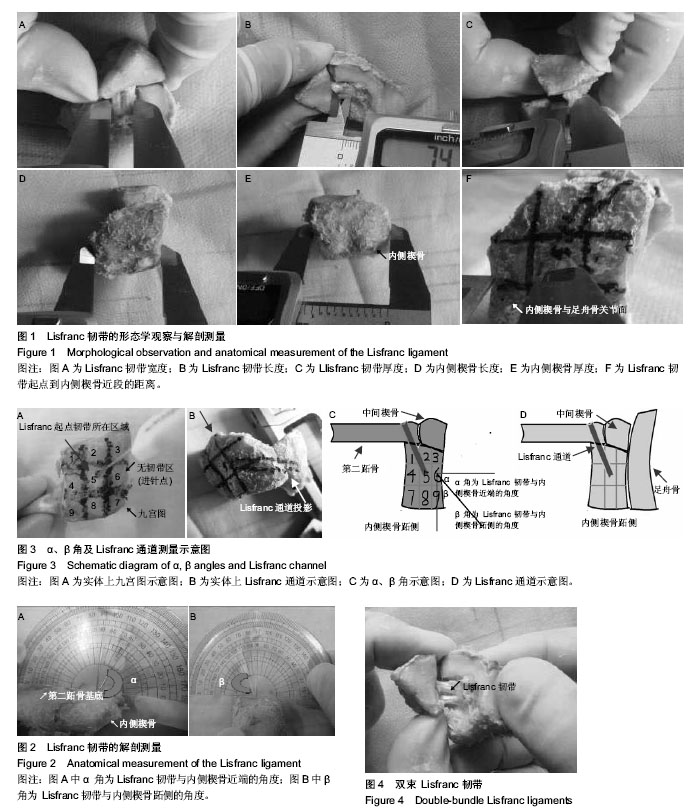
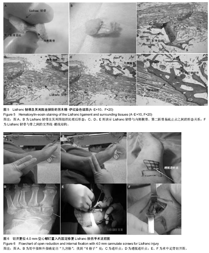
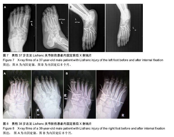
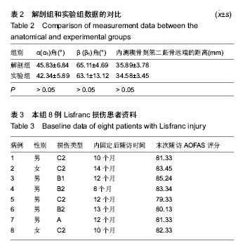
方法:①对18侧成人尸体标本进行解剖,观察Lisfranc韧带的组织形态并进行测量,得到Lisfranc韧带的详细形态参数,作为解剖组,确定Lisfranc韧带的走行和骨性标志;其余14侧成人尸体标本按以上参数用导针固定得出相应Lisfranc韧带相关数据,作为实验组,对比两组测量角度与内侧楔骨到第二跖骨基底长度。②对8例临床上确认的Lisfranc 损伤患者应用解剖学获得的参数和定位法行切开复位螺钉置入内固定治疗,内固定后随访8-14个月,采用美国足踝外科学会踝-后足评分进行功能评估。
结果与结论:①通过解剖观察得出,解剖出的Lisfranc韧带均为2束。解剖组及实验组形态学测量结果比较差异无显著性意义(P > 0.05)。为了方便临床应用,作者提出“Lisfranc通道”这一概念,将Lisfranc通道定义为内侧楔骨(无韧带区)到第二跖骨基底部长(31.65±2.23) mm、宽(8.16±1.37) mm、高(1.69±0.21) mm、与内侧楔骨近端成(45.83±6.84)°角、与内侧楔骨跖侧成(65.11±4.69)°角的一条通道。通过做苏木精-伊红染色后,了解到Lisfranc韧带与周围组织的组织学特性:从层次上分为Lisfranc肌腱、潮线、矿化纤维软骨、骨等4层结构。②8例Lisfranc损伤患者应用解剖学获得的参数和定位法进行切开复位内固定后均获得较高的美国足踝外科学会踝-后足评分,为(80.30±4.85)分。解剖实验获得了Lisfranc关节及韧带详细的形态学参数,为该部位修复提供了解剖学资料,避免盲目置入内固定物,造成Lisfranc韧带起点的损伤,影响Lisfranc韧带修复效果。


中国组织工程研究
杂志出版内容重点:组织构建;骨细胞;软骨细胞;细胞培养;成纤维细胞;血管内皮细胞;骨质疏松组织工程


全文链接:

关键词: 组织构建, 组织工程, 解剖, Lisfranc韧带, 测量, 临床试验, 随访

Abstract:

BACKGROUND: The Lisfranc joint has a complex structure with bony joint surfaces overlapped on the conventional X-ray film, which is diagnosed difficultly. There are less anatomical studies on the Lisfranc ligament, which leads to no accurate anatomical evidence for the repair of Lisfranc ligament injury.

OBJECTIVE: To measure the stable structure of Lisfranc ligament at static state so as to provide anatomical evidence for clinical repair of Lisfranc ligament injury.
METHODS: (1) Eighteen adult corpse specimens were dissected to observe the morphology of Lisfranc ligament and measure relevant parameters as anatomical group. In the anatomical group, the traveling trend and bony landmarks were confirmed. Another 14 adult corpse specimens were taken as experimental group to fix the Lisfranc ligament using a guide pin and detect the corresponding Lisfranc ligament data based on the above-mentioned parameters. Measured angle and length from the entocuneiform to the second metatarsal base were compared between the two groups. (2) Eight patients with clinically confirmed Lisfranc injury were subject to open reduction and internal fixation surgery based on anatomical parameters and positioning method. Patients were followed up for 8-14 months, and AOFAS Ankle Hindfoot Scale was used for functional evaluation.
RESULTS AND CONCLUSION: The Lisfranc ligament had two bundles. There was no difference in the morphological measurement between the anatomical and experimental groups (P > 0.05). “Lisfranc channel” was proposed by the authors, which was defined as: the length, width and height from the entocuneiform (with no ligament) to the second metatarsal base were (31.65±2.23) mm, (8.16±1.37) mm, (1.69±0.21) mm, respectively, and the angles with the proximal entocuneiform and plantar side of the proximal entocuneiform were (45.83±6.84)° and (65.11±4.69)°, respectively. Hematoxylin-eosin staining showed the histological characteristics of the Lisfranc ligament with the surrounding tissues and there was a four-layer structure: Lisfranc tendons, tide lines, mineralized fibrocartilage and bone. (2) After treatment, the AOFAS Ankle Hindfoot Scale scores were improved to (80.30±4.85) points in the eight patients. Morphological parameters of Lisfranc joint and ligament obtained from anatomical experiments provide anatomical evidence for the repair of Lisfranc injury, and avoid blinded internal fixation that can cause the damage to the starting point of the Lisfranc ligament and impact the restorative effects on Lisfranc injury.


中国组织工程研究
杂志出版内容重点:组织构建;骨细胞;软骨细胞;细胞培养;成纤维细胞;血管内皮细胞;骨质疏松组织工程


全文链接:

Key words: Ligaments, Autopsy, Internal Fixators, Follow-Up Studies

中图分类号: