| [1] Mosedale RF. Current indications and methods of periodontal splinting. Dent Update. 2007;34(3):168-170, 173-174, 176-178 passim.
[2] Ma PS, Brudvik JS. Managing the maxillary partially edentulous patient with extensive anterior tooth loss and advanced periodontal disease using a removable partial denture. J Prosthe Dent. 2008;100(4):259-263.
[3] Christensen GJ. When is a full-crown restoration indicated? J Am Dent Assoc. 2007;138(1):101-103.
[4] Bernal G, Garvajal JC, Munoz VCA. A review of the clinical management of mobile teeth. J Contemp Dent Dract. 2002; 15(4):10-22.
[5] Baruch H, Ehrlich J. Splinting a review of the literature. J Refuat Hapeh Vehash Nayim. 2001;18(1):29-40.
[6] Kleinfelder JW, Ludwig K. Maximal bite force in patients with reduced periodontal tissue support with and without splinting. J Periodontol. 2000;73(10):1184-1187.
[7] 曹采方.牙周病学[M].北京:人民卫生出版社,2004.
[8] Forabosco A, Grandi T, Cotti B. The importance of splinting of teeth in the therapy of periodontitis. Minerva Stomatol. 2006; 55(3):87-97.
[9] Rappelli G, Putignano A. Tooth splinting with fiber-reinforced composite materials: achieving predictable aesthetics. Pract Proced Aesthet Dent. 2002;14(6):495-500.
[10] 冯海兰,徐军.口腔修复学[M],第1版.北京:北京大学医学出版社, 2005.
[11] State Council of the People's Republic of China. Administrative Regulations on Medical Institution. 1994-09-01.
[12] Schei O, Waerhaug J, Lovdal A, et al. Alveolar bone loss as related to oral hygiene and age. J Periodontol. 1959;30:7-16.
[13] Tokajuk G, Pawińska M, Stokowska W, et al. The clinical assessment ofmobile teeth stabilization with Fibre-Kor. Adv Med Sci. 2006;51(suppl 1):225-226.
[14] 安娜,欧阳翔英.树脂直接黏接法固定牙周松动前牙的二年效果观察[J].现代口腔医学杂志,2009,23(6):570-573.
[15] Goldberg AJ, FreNch MA. An innovative pre-impregnated glass fiber reinforce composite. Dent Clin North Am. 1999; 43(1):127-133.
[16] 张翼,傅志英.两种牙周夹板在前牙松牙固定术的评价[J].口腔颌面修复学杂志,2003,4(1):33-35.
[17] Rudo DN, Karbhari VM. Physical behaviors of fiber reinforcement as applied to tooth stabilization. Dent Clin North Am.1999;43(1):7-35.
[18] Strassle HE, Serio CL. Esthetic considerations when splinting with fiber-reinforced composites. Dent Clin North Am. 2007; 51(2): 507-524.
[19] Ramols VJ, Runyan DA, Christenaen CC. The effect of plasma-treated polyethylene fiber on the fracture strength of polymethyl methacrylate. J Prosthet Dent. 2001;81(1):94.
[20] Samadzadeh A, Kugel G, Hurley E, et al. Fracture strengths of provisional restorations reinforced with plasms treated woven polyethylene fiber. J Pmthet Dent. 2000;81(4):447-450.
[21] Thomas J, James W, Edith C. Tooth mobility and periodontal therapy. West Soc Periodontol Periodontal Abstr. 2007;55(1): 3-13.
[22] Christensen G. Reinforcement fibers for splinting teeth. J CRA Newsletter. 1997;24(1):1-2.
[23] Strassler HE, Scherer W, Lopresti J, et al. Long term clinical evalution of a Owoven polyethylene ribbon used for tooth stabilitation and splinting. J Isr Orthod Soo. 2002;10(1):11-15.
[24] Nyman S, Lindhe J. A longitudinal study of combined periodontal and p rosthetic treatment of patients with advanced periodontal disease. J Periodontol. 1979;50(4): 163-169.
[25] Bernal G, Carvajal JC, MuÌoz-Viveros CA. A review of the clinical management of mobile teeth. J Contemp Dent Pract. 2002;3(4):10-22.
[26] leinfelder JW, Ludwigt K. Maximal bite force in patients with reduced periodontal tissue support with and without splinting. J Periodontol. 2002;73(10):1184-1187.
[27] Filip Keulemans. The influence of framework design on the load-bearing capacity of laboratory-made inlay-retained fibre-reinforced composite fixed dental prostheses. J Biomech. 2009;42(7):844-849.
[28] Shi L, Fok AS. Structural optimization of the fibre-reinforced composite substructure in a three-unit dental bridge. Dent mate. 2009;25(6):791-801.
[29] 马丽丽.四种方法进行前牙重度牙周炎松牙固定的临床效果评价[J].口腔材料器械杂志,2008,17(4):181-183.
[30] Howard E Strassler, Chery L Serio. Esthetic considerations when splinting with fiber-reinforced composites. Dent Clin North Am. 2007;51(2):507-524. |
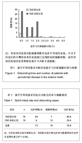
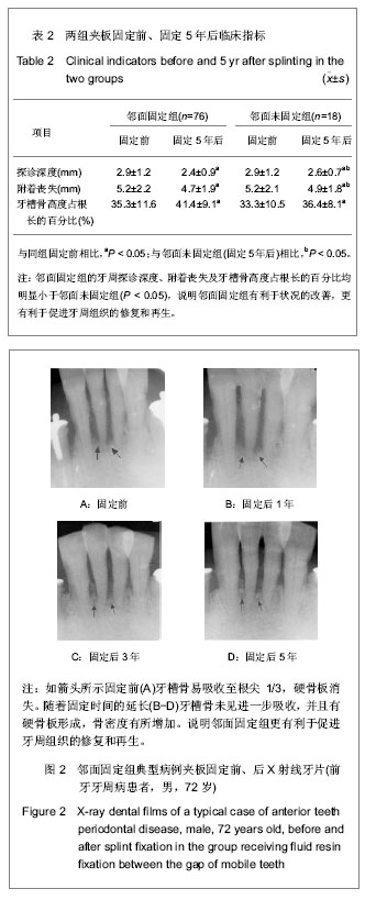
.jpg)
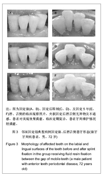
.jpg)
.jpg)