中国组织工程研究 ›› 2013, Vol. 17 ›› Issue (19): 3503-3507.doi: 10.3969/j.issn.2095-4344.2013.19.013

• 干细胞培养与分化 stem cell culture and differentiation • 上一篇    下一篇

大鼠羊膜上皮细胞体外诱导可分化为类神经干细胞

郭 兵,许家军   

  1. 解放军第二军医大学人体解剖学教研室,上海市  200433
  • 收稿日期:2012-12-04 修回日期:2013-03-08 出版日期:2013-05-07 发布日期:2013-05-07
  • 通讯作者: 许家军,博士,教授,解放军第二军医大学人体解剖学教研室,上海市 200433 jjxu@smmu.edu.cn
  • 作者简介:郭兵★,男,1981年生,江苏省人,汉族,2010年解放军第二军医大学毕业,硕士,医师,主要从事大鼠羊膜细胞的体外诱导分化研究。 qiaoweiltd@gmail.com
  • 基金资助:

    国家重点基础研究发展计划(973计划,No. 2005CB724302)

Rat amniotic epithelial cells are induced to differentiate into neural-like stem cells in vitro

Guo Bing, Xu Jia-jun   

  1. Department of Human Anatomy, Second Military Medical University of PLA, Shanghai 200433, China
  • Received:2012-12-04 Revised:2013-03-08 Online:2013-05-07 Published:2013-05-07
  • Contact: Xu Jia-jun, M.D., Professor, Department of Human Anatomy, Second Military Medical University of PLA, Shanghai 200433, China jjxu@smmu.edu.cn
  • About author:Guo Bing★, Master, Physician, Department of Human Anatomy, Second Military Medical University of PLA, Shanghai 200433, China qiaoweiltd@gmail.com
  • Supported by:

     the Major State Basic Research Development Program of China (973 Program), No. 2005CB724302*

摘要:

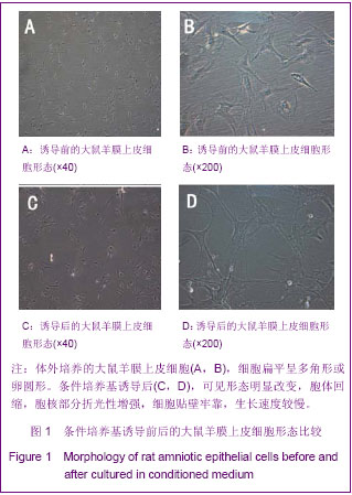
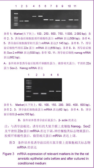
背景:以往研究发现修复神经损伤的细胞来源主要有许旺细胞、嗅鞘细胞、神经干细胞、激活的巨噬细胞等,但这些细胞存在着来源困难、有成瘤性、异体排斥等缺点。而羊膜上皮细胞不存在以上缺陷,且具有多向分化潜能,经诱导后可向心肌样细胞、神经干细胞、肝细胞、成骨和软骨细胞等分化。
目的:体外培养大鼠羊膜上皮细胞,并诱导其向类神经干细胞方向分化。
方法:取妊娠晚期大鼠,采用胰酶消化法获得羊膜上皮细胞,用无血清的神经干细胞条件培养基对细胞进行诱导分化,并用免疫细胞化学法和RT-PCR对诱导前后的细胞相关标志物进行鉴定。
结果与结论:形态学观察结果显示,条件培养基诱导后的羊膜上皮细胞胞体回缩,胞核部分折光性增强,出现类似于树突及轴突样结构,这些突起可交织成网状,细胞贴壁牢靠。免疫细胞化学检测结果显示,与诱导前相比,条件培养基诱导后的羊膜上皮细胞中巢蛋白、胶质纤维酸性蛋白荧光强度较强,而特异性胚胎抗原4、波形蛋白荧光强度较弱。RT-PCR检测显示,与诱导前相比,条件培养基诱导后的羊膜上皮细胞的巢蛋白、胶质纤维酸性蛋白、微管相关蛋白mRNA的表达增强,而平滑肌22α、Sox-2及Nanog mRNA的表达减弱。说明体外诱导的大鼠羊膜上皮细胞可成功分化为类神经干细胞。

关键词: 干细胞, 干细胞培养与分化, 羊膜上皮细胞, 诱导, 分化, 类神经干细胞, 巢蛋白, 胶质纤维酸性蛋白, 微管相关蛋白, 973项目, 干细胞图片文章

Abstract:

BACKGROUND: Previous studies have found that Schwann cells, olfactory ensheathing cells, neural stem cells and activated macrophages may repair nerve injuries, but these cells have some deficiencies, such as source restrictions, tumorigenicity, and allograft rejection. Amniotic epithelial cells have no above-mentioned limits, and possess multiple-differentiation potential. After the induction, amniotic epithelial cells may differentiate into cardiomyocyte-like cells, neural stem cells, liver cells, osteoblasts and chondrocytes.
OBJECTIVE: To culture rat amniotic epithelial cells which can be induced to differentiate into neural-like stem cells.
METHODS: Rat amniotic epithelial cells were collected from late pregnancy rats using digestion method. Serum-free medium of nerve stem cells was used to induce rat amniotic epithelial cells into neural-like stem cells. Normal and induced cells were identified by immunocytochemistry and reverse transcription-PCR methods.
RESULTS AND CONCLUSION: The morphological observation showed that amniotic epithelial cells cultured in the conditioned medium presented with cell body retraction and part of nuclei showed refractive enhancement; dendrite- and axon-like structures appeared, and these processes were woven into a mesh and adhered firmly. Immunocytochemistry results showed that the induced amniotic epithelial cells had a slightly stronger expression of Nestin and glial fibrillary acidic protein, while the pluripotent markers SSEA-4 and Vimentin expressions were weakened. Reverse transcription-PCR test showed that the induced amniotic epithelial cells had a higher mRNA expression of Nestin, glial fibrillary acidic protein and microtubule-associated protein, while the mRNA expression of mesodermal smooth muscle cell marker smooth muscle-22α and pluripotent markers Sox-2 and Nanog declined. These findings suggest that cultured rat amniotic epithelial cells can be induced to differentiate into neural-like stem cells.

Key words: stem cells, stem cell culture and differentiation, amniotic epithelial cells, induction, differentiation, neural-like stem cells, Nestin, glial fibrillary acidic protein, microtubule-associated protein, National “973&rdquo, Program of China, stem cell photographs-containing paper

中图分类号: