中国组织工程研究 ›› 2013, Vol. 17 ›› Issue (3): 465-471.doi: 10.3969/j.issn.2095-4344.2013.03.014

• 材料力学及表面改性 material mechanics and surface modification • 上一篇    下一篇

三种方法构建去细胞肌肉生物支架:组织学和生物力学比较

魏祥科1,文益民2,张 涛1,李 含1   

  1. 1兰州大学第二临床医学院,甘肃省兰州市 730018
    2解放军兰州军区兰州总医院骨科中心,甘肃省兰州市 730050
  • 收稿日期:2012-06-13 修回日期:2012-07-11 出版日期:2013-01-15 发布日期:2013-01-15
  • 通讯作者: 文益民,主任医师,硕士生导师,解放军兰州军区兰州总医院骨科中心,甘肃省兰州市 730050 wenyimin007@163.com
  • 作者简介:魏祥科★,男,1986年生,四川省成都市人,兰州大学在读硕士,主要从事脊髓损伤与修复研究。 792783647@qq.com

Construction of acellular muscle bioscaffolds using three approaches: Histological and biomechanical comparison

Wei Xiang-ke1, Wen Yi-min2, Zhang Tao1, Li Han1   

  1. 1 The Second Clinic College of Medicine of Lanzhou University, Lanzhou 730018, Gansu Province, China
    2 Orthopedics Center, Lanzhou General Hospital of Lanzhou Military Region, Lanzhou 730050, Gansu Province, China
  • Received:2012-06-13 Revised:2012-07-11 Online:2013-01-15 Published:2013-01-15
  • Contact: Wen Yi-min, Chief physician, Master’s supervisor, Orthopedics Center, Lanzhou General Hospital of Lanzhou Military Region, Lanzhou 730050, Gansu Province, China wenyimin007@163.com
  • About author:Wei Xiang-ke★, Studying for master’s degree, the Second Clinic College of Medicine of Lanzhou University, Lanzhou 730018, Gansu Province, China 792783647@qq.com

摘要:

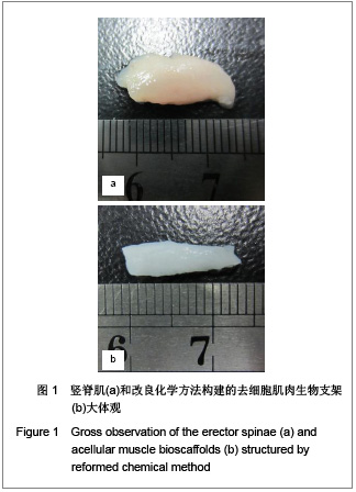
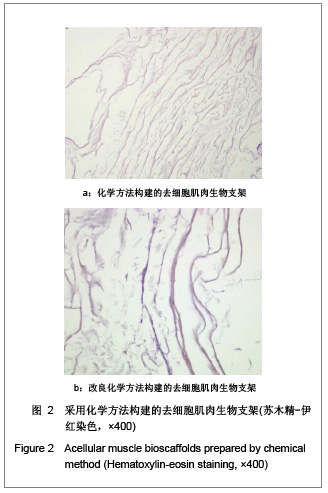
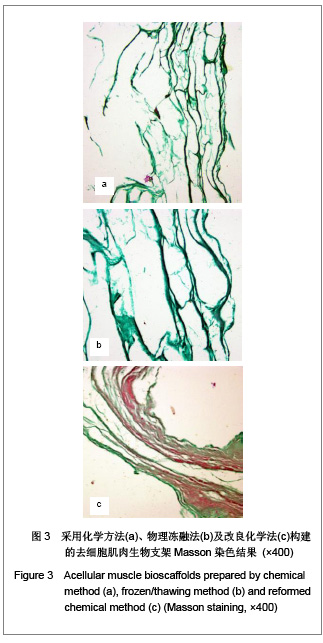
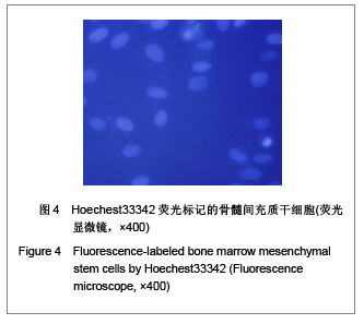
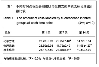
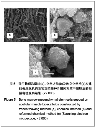
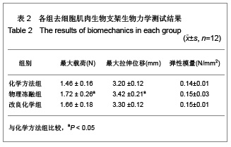
背景:目前构建去细胞肌肉生物支架的方法有化学方法、物理冻融法和冷冻法。
目的:比较化学方法、物理冻融法和改良化学方法构建去细胞肌肉生物支架组织学和生物力学差异。
方法:将6只SD大鼠竖脊肌截成36段,随机分3组(n=12),分别采用化学方法、物理冻融方法、改良化学方法构建去细胞肌肉生物支架。将荧光标记的骨髓间充质干细胞接种于3组支架上进行组织学与生物力学检测。
结果与结论:苏木精-伊红染色显示3组支架均可见平行排列结构,化学方法组连续性较物理冻融组、改良化学组差;Masson染色显示3组支架结构主要为胶原纤维,物理冻融组残留一些肌纤维,间隙不均。物理冻融组第7天荧光标记种子细胞计数明显少于化学方法组、改良化学组(P < 0.01),第14天物理冻融组少于改良化学组(P < 0.05);扫描电镜可见细胞贴附各组支架生长、大量存活;物理冻融组最大载荷高于化学方法组(P < 0.05),与改良化学组无明显差别,各组弹性模量无差异;物理冻融组孔隙率低于化学方法组、改良化学组(P < 0.05)。表明改良化学方法可以兼顾支架的组织学和生物力学特性,彻底清除肌细胞,较多保留更完整的细胞外基质。

关键词: 生物材料, 材料力学及表面改性, 去细胞肌肉生物支架, 骨髓间充质干细胞, 化学方法, 支架构建, 组织学, 生物力学, 物理冻融, 其他基金, 生物材料图片文章

Abstract:

BACKGROUND: The methods of constructing acellular muscle bioscaffolds include chemical method, frozen/thawing method and freezing method.OBJECTIVE: To study the histological and biomechanical differences among the acellular muscle bioscaffolds prepared using chemical method, frozen/thawing method and reformed chemical method.
METHODS: Totally 36 segments of the erector spinae from six Sprague-Dawley rats were randomized into three groups by different preparation methods (n=12): chemical method group (group A), frozen/thawing group (group B) and reformed chemical method group (group C). After sterilization and seeding of bone mesenchymal stem cells, the scaffolds from three groups were compared on histology and biomechanics.
RESULTS AND CONCLUSION: Hematoxylin-eosin staining showed the internal structure was in parallel rows in the three group, but the continuity of scaffold structure in group A was less than groups B and C. Masson staining confirmed essential component of scaffolds was collagen fibers in the three group and some residual muscle fibers in group B with asymmetry clearance. The number of living seed cells labeled by fluorescence in group B was significantly less than that in groups A and C at 7 days (P < 0.01) and less than that in group C at 14 days (P < 0.05). Lots of cells attaching to each group scaffolds were flourished confirmed by scanning electron microscope. Group B exceeded group A in the maximum load of scaffold (P < 0.05) but not group C. There was no difference among three groups in modulus of elasticity. Porosity of group B was lower than that of groups A and C (P < 0.05). These findings indicate that the reformed chemical method is more ideal, which can balance the histology and biomechanics of acellular muscle bioscaffolds, remove muscle cells more thoroughly and maintain more extracellular matrix.

Key words: biomaterials, material mechanics and surface modification, acellular muscle bioscaffolds, bone marrow mesenchymal stem cells, chemical method, scaffold construction, histology, biomechanics, frozen/thawing method, other grants-supported paper, biomaterial photographs-containing paper

中图分类号: