中国组织工程研究 ›› 2019, Vol. 23 ›› Issue (34): 5425-5429.doi: 10.3969/j.issn.2095-4344.1952

• 药物控释材料 drug delivery materials • 上一篇    下一篇

磁靶向维拉帕米纳米颗粒促进周围神经再生的实验研究

宗  强1,徐亚楠2,曲天一2,李立军3,海米提·阿布都艾尼3,倪东馗3
  

  1. 滨州医学院烟台附属医院,1创伤骨科,2急诊科,山东省烟台市 264100;3天津医科大学第二医院骨科,天津市  300211
  • 收稿日期:2019-05-20 出版日期:2019-12-08 发布日期:2019-12-08
  • 作者简介:宗强,男,1989年生,山东省滨州市人,汉族,2016年天津医科大学毕业,硕士,医师,主要从事创伤骨科研究。
  • 基金资助:

    滨州医学院附属医院、教学医院科研计划项目(BY2016KJ37),项目负责人:宗强;天津市卫计委科技基金(2015KZ094),项目负责人:李立军

Magnetic verapamil nanoparticles promote peripheral nerve regeneration

Zong Qiang1, Xu Yanan2, Qu Tianyi2, Li Lijun3, Hai Miti·Abuduaini3, Ni Dongkui3
  

  1. 1Department of Traumatology, 2Department of Emergency, Affiliated Yantai Hospital of Binzhou Medical University, Yantai 264100, Shandong Province, China; 3Department of Orthopedics, Second Hospital of Tianjin Medical University, Tianjin 300211, China
  • Received:2019-05-20 Online:2019-12-08 Published:2019-12-08
  • About author:Zong Qiang, Master, Physician, Department of Traumatology, Affiliated Yantai Hospital of Binzhou Medical University, Yantai 264100, Shandong Province, China
  • Supported by:

    the Scientific Research Foundation of Affiliated Yantai Hospital of Binzhou Medical University, No. BY2016KJ37 (to ZQ); Tianjin Health and Family Planning Commission Foundation, No. 2015KZ094 (to LLJ)

摘要:

文章快速阅读:


文题释义:
聚乳酸-羟基乙酸共聚物:由两种单体即乳酸和羟基乙酸聚合而成,是一种可降解的功能高分子有机化合物,具有无毒、制备方便、生物相容性良好等特点,可作为多种药物的缓释载体,目前被广泛应用于制药、医用工程材料和现代化工业领域。
维拉帕米:作为钙离子通道阻滞剂常被用于治疗心血管疾病,目前研究发现其在抑制组织纤维化中也具有一定的意义,其可通过调节成纤维细胞内外钙离子浓度来影响细胞外基质的合成和代谢过程,尤其是可减少胶原的合成和分泌,在术后粘连、增生性瘢痕和纤维化疾病治疗中有明显的效果。
 
 
背景:研究发现维拉帕米可有效抑制纤维化、减少瘢痕的形成,但其在神经瘢痕中的使用相对较局限。
目的:探索磁靶向维拉帕米纳米颗粒修复大鼠坐骨神经损伤的效果。
方法:以维拉帕米为模型药物、聚乳酸-羟基乙酸共聚物为包覆材料,制备磁靶向维拉帕米纳米颗粒,并进行表征。取SPF级SD大鼠(购自天津奥晨实验动物有限公司)45只,建立大鼠右侧坐骨神经损伤模型,利用随机数字表法分为3组:A、B组每周尾静脉注射 1次磁靶向维拉帕米纳米颗粒悬浮液,A组右下肢施加外磁场2 h,B组不施加外磁场,C组每周尾静脉注射维拉帕米溶液,3组中注射维拉帕米的量相同。注射药物后,通过MRI检查明确维拉帕米分布情况;药物注射8周,神经电生理检查右侧近、远侧坐骨神经干复合肌动作电位,苏木精-伊红染色观察右侧坐骨神经再生及瘢痕形成情况。实验已通过滨州医学院烟台附属医院医学伦理委员会批准,批准号:F-KY-0022-20161201-01。
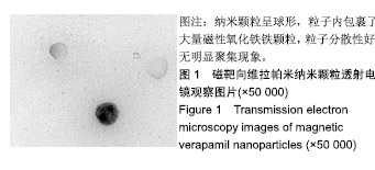
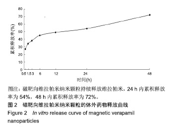
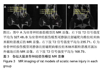
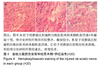
结果与结论:①磁靶向维拉帕米纳米颗粒呈球形,粒径为(208.3±0.8) nm,包封率为(70.21±3.25)%,载药量为 5.23%,具有良好的体外缓慢释放药物性能;②坐骨神经损伤模型大鼠右下肢T2信号强度平均为327.48,B组右下肢T2信号强度平均为235.71,A组右下肢T2信号强度平均为168.79;③A组神经传导速度高于 B、C 组(P < 0.05),B、C 组神经传导速度比较差异无显著性意义(P > 0.05);④苏木精-伊红染色显示,A 组吻合处神经纤维排列较整齐,瘢痕较少,B、C组吻合处神经纤维较紊乱、密度较多;⑤结果表明,磁靶向维拉帕米纳米颗粒可促进坐骨神经损伤的修复。

关键词: 磁靶向维拉帕米纳米颗粒, 维拉帕米, 聚乳酸-羟基乙酸共聚物, 纳米微粒, 外磁场, 磁靶向, 磁共振成像, 坐骨神经损伤, 神经修复

Abstract:

BACKGROUND: Verapamil has been shown to effectively inhibit fibrosis and reduce scar. But its application is relatively limited in the repair of nerve scar.
OBJECTIVE: To investigate the effect of magnetic verapamil nanoparticles in the repair of sciatic nerve injury in rats.
METHODS: Verapamil was used as the model drug and poly(lactic-co-glycolic acid) as drug carrier to prepare verapamil nanoparticles and its physicochemical properties were characterized. Forty-five Sprague-Dawley rats (bought from Tianjin Aochen Laboratory Animals Co., Ltd., China) of specific-pathogen-free grade were chosen to establish rat models of the right sciatic nerve injury. Then these models were randomly divided into three groups: rats in the groups A and B were injected with magnetic verapamil nanoparticles via the tail vein once a week and their right lower limbs were exposed to the magnetic field for 2 hours. Rats in the group B received injection of magnetic verapamil nanoparticles via the tail vein once a week without exposure to the outside magnetic field. Rats in the group C received tail vein injection of verapamil solution once a week. The amount of verapamil injected was the same in the three groups. After 8 weeks of drug injection, electrophysiological examination of the proximal and distal sciatic nerve trunks was performed to measure the compound muscle action potential, and hematoxylin-eosin staining was also performed to observe right sciatic nerve regeneration and scar formation. This study was approved by the Medical Ethics Committee of Affiliated Yantai Hospital of Binzhou Medical University (approval No. F-KY-0022-20161201-01).
RESULTS AND CONCLUSION: Magnetic verapamil nanoparticles were spherical with the mean diameter of 208.3±0.8 nm, encapsulation efficiency of (70.21±3.25)%, and drug loading rate of 5.23%, respectively. They exhibited good release effect in vitro. MRI showed that the mean T2 value of the right lower limbs of rat models of sciatic nerve injury was 327.48, and it was 235.71 in the group B and 168.79 in the group A. The nerve conduction velocity in the group A was significantly greater than that in the groups B and C (P < 0.05), and there was no significant difference in nerve conduction velocity between groups B and C (P > 0.05). Hematoxylin-eosin staining revealed that in the group A, the nerve fibers in the anastomosis site were arranged neatly and there were few scars. In the groups B and C, the nerve fibers in the anastomosis site were poorly arranged and there were many scars. These results suggest that magnetic verapamil nanoparticles can promote the recovery of rat sciatic nerve injury.

Key words: magnetic verapamil nanoparticles, verapamil, poly(lactic-co-glycolic acid), nanoparticle, outside magnetic field, magnetic targeting, magnetic resonance imaging, sciatic nerve injury, nerve repair

中图分类号: