| [1]陆洋, 董青.下颌第1乳磨牙近中根管分析[J]. 牙体牙髓牙周病学杂志,2016,26(11):682-684.[2]Ozcan G,Sekerci AE,Kocoglu F.C-shaped mandibular primary first molar diagnosed with cone beam computed tomography: A novel case report and literature review of primary molars' root canal systems. J Indian Soc Pedod Prev Dent. 2016;34(4):397-404.[3]Yue W,Kim E.Nonsurgical Endodontic Management of a Molar-Incisor Malformation-affected Mandibular First Molar: A Case Report. J Endod. 2016;42(4):664-668.[4]D'Souza KM,Aras MA.Three-dimensional finite element analysis of the stress distribution pattern in a mandibular first molar tooth restored with five different restorative materials. J Indian Prosthodont Soc. 2017;17(1):53-60.[5]Albatayneh OB,Shaweesh AI,Alsoreeky ES.Timing and sequence of emergence of deciduous teeth in Jordanian children. Arch Oral Biol. 2015;60(1):126-133.[6]Asgary S,Nikneshan S,Akbarzadehbagheban A,et al.Evaluation of diagnostic accuracy and dimensional measurements by using CBCT in mandibular first molars. J Clin Exp Dent. 2016;8(1):e1-8 [7]Mokhtari H,Niknami M,Zonouzi HR,et al.Accuracy of Cone-Beam Computed Tomography in Determining the Root Canal Morphology of Mandibular First Molars. Iran Endod J. 2016 ;11(2): 101-105. [8]Kamburo?lu K,Sönmez G,Berkta? ZS,et al.Effects of various cone-beam computed tomography settings on the detection of recurrent caries under restorations in extracted primary teeth. Imaging Sci Dent. 2017;47(2):109-115. [9]Kashyap RR,Beedubail SP,Kini R,et al.Assessment of the number of root canals in the maxillary and mandibular molars: A radiographic study using cone beam computed tomography.J Conserv Dent. 2017;20(5):288-291.[10]Mahesh BS, P Shastry S, S Murthy P, et al.Role of Cone Beam Computed Tomography in Evaluation of Radicular Cyst mimicking Dentigerous Cyst in a 7-year-old Child: A Case Report and Literature Review. Int J Clin Pediatr Dent. 2017;10(2):213-216.[11]Dhingra A,Manchanda N.Modifications in Canal Anatomy of Curved Canals of Mandibular First Molars by two Glide Path Instruments using CBCT. J Clin Diagn Res. 2014;8(11):ZC13-7. [12]张贤华.CBCT在重度牙周病诊断和辅助制定治疗计划的应用[J].中华老年口腔医学杂志,2014 ,12(1):4325-4327.[13]朱敏,林梓桐,文珊辉,等.CBCT根管形态三维容积重建可视化技术的研究[J].口腔医学研究,2015,35(6):601-603.[14]Madani ZS, Mehraban N,Moudi E,et al.Root and Canal Morphology of Mandibular Molars in a Selected Iranian Population Using Cone-Beam Computed Tomography. Iran Endod J. 2017; 12(2):143-148.[15]王琨,刘波,冯汝舟,等.乳磨牙根管系统的研究进展[J]. 医学综述, 2018,24(1):112-116.[16]张丹,陈俊宏,兰贵华,等.中国人下颌第一前磨牙牙根及根管形态的研究[J].第三军医大学学报,2016,38(10):1188-1194.[17]Ni N,Cao S,Han L,et al.Cone-beam computed tomography analysis of root canal morphology in mandibular first molars in a Chinese population: a clinical study. Evidence-Based Endodontics. 2018,3(1):1-4[18]刘钦捷,罗明,梁衍平,等.下颌第一前磨牙3牙根3根管临床报告及文献回顾[J].口腔疾病防治,2017, 25(10):656-660.[19]Self CJ.Tooth Roots and the Periodontal Ligament: Morphology, Modeling and Behavior.2015; 243(24):56-58.[20]Baziar H,Daneshvar F,Mohammadi A,et al.Endodontic management of a mandibular first molar with four canals in a distal root by using cone-beam computed tomography: a case report. J Oral Maxillofac Res. 2014;5(1):e5.[21]Harlamb S.Management of incompletely developed teeth requiring root canal treatment. Aust Dent J. 2016;61 Suppl 1:95-106.[22]Madero-Ayora MJ,Reina-Tosina J,Crespo-Cadenas C.Characterization of mandibular molar root and canal morphology using cone beam computed tomography and its variability in Belgian and Chilean population samples. Imaging Sci Dent. 2015;45(2):95-101.[23]Nur BG,Ok E,Altunsoy M,et al.Evaluation of the root and canal morphology of mandibular permanent molars in a south-eastern Turkish population using cone-beam computed tomography.Eur J Dent. 2014;8(2):154-159. |
.jpg) 文题释义:
乳磨牙的根管系统:与恒牙相比更为复杂,侧支根管多而乱;髓底多见副根管;根管分布存在变异,形态复杂。了解乳磨牙根管系统的组成有助于指导临床医师在对乳磨牙行根管治疗时避免遗漏根管,提高根管治疗的成功率。
根管治疗(root canal therapy):是牙髓病和根尖周病的国际上最常用的有效治疗方法。根管治疗术的原理是通过机械和化学方法去除根管内的大部分感染物,并通过充填根管、封闭冠部,防止发生根尖周病变或促进已经发生的根尖周病变的愈合。
文题释义:
乳磨牙的根管系统:与恒牙相比更为复杂,侧支根管多而乱;髓底多见副根管;根管分布存在变异,形态复杂。了解乳磨牙根管系统的组成有助于指导临床医师在对乳磨牙行根管治疗时避免遗漏根管,提高根管治疗的成功率。
根管治疗(root canal therapy):是牙髓病和根尖周病的国际上最常用的有效治疗方法。根管治疗术的原理是通过机械和化学方法去除根管内的大部分感染物,并通过充填根管、封闭冠部,防止发生根尖周病变或促进已经发生的根尖周病变的愈合。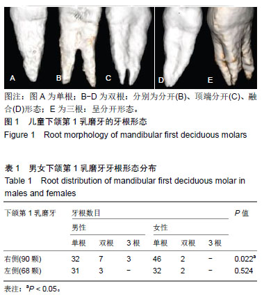