中国组织工程研究 ›› 2019, Vol. 23 ›› Issue (19): 2986-2990.doi: 10.3969/j.issn.2095-4344.1244
• 口腔组织构建 oral tissue construction • 上一篇 下一篇
锥形束CT评价成人安氏Ⅱ1类拔牙后上前牙内收前后牙槽骨变化
姜委杰1,2,刘 珺2,刘昌翠3,张 晨1,2
- (1青岛大学口腔医学院,山东省青岛市 266000;2青岛大学附属医院,山东省青岛市 266000;3泰安市口腔医院,山东省泰安市 271000)
Alveolar bone change of the maxillary anterior region after retraction in adult patients with class II1 malocclusion using cone-beam CT
Jiang Weijie1, 2, Liu Jun2, Liu Changcui3, Zhang Chen1, 2
- (1School of Stomatology, Qingdao University, Qingdao 266000, Shandong Province, China; 2Affiliated Hospital of Qingdao University, Qingdao 266000, Shandong Province, China; 3School of Stomatology of Taian, Taian 271000, Shandong Province, China)
摘要:
文章快速阅读:
.jpg) 文题释义:
牙槽骨:指上颌骨下缘、下颌骨的上缘镶嵌牙根的部位。上、下颌骨的牙槽突部分,由骨皮质、骨松质和固有牙槽骨构成,又称牙槽突,包围牙根部的颌骨突起。容纳牙齿的窝称为牙槽窝,两牙之间的牙槽骨称牙槽间隔,多根牙的根间牙槽骨称根间骨隔。牙槽骨结构和其他骨骼基本一致,有骨皮质和骨松质。其内壁称固有牙槽骨,如多孔的骨板,又叫筛状板。牙周膜的血管神经穿过筛状板的小孔与骨髓相连。为骨骼中变化最活跃的部分,随着牙齿的发育、乳牙的替换、恒牙的移动而改建的。
锥形束CT:计算机断层成像技术(CT)自诞生以来,CT扫描方式发生了巨大的变化,二维CT从单一探测器的平行束递增发展到多探测器扇形束旋转扫描;三维CT则从最早的单排螺旋CT发展为多排探测器扫描同时给出8-16层数据的多层螺旋CT,近几年使用面阵探测器的锥形束CT也逐渐进入实用阶段。相对于其他的几何结构,锥形束CT系统具有空间分辨率高、 数据采集时间短、射线利用效率高等显著优势。
文题释义:
牙槽骨:指上颌骨下缘、下颌骨的上缘镶嵌牙根的部位。上、下颌骨的牙槽突部分,由骨皮质、骨松质和固有牙槽骨构成,又称牙槽突,包围牙根部的颌骨突起。容纳牙齿的窝称为牙槽窝,两牙之间的牙槽骨称牙槽间隔,多根牙的根间牙槽骨称根间骨隔。牙槽骨结构和其他骨骼基本一致,有骨皮质和骨松质。其内壁称固有牙槽骨,如多孔的骨板,又叫筛状板。牙周膜的血管神经穿过筛状板的小孔与骨髓相连。为骨骼中变化最活跃的部分,随着牙齿的发育、乳牙的替换、恒牙的移动而改建的。
锥形束CT:计算机断层成像技术(CT)自诞生以来,CT扫描方式发生了巨大的变化,二维CT从单一探测器的平行束递增发展到多探测器扇形束旋转扫描;三维CT则从最早的单排螺旋CT发展为多排探测器扫描同时给出8-16层数据的多层螺旋CT,近几年使用面阵探测器的锥形束CT也逐渐进入实用阶段。相对于其他的几何结构,锥形束CT系统具有空间分辨率高、 数据采集时间短、射线利用效率高等显著优势。摘要
背景:安氏Ⅱ类错患者临床上常采用拔牙矫治的方案以获得正常的覆、覆盖,改善患者面型及侧貌,此类型患者拔牙后矫治效果与前牙内收量及相应牙槽骨改建量有直接关系。
目的:观察安氏Ⅱ1类成人患者上前牙区内收前后牙槽骨变化。
方法:选取30例安氏Ⅱ1类成年拔牙患者,双颌排齐整平更换不锈钢方丝后稳定1个月(T0期),采用Ⅱ类牵引滑动内收上前牙,调整前牙覆、覆盖至正常范畴后固定1个月(T1期)。在T0期、T1期拍摄锥形束CT,对上前牙内收量、牙槽骨厚度及高度测量分析。
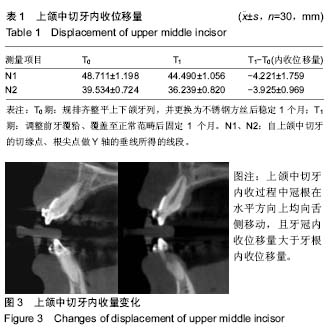
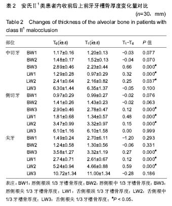
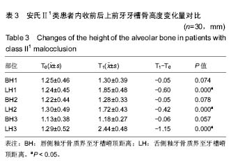
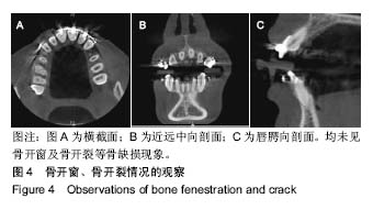
结果与结论:①内收后,上中切牙冠根均舌向移动,内收量牙冠大于牙根;②除根尖区外,前牙区牙槽骨厚度在唇侧增大,腭侧减小(P < 0.05),根尖区变化相反;③唇、舌侧牙槽嵴根向降低,腭侧差异有显著性(P < 0.05);④结果说明,成人安氏Ⅱ1类拔牙患者内收过程中上前牙做倾斜移动,内收时上前牙牙槽骨改建与牙齿移动并非一致,唇腭侧均发生活跃的骨改建,但总体表现为骨吸收大于骨增生。
中国组织工程研究杂志出版内容重点:组织构建;骨细胞;软骨细胞;细胞培养;成纤维细胞;血管内皮细胞;骨质疏松;组织工程
ORCID: 0000-0002-1808-9467(姜委杰)
中图分类号:
.jpg)
.jpg)
.jpg) 文题释义:
牙槽骨:指上颌骨下缘、下颌骨的上缘镶嵌牙根的部位。上、下颌骨的牙槽突部分,由骨皮质、骨松质和固有牙槽骨构成,又称牙槽突,包围牙根部的颌骨突起。容纳牙齿的窝称为牙槽窝,两牙之间的牙槽骨称牙槽间隔,多根牙的根间牙槽骨称根间骨隔。牙槽骨结构和其他骨骼基本一致,有骨皮质和骨松质。其内壁称固有牙槽骨,如多孔的骨板,又叫筛状板。牙周膜的血管神经穿过筛状板的小孔与骨髓相连。为骨骼中变化最活跃的部分,随着牙齿的发育、乳牙的替换、恒牙的移动而改建的。
锥形束CT:计算机断层成像技术(CT)自诞生以来,CT扫描方式发生了巨大的变化,二维CT从单一探测器的平行束递增发展到多探测器扇形束旋转扫描;三维CT则从最早的单排螺旋CT发展为多排探测器扫描同时给出8-16层数据的多层螺旋CT,近几年使用面阵探测器的锥形束CT也逐渐进入实用阶段。相对于其他的几何结构,锥形束CT系统具有空间分辨率高、 数据采集时间短、射线利用效率高等显著优势。
文题释义:
牙槽骨:指上颌骨下缘、下颌骨的上缘镶嵌牙根的部位。上、下颌骨的牙槽突部分,由骨皮质、骨松质和固有牙槽骨构成,又称牙槽突,包围牙根部的颌骨突起。容纳牙齿的窝称为牙槽窝,两牙之间的牙槽骨称牙槽间隔,多根牙的根间牙槽骨称根间骨隔。牙槽骨结构和其他骨骼基本一致,有骨皮质和骨松质。其内壁称固有牙槽骨,如多孔的骨板,又叫筛状板。牙周膜的血管神经穿过筛状板的小孔与骨髓相连。为骨骼中变化最活跃的部分,随着牙齿的发育、乳牙的替换、恒牙的移动而改建的。
锥形束CT:计算机断层成像技术(CT)自诞生以来,CT扫描方式发生了巨大的变化,二维CT从单一探测器的平行束递增发展到多探测器扇形束旋转扫描;三维CT则从最早的单排螺旋CT发展为多排探测器扫描同时给出8-16层数据的多层螺旋CT,近几年使用面阵探测器的锥形束CT也逐渐进入实用阶段。相对于其他的几何结构,锥形束CT系统具有空间分辨率高、 数据采集时间短、射线利用效率高等显著优势。